Embed Size (px)
Citation preview

Cardiovascular System = Heart, blood vessels, and blood
-Transports H2O, nutrients, waste, O2, CO2, hormones, and immune cells.
-Regulates heat, pH, and pressure.
-Associated with lymphatic system. Lymphatic System = Lymphatic
vessels, lymphatic hearts, & lymph
Circulatory System
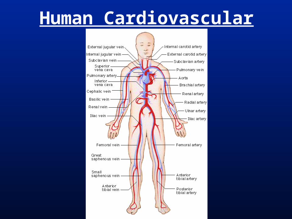
Human Cardiovascular

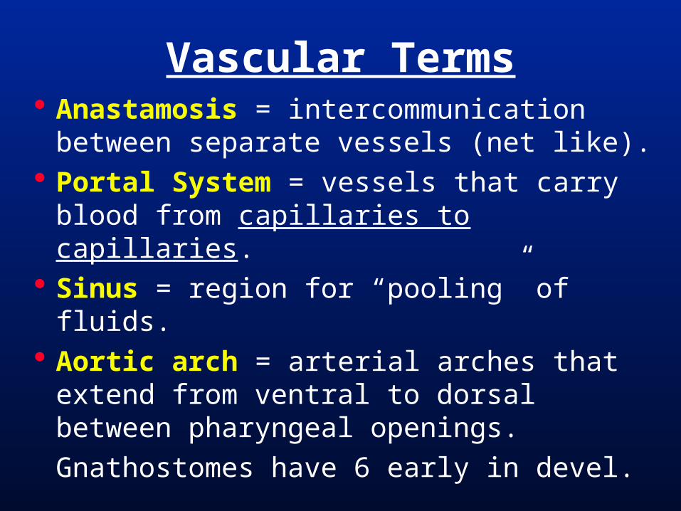
Anastamosis = intercommunication between separate vessels (net like).
Portal System = vessels that carry blood from capillaries to capillaries.
Sinus = region for “pooling” of fluids. Aortic arch = arterial arches that
extend from ventral to dorsal between pharyngeal openings.
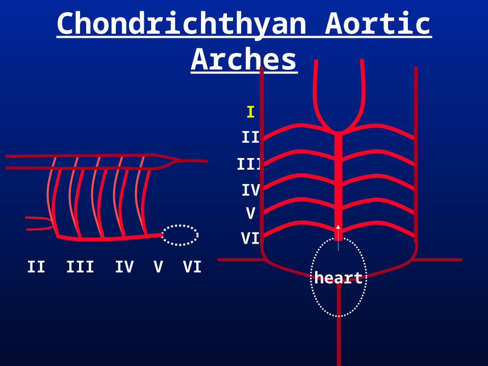
Gnathostomes have 6 early in devel.
Vascular Terms

Arteries = Large; carry blood away from heart
Arterioles = Smaller; carry blood away from heart
Capillaries = Very small; gas & nutrient exchange occur here
Veinules = Smaller; carry blood to heart
Veins = Large; carry blood to heart
Vascular

Blood = connective tissue; cells (in vertebrates) in a water-based matrix.
Plasma = matrix around blood cells. Transports: H2O, nutrients, waste, CO2, hormones
Erythrocytes = Contain hemoglobin; transport O2; No nuclei in mammals.
Leucocytes = Immune cells. Platelets = Cell fragments, involved in
clotting after damage.
Vertebrate Blood

Plasma
Vertebrate Blood
Hematopoesis – Blood Devel.
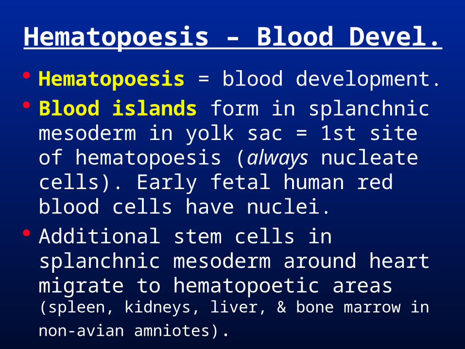
Hematopoesis = blood development. Blood islands form in splanchnic
mesoderm in yolk sac = 1st site of hematopoesis (always nucleate cells). Early fetal human red blood cells have nuclei.
Additional stem cells in splanchnic mesoderm around heart migrate to hematopoetic areas (spleen, kidneys, liver,
& bone marrow in non-avian amniotes).
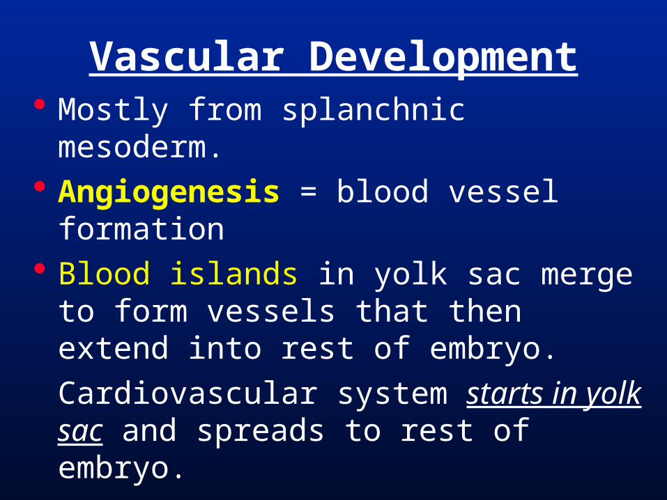
Mostly from splanchnic mesoderm. Angiogenesis = blood vessel
formation Blood islands in yolk sac merge to form
vessels that then extend into rest of embryo.
Cardiovascular system starts in yolk sac and spreads to rest of embryo.
Vascular Development

Human Blood Islands

Basic Chordate Arterial Circ. Blood moves from posterior and
ventral to the pharynx (heart/sinus venosus)
Anteriorly under the pharynx (ventral aorta)
Dorsolaterally up through the pharynx (pharyngeal/aortic arches)
Then to the body anteriorly and posteriorly from the dorsal pharynx (dorsal aorta).

Lancelet Arterial Circulation
ventral aorta (median)
sinus venosus (median)
dorsal aorta (paired - median)

Lancelet Venous Circulation
hepatic vein (median)
Anterior & posterior cardinal veins (paired)
sinus venosus (median)
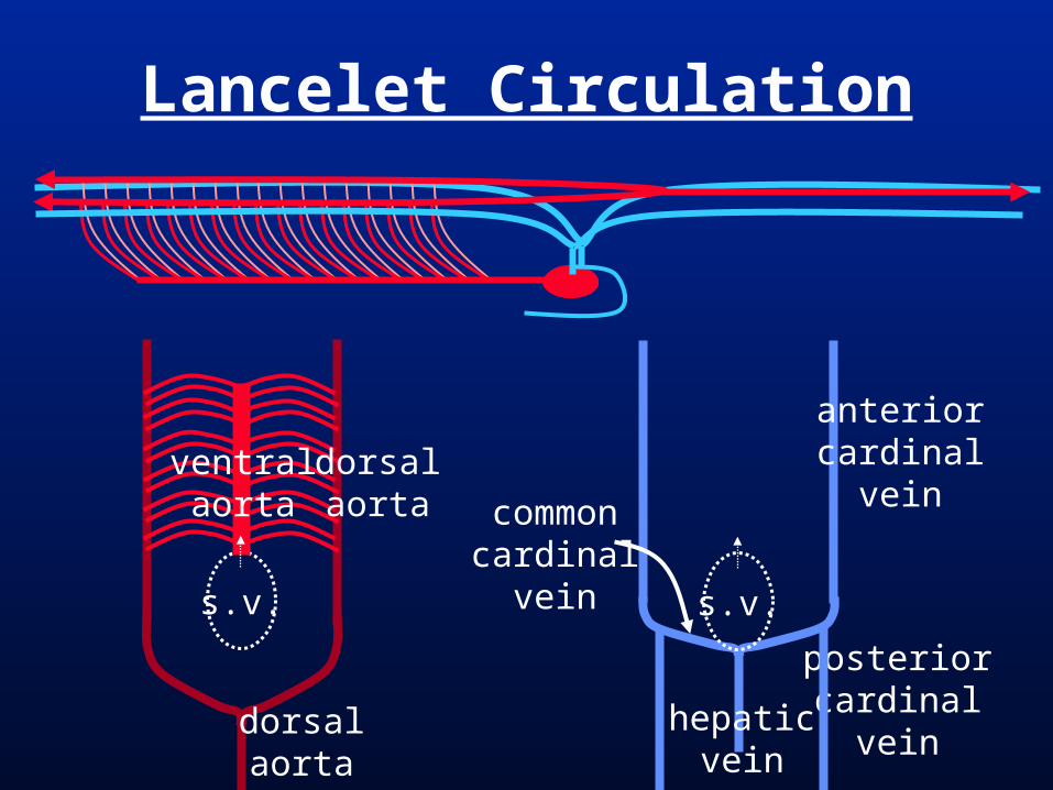
Lancelet Circulation
s.v.
dorsalaorta
dorsalaorta
ventralaorta
s.v.
anteriorcardinal
vein
posteriorcardinal
veinhepatic
vein
commoncardinal
vein

Basic Amniote Circulation
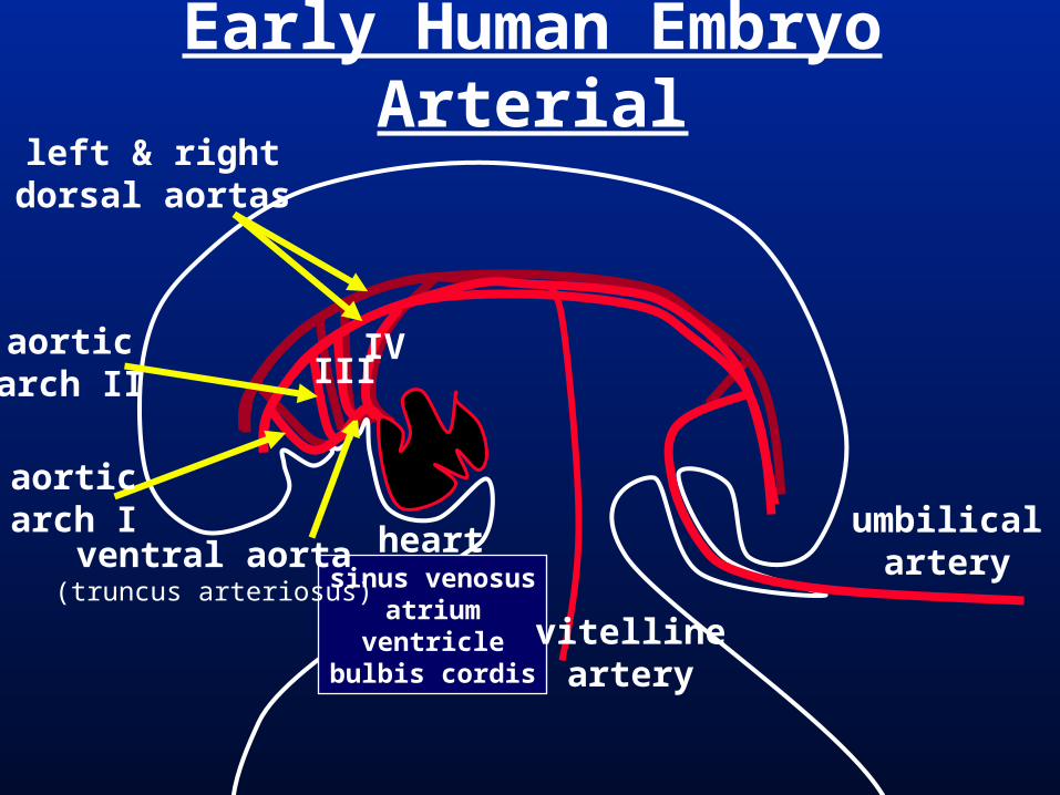
Early Human Embryo Arterial
heartsinus venosus
atriumventricle
bulbis cordis
umbilicalarteryventral aorta
(truncus arteriosus)
left & rightdorsal aortas
aorticarch II
aorticarch I
IIIIV
vitellineartery

Early Human Embryo Arterial
heartumbilical
arteryventral aorta(truncus arteriosus)
left & rightdorsal aortas
IIIIV VI
vitellineartery
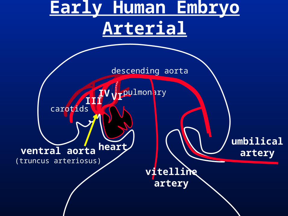
Early Human Embryo Arterial
heartumbilical
arteryventral aorta(truncus arteriosus)
IIIIV VI
carotids
pulmonary
descending aorta
vitellineartery

Early Human Embryo Arterial

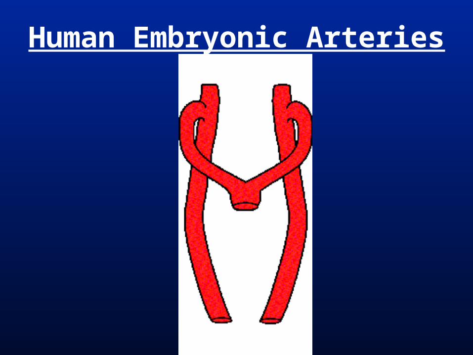
Embryonic Arteries
III
IIIIV
VI
I II III IV V VI
V
ventral aorta
dorsal aorta
HEART

Human Embryonic Arteries
III
IIIIV
VI
HEART
external carotid
internal carotidcommon carotid
descending aorta
aortic arch
subclavian
pulmonary artery ductus arteriosus(before birth)
Human Embryonic Arteries

Human Aortic Arches
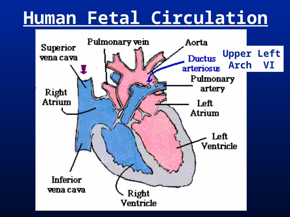
Human Fetal Circulation
Upper LeftArch VI

Early Human Embryo Circulation
heartsinus venosus
atriumventricle
bulbis cordis
ventral aorta(truncus arteriosus)
allantoic (umbilical) veins
vitelline(hepatic)
veins
left & right anterior cardinal veins
left & right posterior cardinal veins
left & right common cardinal
veins

Early Human Embryo Circulation
heart
allantoic (umbilical) veins
vitelline(hepatic)
veins
left & right anterior cardinal veins
left & right posterior cardinal veins
azygous vein
jugular veins
superiorvenacava
inferiorvenacava
hepaticportalveinhepatic
vein
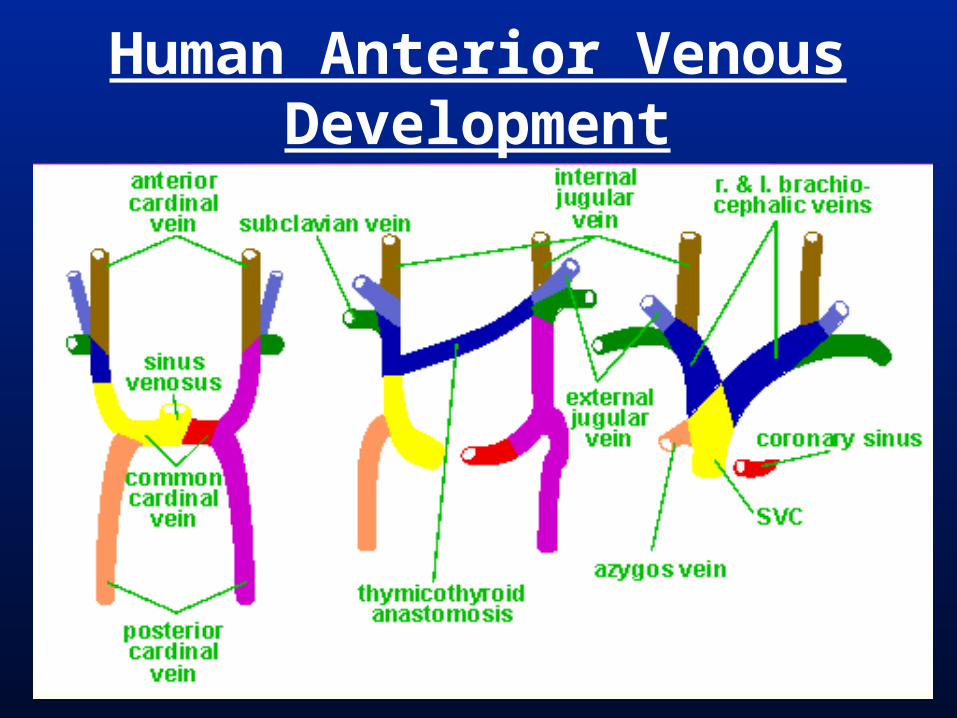
Human Anterior Venous Development

Basic Vertebrate Circulation

Endothelium = simple squamous epithelium lining inside of blood vessels (tunica intima)
Tunica externa = fibrous connective tissue surrounding the outside of the vessel
Tunica media = between the tunica intima and tunica externa; elastic connective tissue and/or smooth muscle.
Blood Vessel Structure
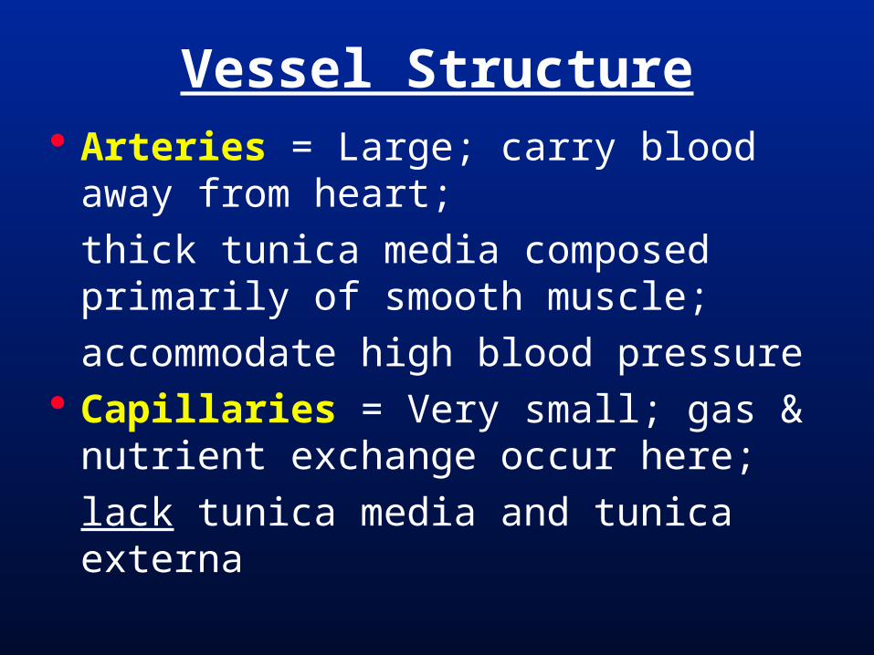
Arteries = Large; carry blood away from heart;
thick tunica media composed primarily of smooth muscle;
accommodate high blood pressure Capillaries = Very small; gas &
nutrient exchange occur here;
lack tunica media and tunica externa
Vessel Structure
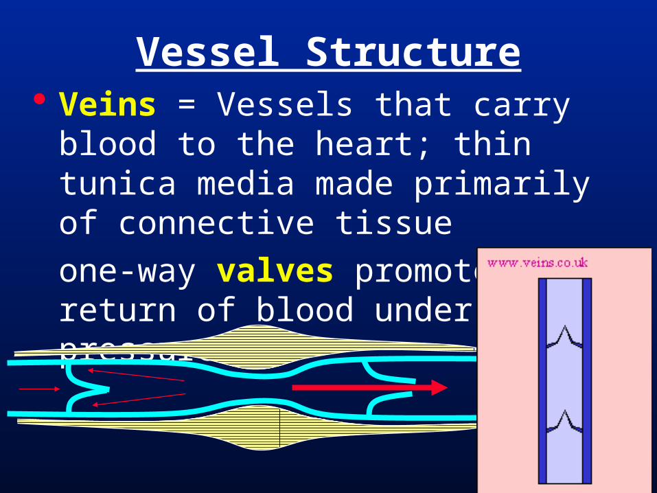
Veins = Vessels that carry blood to the heart; thin tunica media made primarily of connective tissue
one-way valves promote the return of blood under low pressure
Vessel Structure
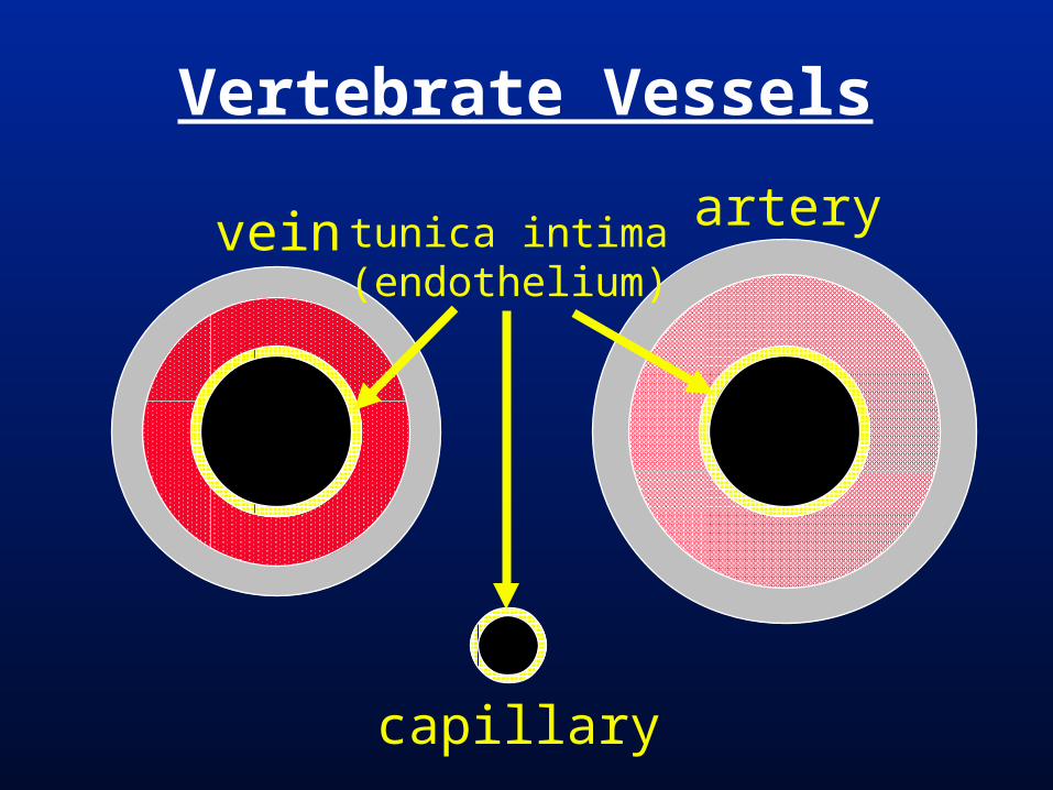
Vertebrate Vessels
vein arterytunica intima(endothelium)
capillary

Adult Human Arterial system

Adult Human Arterial system
deep brachial
subclavianaxillary
brachial
radialulnar
internal thoracichumeral circumflex
lateral thoracic
common iliacexternal iliacinternal iliac
deep femoralfemoral circumflex
femoral
poplitealanterior tibialposterior tibial
external carotidinternal carotid
common carotidvertebral
aorta*coeliac*
superior mesenteric*
inferior mesenteric*
renalgonadal
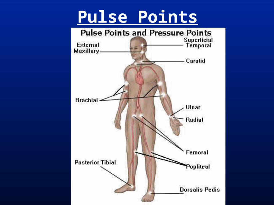
Pulse Points

Carotid & Radial Pulse Points

Human Venous System
Azygous vein
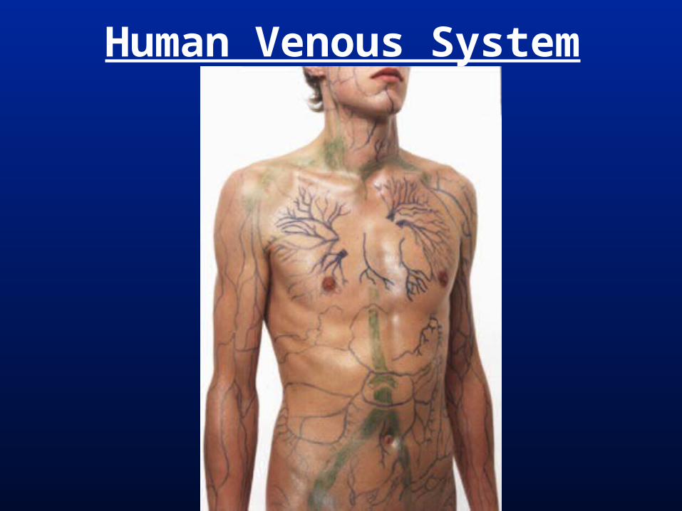
Human Venous System

Human Arm Venous System
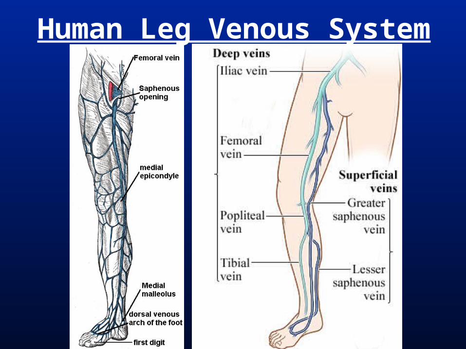
Human Leg Venous System
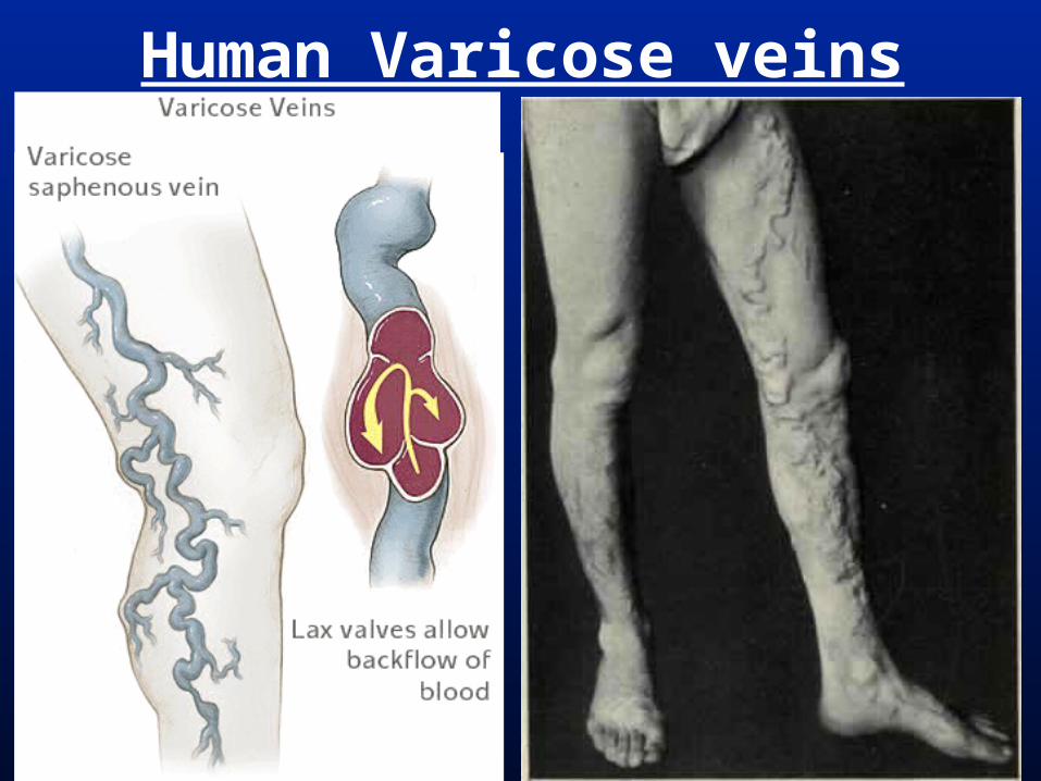
Human Varicose veins


Hematopoesis In non-amniotes occurs primarily in
the spleen, kidneys, and liver.
Especially the spleen. Hagfishes, lampreys, & lungfishes =
no spleen In amniotes occurs in bone marrow
(especially in humans and other mammals) as well as in other tissues. No bone marrow in birds.

Subphylum Vertebrata
hagfis
hes
lam
preys
Chondrichth
ys
Actin
optery
gii
coel
acan
th
lungfis
h
amphib
ians
Mam
mal
ia
Reptil
ia
hemato-poesis inmarrow
blood cells *erythrocytes lack nuclei
*
discrete spleen
sple
enlo
st

Lancelet Circulation

Vertebrate Circulation
heart
ventralaorta
externalcarotid
dorsalaorta
Iliacartery
subclavianartery

Vertebrate Circulationanterior & posterior
cardinal veins renal portal
iliacvein
lateralabdominal
veinsubclavian
vein
heart hepaticportal
jugulars

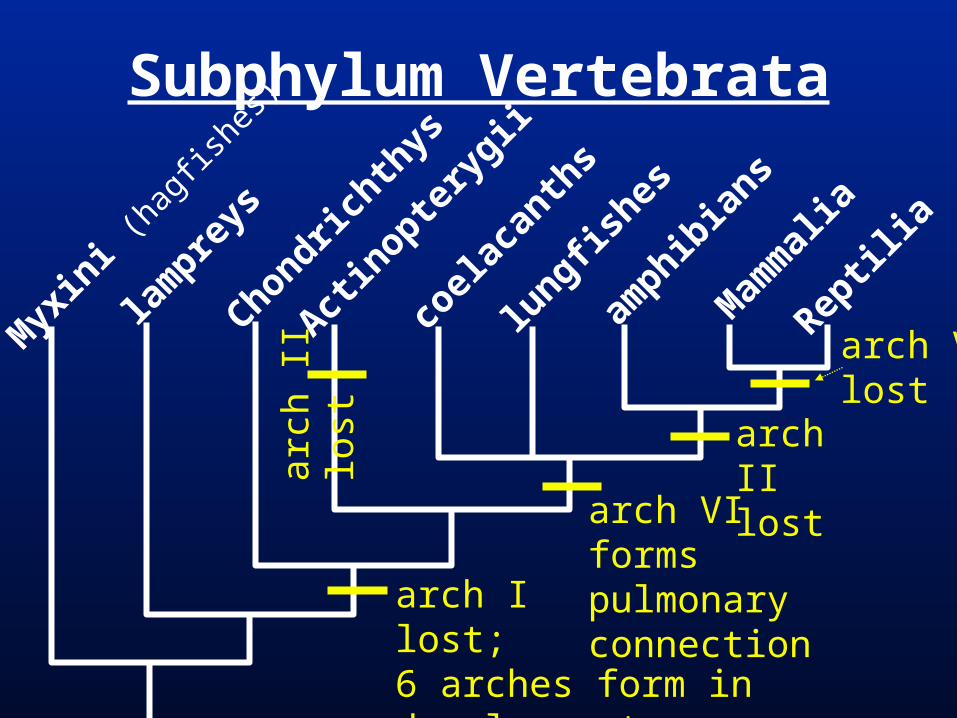
Gnathostomes = Arch I lost in adults; 6 arches early in development
Sarcopterygiians = Pulmonary arteries from arch VI
Tetrapods & Actinopts. = Arch II lost Amniotes = Arch V lost Mammals = left Arch IV forms aorta;
right Arch IV forms base of subclavian Birds = right Arch IV forms aorta
Aortic Arches

Gnathostome Embryo Aortic Arches
heart
I
II
III
IV
V
VI
internalcarotid
externalcarotid
dorsalaorta
ventralaorta
dorsalaorta
I II III IV V VI
heart
Chondrichthyan Aortic Arches
I
II
III
IV
V
VI
I II III IV V VI
heart
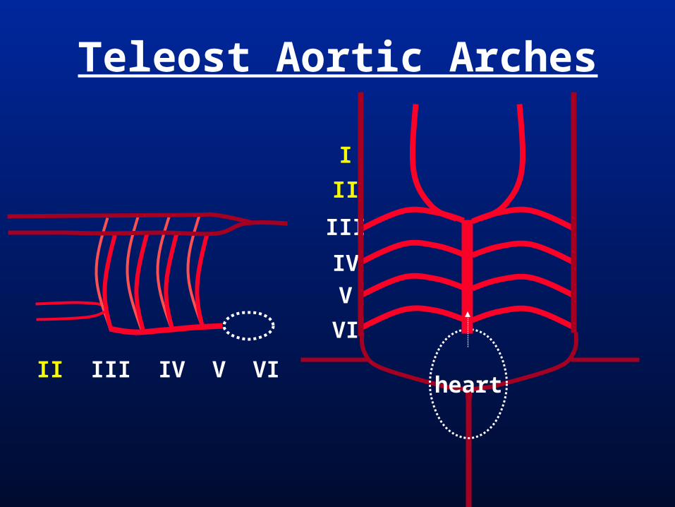
Teleost Aortic Arches
I
II
III
IV
V
VI
I II III IV V VI

Lungfish Aortic Arches
pulmonaryartery
heart
I
II
III
IV
V
VI
I II III IV V VI
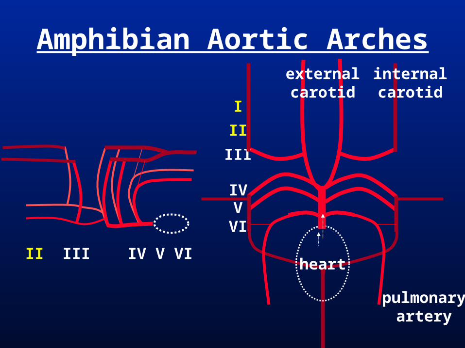
Amphibian Aortic Arches
heart
I
II
III
IVVVI
internalcarotid
externalcarotid
pulmonaryartery
I II III IV V VI
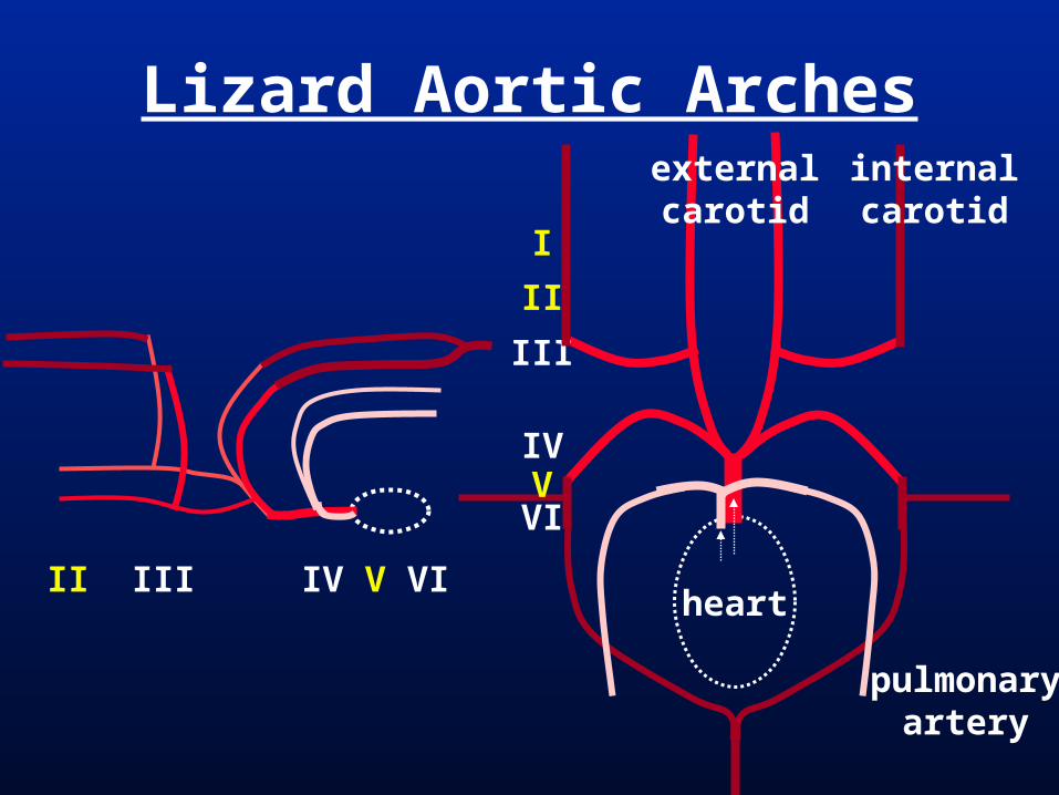
Lizard Aortic Arches
heart
I
II
III
IVVVI
internalcarotid
externalcarotid
pulmonaryartery
I II III IV V VI
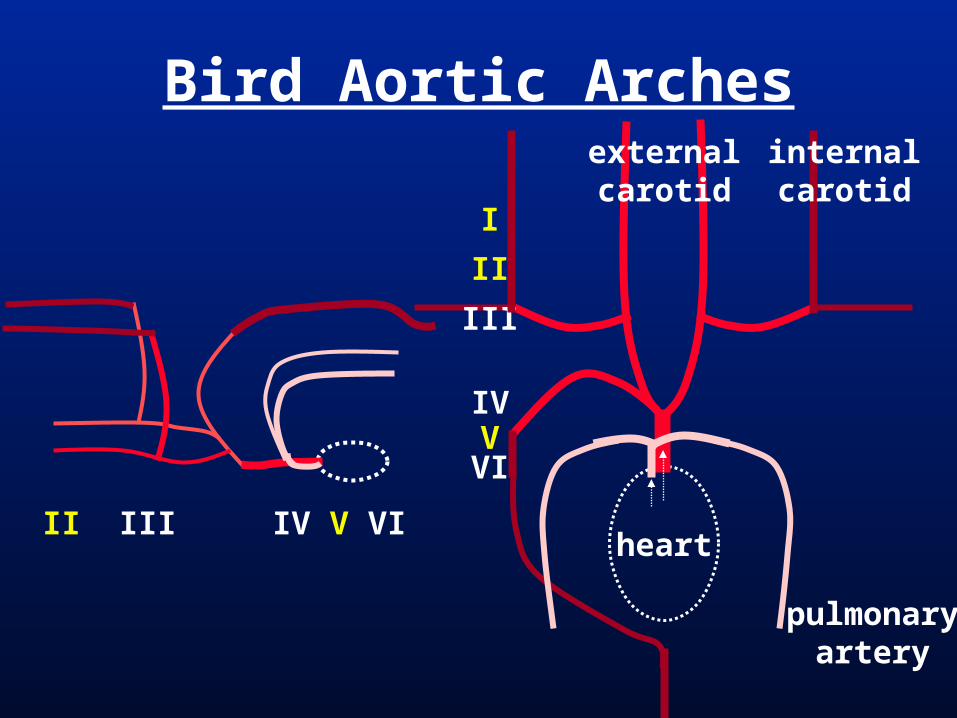
Bird Aortic Arches
heart
I
II
III
IVVVI
internalcarotid
externalcarotid
pulmonaryartery
I II III IV V VI
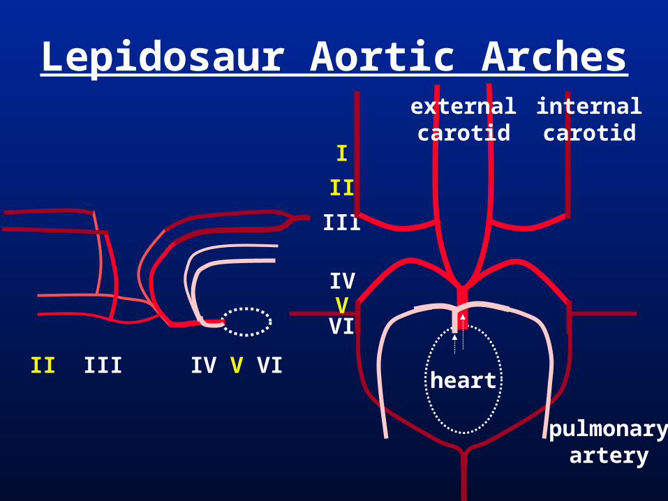
Lepidosaur Aortic Arches
heart
I
II
III
IVVVI
internalcarotid
externalcarotid
pulmonaryartery
I II III IV V VI
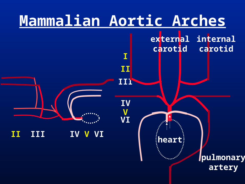
Mammalian Aortic Arches
heart
I
II
III
IVVVI
internalcarotid
externalcarotid
pulmonaryartery
I II III IV V VI
Subphylum Vertebrata
Myx
ini (
hagf
ishes
)
lam
preys
Chondrichth
ys
Actin
optery
gii
coel
acan
ths
lungfis
hes
amphib
ians
Mam
mal
ia
Reptil
ia
arch Vlost
arch VIforms pulmonaryconnection arch I
lost; 6 arches form in development
arch IIlost ar
ch II
lost
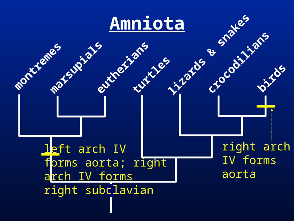
Amniota
montre
mes
mar
supia
ls
euth
eria
ns
turtl
es
lizar
ds & s
nakes
croco
dilian
s
birds
right archIV formsaorta
left arch IVforms aorta; rightarch IV formsright subclavian
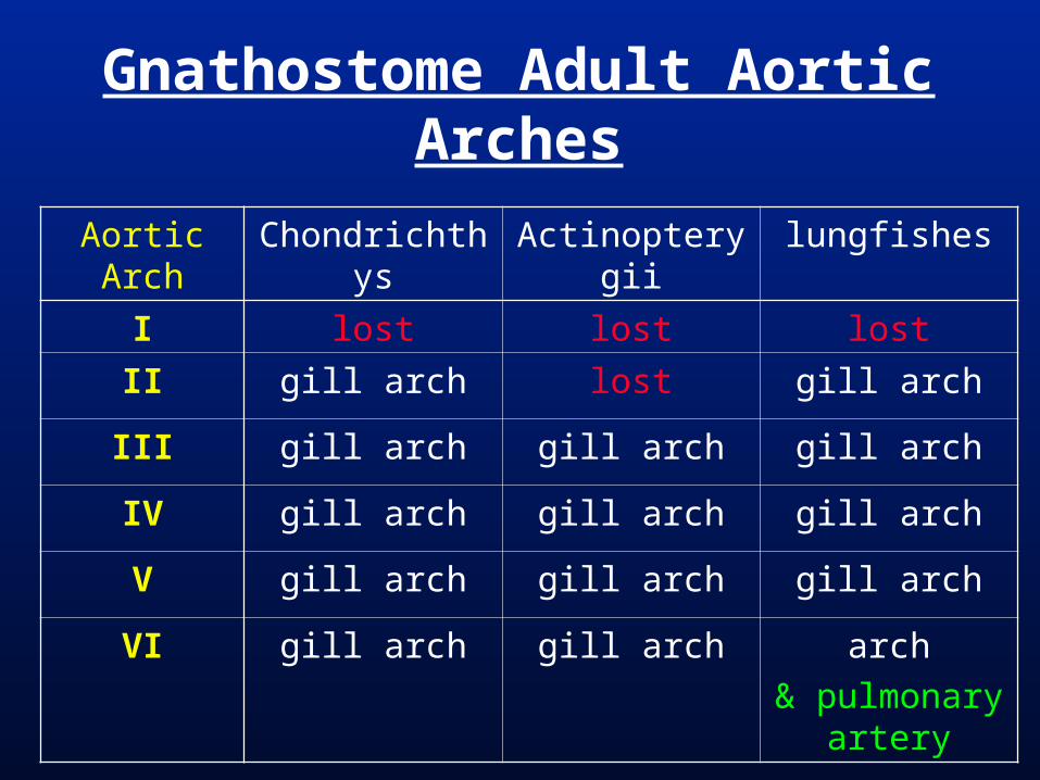
Gnathostome Adult Aortic Arches
Aortic Arch Chondrichthys Actinopterygii lungfishes
I lost lost lost
II gill arch lost gill arch
III gill arch gill arch gill arch
IV gill arch gill arch gill arch
V gill arch gill arch gill arch
VI gill arch gill arch arch
& pulmonary artery

Gnathostome Adult Aortic Arches
Aortic Arch amphibians turtles lizards
I lost lost lost
II lost lost lost
III common carotids
common carotids
common carotids
IV l. and r. aortic arches
l. and r. aortic arches
l. and r. aortic arches
V aortic arch(lost in frogs)
lost lost
VI aortic arch & pulmonary
artery
pulmonary artery
pulmonary artery

Gnathostome Adult Aortic Arches
Aortic Arch crocodilians birds Mammalia
I lost lost lost
II lost lost lost
III common carotids
com. carotids / subclavian
bases
common carotids & ext. carotid base
IV l. and r. aortic arches
l. lost
r. aorta
l. aorta
r. subclavian
V lost lost lost
VI pulmonary artery
pulmonary artery
pulmonary artery

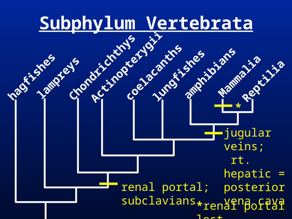
Gnathostomes = renal portal vein Tetrapods = inferior vena cava from
right hepatic; anterior cardinal veins form internal & external jugular veins
Mammals = inferior vena cava connects to & replaces renal portal
Some Mammals (humans, cats, etc.) = lose the left anterior cardinal vein (the right anterior cardinal vein = superior vena cava)
Venous System

Venous System
sinusvenosus
hepaticvein
hepaticportal
anteriorcardinal
posteriorcardinal
commoncardinal
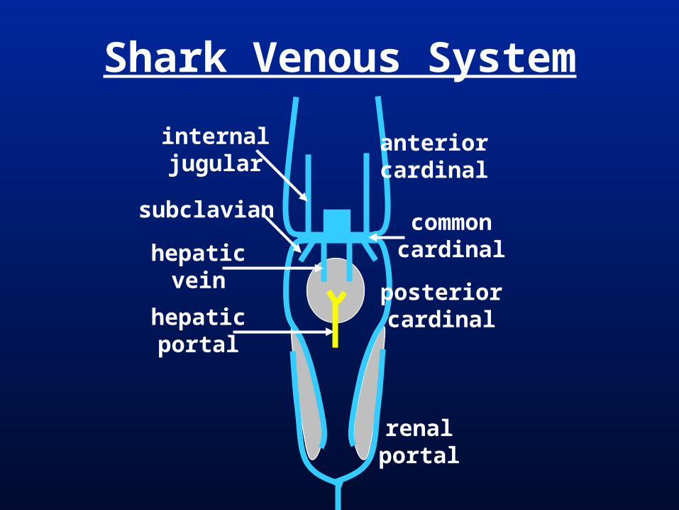
Shark Venous System
anteriorcardinal
posteriorcardinal
commoncardinal
renalportal
internaljugular
subclavian
hepaticvein
hepaticportal

posteriorcardinal
Amphibian Venous System
anteriorcardinalcommoncardinal
renalportal
hepaticvein
hepaticportal
subclavian
internaljugular
externaljugular
posteriorvena cava
iliac
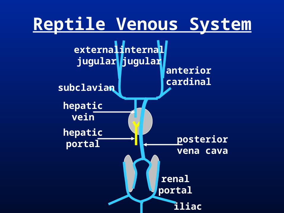
Reptile Venous System
anteriorcardinal
renalportal
hepaticvein
hepaticportal
internaljugular
externaljugular
posteriorvena cava
iliac
subclavian

Mammal Venous System
anteriorcardinal
hepaticvein
hepaticportal
internaljugular
externaljugular
posteriorvena cava
iliac
subclavian
renalvein
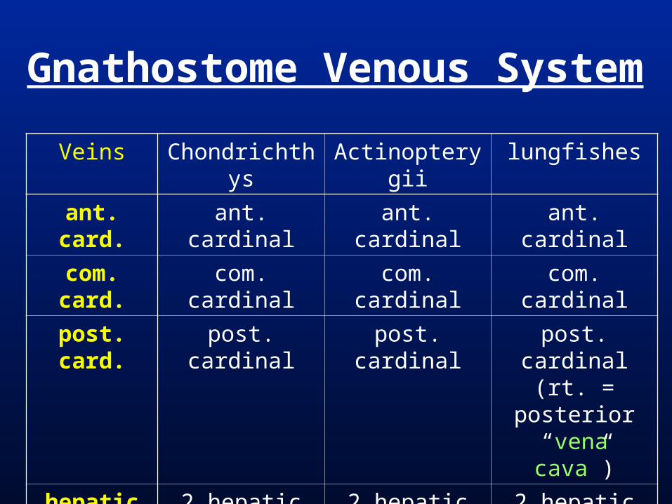
Gnathostome Venous System
Veins Chondrichthys Actinopterygii lungfishes
ant. card. ant. cardinal ant. cardinal ant. cardinal
com. card. com. cardinal com. cardinal com. cardinal
post. card. post. cardinal post. cardinal post. cardinal (rt. = posterior “vena cava”)
hepatic 2 hepatic veins 2 hepatic veins 2 hepatic veins
hep. portal hepatic portal hepatic portal hepatic portal
renal port. renal portal renal portal renal portal

Gnathostome Venous SystemVeins amphibians turtles Lepidosauria
ant. card. ant. cardinal (jugular vein &
brachiocephalics)
ant. cardinal (jugular vein &
brachiocephalics)
ant. cardinal (jugular vein &
brachiocephalics)
com. card. com. cardinal (s. vena cavas)
com. cardinal(s. vena cavas)
com. cardinal(s. vena cavas)
post. card. post. cardinal l. lostr. azygous vein
l. lostr. azygous vein
hepatic l. hepatic vein
r. p. vena cava
l. hepatic vein
r. p. vena cava
l. hepatic vein
r. p. vena cava
hep. portal hepatic portal hepatic portal hepatic portal
renal port. renal portal renal portal renal portal

Gnathostome Venous SystemVeins crocodilians birds Mammalia
ant. card. ant. cardinal (jugular vein &
brachiocephalics)
ant. cardinal (jugular vein &
brachiocephalics)
ant. cardinal (jugular vein &
brachiocephalics)
com. card. com. cardinal(s. vena cavas)
com. cardinal(s. vena cavas)
com. cardinal (s. vena cavas
-left s. v. c. lost in some)
post. card. l. lostr. azygous vein
l. lostr. azygous vein
l. lostr. azygous vein
hepatic l. hepatic vein
r. i. vena cava
l. hepatic vein
r. i. vena cava
l. hepatic vein
r. i. vena cava
hep. portal hepatic portal hepatic portal hepatic portal
renal port. renal portal renal portal lost

gut
Vein from yolk sac to heart (vitelline vein) impinged on by hepatic diverticulum and forms hepatic capillaries.
Hepatic Portal Vein
heart
vitelline vein (from yolk sac)
liver (with hepatic veins)
hepatic portal vein
Subphylum Vertebrata
hagfis
hes
lam
preys
Chondrichth
ys
Actin
optery
gii
coel
acan
ths
lungfis
hes
amphib
ians
Mam
mal
ia
Reptil
ia
renal portal; subclavians
jugular veins; rt. hepatic = posterior vena cava
*renal portal lost
*

Heart = Muscular pump, moves blood via rhythmic contraction.
Contraction involuntary & initiated within the heart. (not-neuronal)Modified branching (cardiac) muscle cells transmit contraction signal.
Chamber = Cavity for blood collection separated by valves
Valves = Flaps of tissue that prevent back-flow of fluid (in this case blood)
Heart

Human Heart Development Formed from splanchnic mesoderm. Endocardial tissue = forms simple
squamous epithelium that lines the heart chambers and forms the heart valves
Myocardial = epithelium forms heart muscle Hollow endocardial primordia form by
splanchnic mesoderm ingression, then fuse to form a the endocardium (endothelium).
Epithelial splanchnic mesoderm surrounds the endocardium forming the cardiac muscle of the myocardium.

Human Heart Development
endocardium
myocardium

Human Heart Development
Sinus Venosus
(ventral aorta)
Sinus venosus = slightly muscular chamber; receives blood from cardinal veins = sinoatrial node (“pacemaker”) in amniotes.
Sinoatrial valve = between s.v. & atrium.
Atrium = slightly muscular chamber.
Atrioventricular valve = between atrium & ventricle.
Ventricle = highly muscular chamber.
Conus arteriosus/ Bulbus arteriosus = chamber after ventricle (muscular with valves = conus; elastic, no valves = bulbus ; embryonic = bulbis cordis)
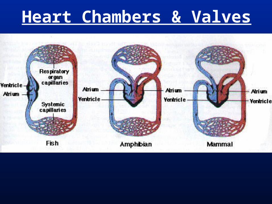
Heart Chambers & Valves

Human Heart Development

Sinus Venosus lost except some cells that become the sinoatrial node (“pacemaker”)
Sinoatrial valve lost. Atrium divides into right and left. Atrioventricular valve divides to form
(right) tricuspid and (left) bicuspid/mitral valves
Ventricle divides into right and left.
Human Heart Development
Bulbis cordis lost. Ventral aorta (truncus arteriosus)
divides to become
- (left) ascending aorta that will maintain connection to aortic arches III & IV and
- (right) pulmonary trunk artery that will maintain connection to aortic arch VI.
Human Heart Development
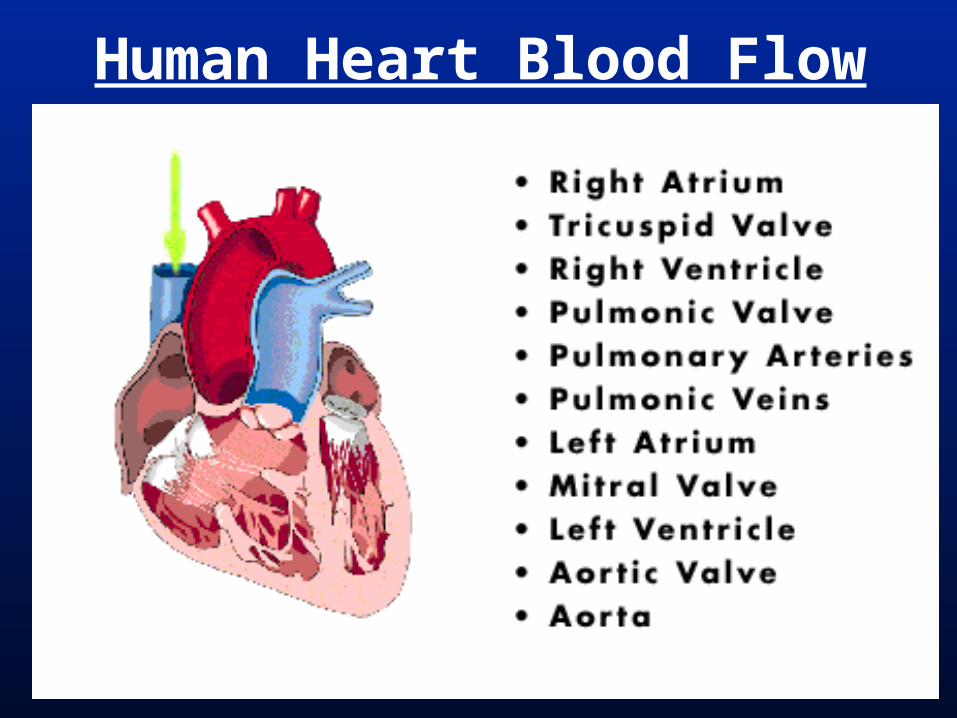
Human Heart Blood Flow
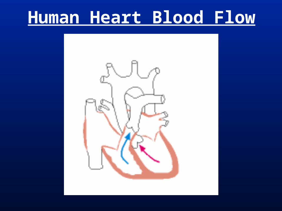
Human Heart Blood Flow
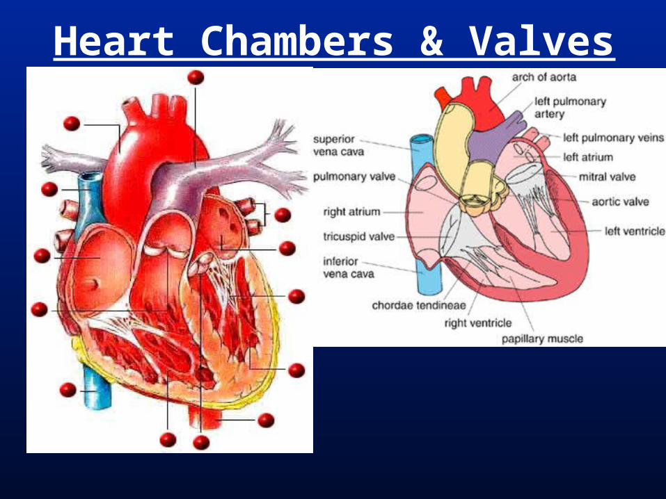
Heart Chambers & Valves

Heart Position/Stethoscope

Blood Flow From Heart
low oxygen
high oxygen

Heart Chambers & Valves
Teleost

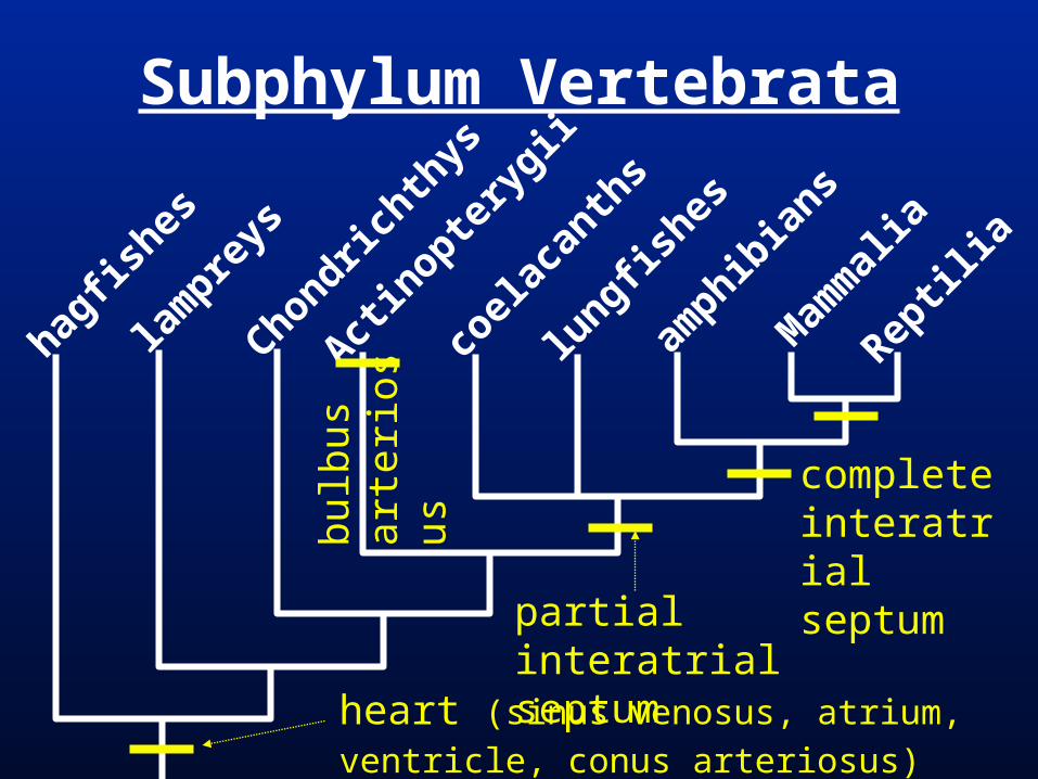
Sarcopterygiians = at least partially developed interatrial septum
Tetrapods = complete interatrial septum
Amniotes = conus arteriosus lost during devel; sinus venosus incorp. into right atrium; at least partially developed interventricular septum
Mammals & Archosaurs = complete interventricular septum
Heart Chambers
Heart Chambers & Valves

Heart Chambers & Valves
bulb
us
arte
riosu
s
Subphylum Vertebrata
hagfis
hes
lam
preys
Chondrichth
ys
Actin
optery
gii
coel
acan
ths
lungfis
hes
amphib
ians
Mam
mal
ia
Reptil
ia
partial interatrialseptum
heart (sinus venosus, atrium, ventricle,
conus arteriosus)
completeinteratrialseptum

Amniota
montre
mes
mar
supia
ls
euth
eria
ns
turtl
es
lizar
ds & s
nakes
croco
dilian
s
birds
conus arteriosus lost in adult;incomplete interventricular septum;sinus venosus lost (in rt. atrium)
completeinterventricularseptum
completeinterventricularseptum

Gnathostome Heart
Chambers Chondrichthys Actinopterygii lungfishes
Sinus Venosus
sinus venosus sinus venosus sinus venosus
Atrium atrium atrium atrium (partially l.-r. divided)
Ventricle ventricle venticle ventricle
Conus Arteriosus
conus arteriosus
bulbus arteriosus (not
muscular)
conus arteriosus

Gnathostome Heart
Chambers amphibians lizards turtles
Sinus Venosus
sinus venosus sinoatrial node (incorp. in rt. atrium)
sinoatrial node (incorp. in rt. atrium)
Atrium left atrium
right atrium
left atrium
right atrium
left atrium
right atrium
Ventricle ventricle ventricle (partial. left-right divided)
ventricle (partial. left-right divided)
Conus Arteriosus
conus arteriosus
lost lost

Gnathostome Heart
Chambers crocodilians birds Mammalia
Sinus Venosus
sinoatrial node (incorp. in rt. atrium)
sinoatrial node (incorp. in rt. atrium)
sinoatrial node (incorp. in rt. atrium)
Atrium left atrium
right atrium
left atrium
right atrium
left atrium
right atrium
Ventricle left ventricle
right ventricle(connected by
foramen of Panizza)
left ventricle
right ventricle
left ventricle
right ventricle
Conus Arteriosus
lost lost lost


Plasma in capillaries is forced into the tissues by blood pressure.
Edema = swelling of tissues due to accumulation of fluid (plasma/lymph).
Usually, edema is avoided because…
plasma in tissues diffuses into blind lymphatic capillaries and is called lymph.
Lymphatic System
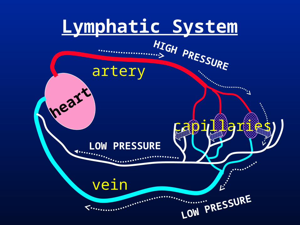
Lymphatic SystemHIGH PRESSURE
heart
artery
vein
capillaries
LOW PRESSURE
LOW PRESSURE

Lymphangitis = inflamation of a lymph vessel. (ROOT WORDS)
Lymphangitis of the vessel indicated by the arrow caused the edema in the middle finger.
Lymphatic System

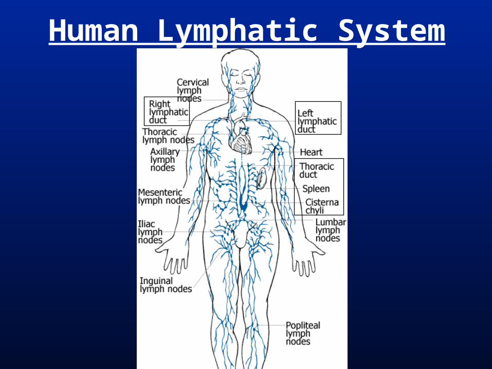
Lymph vessels = thin walled vessels with valves (similar in structure to veins, but with weak contractile ability); return lymph to venous system.
Lymph nodes (lymphatic cisterns) = expanded lymph vessels filled with connective tissue and leukocytes. Immunological filters.
Lacteals = lymph vessels in villi of the jejuno-ileum (small intestine) absorb fats.
Lymphatic System

Lymph Vessel

Lymph Node & Lg. Vessel(in mesentary proper)

Human Cephalic Lymph System

Human Lymphatic System
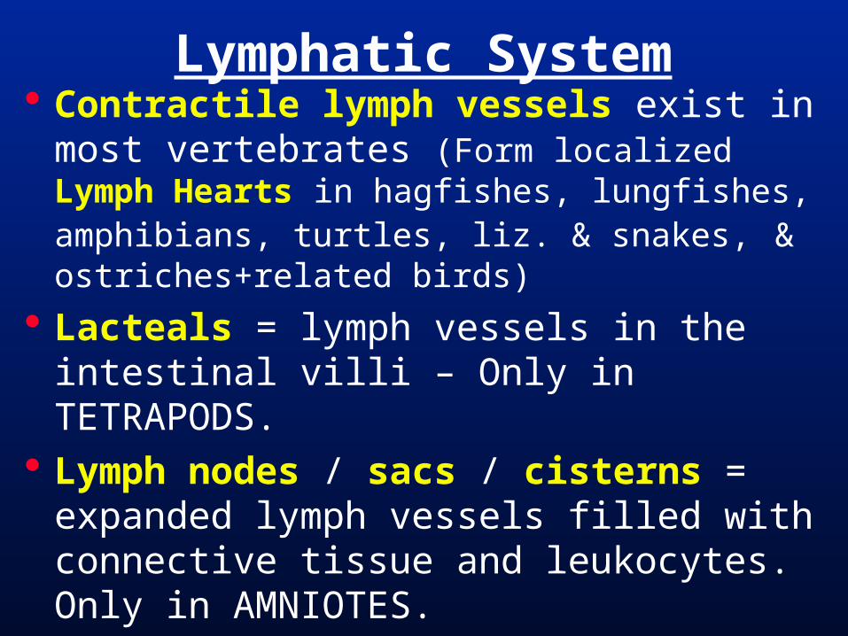
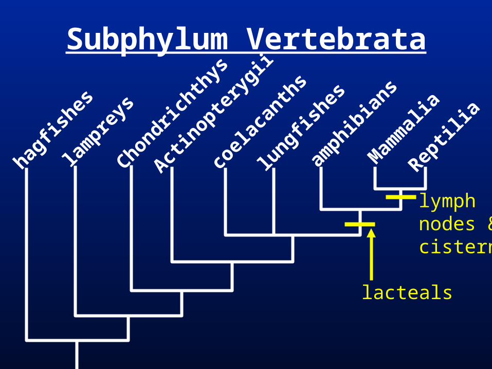
Contractile lymph vessels exist in most vertebrates (Form localized Lymph Hearts in hagfishes, lungfishes, amphibians, turtles, liz. & snakes, & ostriches+related birds)
Lacteals = lymph vessels in the intestinal villi – Only in TETRAPODS.
Lymph nodes / sacs / cisterns = expanded lymph vessels filled with connective tissue and leukocytes. Only in AMNIOTES.
Lymphatic System
Human Lymphatic System
Subphylum Vertebrata
hagfis
hes
lam
preys
Chondrichth
ys
Actin
optery
gii
coel
acan
ths
lungfis
hes
amphib
ians
Mam
mal
ia
Reptil
ia
lacteals
lymphnodes &cisterns

Ventilation = movement of air or water across a respiratory surface.
Apnea = cessation of ventilation Unidirectional ventilation = air/water
moves 1 direction over resp. surface Bidirectional ventilation = air/water
moves 2 directions over resp. surface. (tidal)
Concurrent & Counter current exchange
General Respiratory Systems

Concurrent
Same Direction Current
to body
blood
water or air
from body (heart)
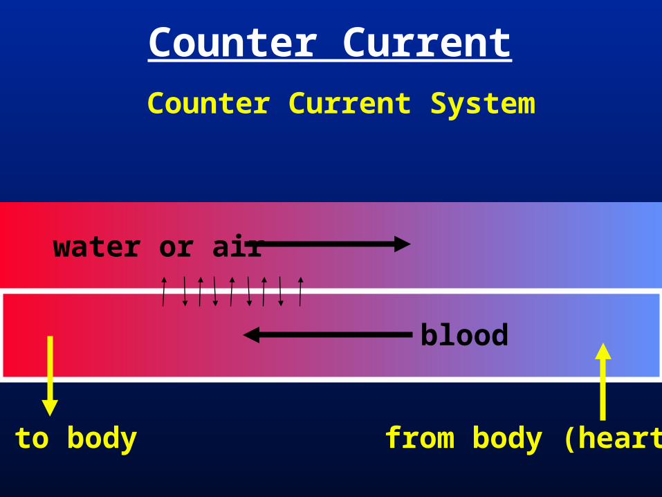
Counter Current
Counter Current System
to body
blood
water or air
from body (heart)
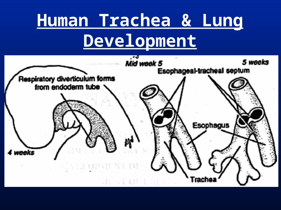
Human Trachea & Lung Development

Human Respiratory System
nasal cavitypharynxlarynx
tracheabronchusbronchiolealveolus
inlung

Human Respiratory System
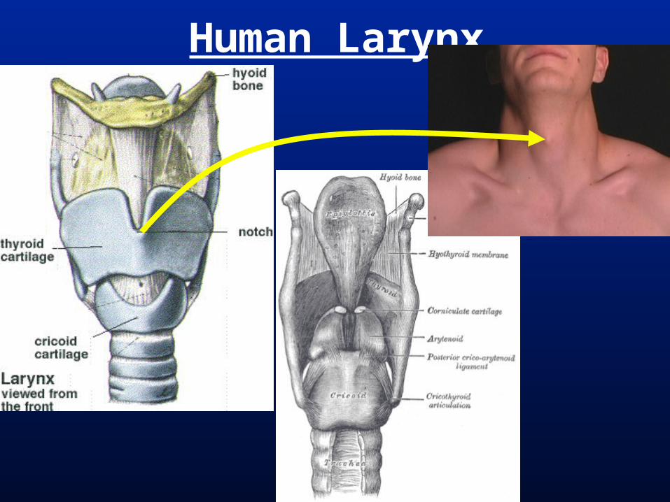
Human Larynx
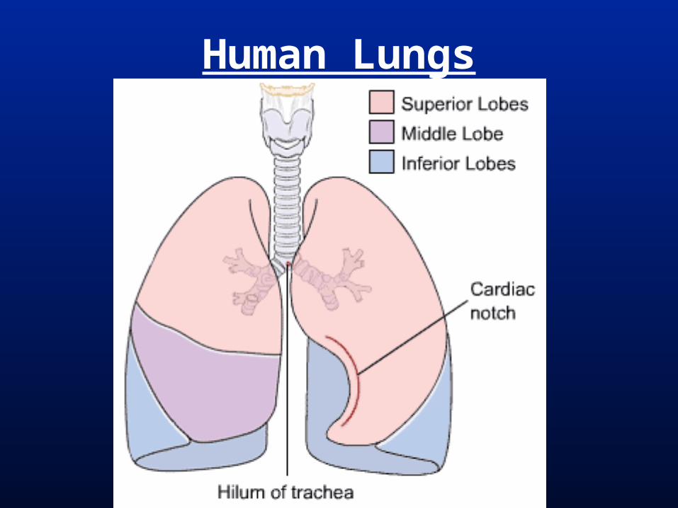
Human Lungs

Human Ventilation
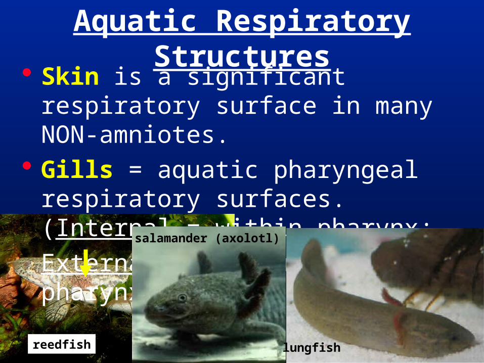
Skin is a significant respiratory surface in many NON-amniotes.
Gills = aquatic pharyngeal respiratory surfaces. (Internal = within pharynx;
External = protrude from pharynx)
Aquatic Respiratory Structures
salamander (axolotl)
lungfishreedfish

Internal GillsChondrichthys
Actinopterygii

Gills Gill = bony pharyngeal arch and two sets
of filaments. Absent in Amniotes and adult
Lissamphibians Each filament bears many lamellae
(small, flat projections) & blood passes through capillaries in the lamellae.
Blood movement through lamellae sets up a counter current system.
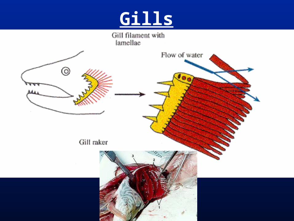
Gills
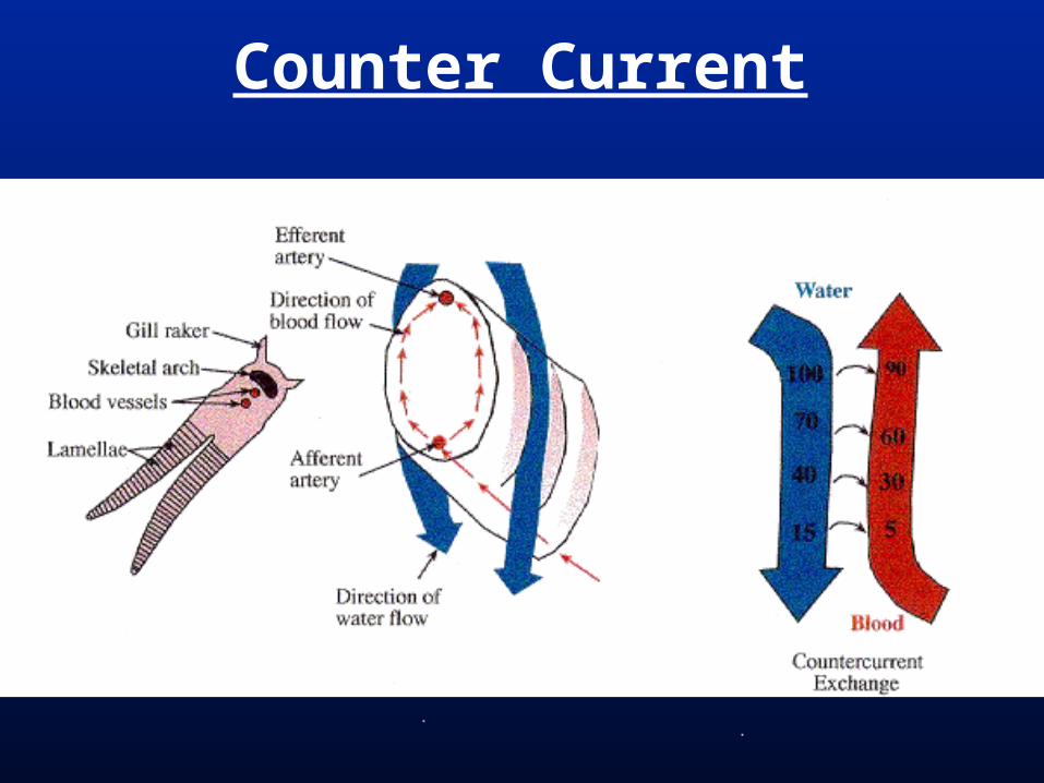
Counter Current
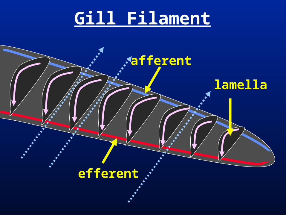
Gill Filament
lamella
efferent
afferent
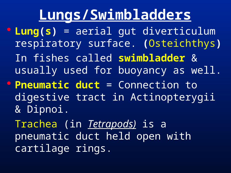
Lungs/Swimbladders Lung(s) = aerial gut diverticulum
respiratory surface. (Osteichthys)
In fishes called swimbladder & usually used for buoyancy as well.
Pneumatic duct = Connection to digestive tract in Actinopterygii & Dipnoi.
Trachea (in Tetrapods) is a pneumatic duct held open with cartilage rings.

Lungs/Swimbladders Respiratory lungs/swimbladders are
often compartmentalized. Faveoli = non-mammal lung chambers. Alveoli = rounded mammal lung
chambers.
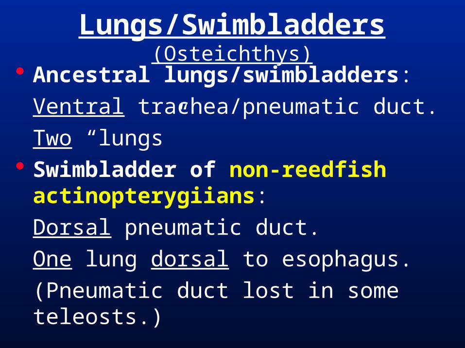
Ancestral lungs/swimbladders:
Ventral trachea/pneumatic duct.
Two “lungs” Swimbladder of non-reedfish
actinopterygiians:
Dorsal pneumatic duct.
One lung dorsal to esophagus.
(Pneumatic duct lost in some teleosts.)
Lungs/Swimbladders (Osteichthys)

Lungs/Swimbladders
Actinopterygii
Polypterus
lungfish
salamanderbass
carp
Amia calva

Craniate Respiration
hagfis
hes
lam
preys
Chondrichth
ys
Actin
optery
gii
coel
acan
ths
lungfis
hes
amphib
ians
Mam
mal
ia
Reptil
ia
lungs(swimbladder)
gillfilamentslost
respiratory gills with filaments
trachea(withcartilage“rings”)
alveoli

sturg
eons
& pad
dlefis
h
gars bowfin
Amia
cal
va
TELEOSTS
reed
fishes
Class Actinopterygii
single dorsal lung withdorsal connection todigestive tract

Aerial Respiratory Modifications
Ancestrally - amphibians & “fishes”
- “Swallow” air – push into lungs
- Elastic lungs recoil to push air out. Prehepatic diaphragm (mammals)
- Negative pressure pulls air into lungs
- Body cavity contraction pushes air out. Posthepatic diaphragm (crocodilians)
- Negative pressure pulls air into lungs
- Liver pushes air out of lungs.

Ventilation“swallow” air
posthepatic
prehepatic

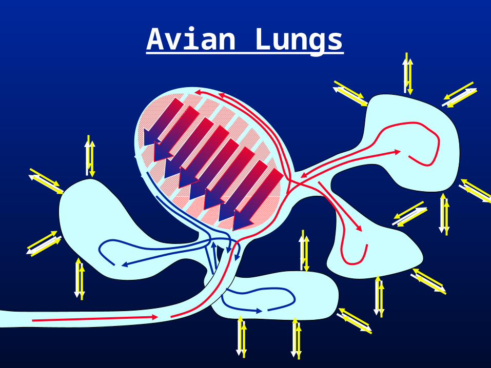
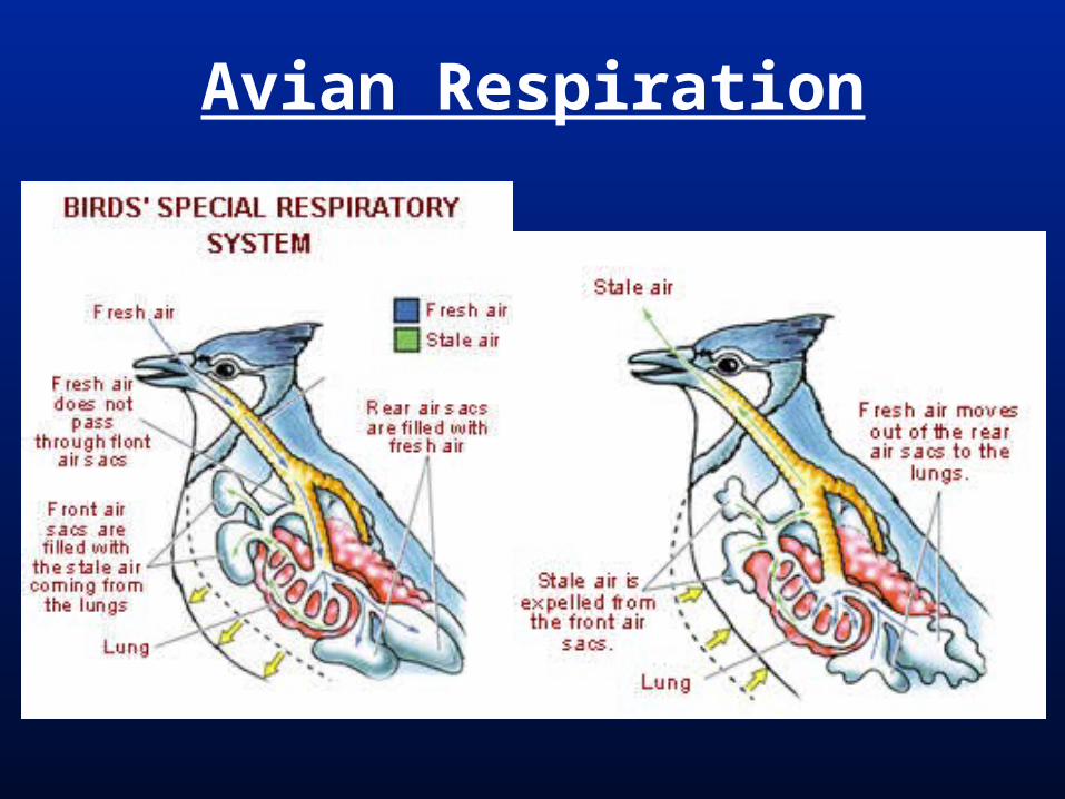
Parabronchi (birds) = one-way, passageways through the lung faveoli.
Lungs not very compressible. Air Sacs (birds) = membranous sacs
for containing air (9 off of each lung) Air moved by expansion & contraction
of air sacs. Countercurrent, constant flow of O2 rich
air over capillaries even when exhaling.
Bird Respiration
Avian Lungs

Avian Inhalation

Avian Exhalation
Avian Respiration

Amniota
monotre
mes
mar
supia
ls
euth
eria
ns
turtl
es
lizar
ds & s
nakes
croco
dilian
s
birds
prehepaticdiaphragm
air sacs;parabronchi
* posthepatic diaphragm
*