Embed Size (px)
Citation preview
Title: A systematic review of dynamic cerebral and peripheral endothelial function in
lacunar stroke versus controls.
Authors: Susan F Stevenson, BSc (Hons)
Fergus N Doubal, MB ChB, MRCP
Kirsten Shuler, BSc
Joanna M Wardlaw, FRCP, FRCR, FMedSci
Affiliation: SINAPSE Collaboration, SFC Brain Imaging Research Centre, Division
of Clinical Neurosciences, University of Edinburgh, Edinburgh, UK.
Correspondence: Prof J Wardlaw, SINAPSE Collaboration, SFC Brain Imaging
Research Centre, Division of Clinical Neurosciences, Western General Hospital,
Crewe Rd, Edinburgh, EH4 2XU, UK.
Tel: +44 131 537 2943
Fax: +44 131 332 5150
Email: [email protected]
1
Acknowledgements and Source(s) of Funding
The work was undertaken as part of a Special Study Module, College of Medicine
and Veterinary Medicine, University of Edinburgh, UK. FD was funded by a
Wellcome Trust Research Training Fellowship (075611). JMW is funded by the
Scottish Funding Council and Chief Scientist Office as part of the SINAPSE (Scottish
Imaging Network - Platform for Scientific Excellence, www.sinapse.ac.uk)
Collaboration.
Conflict(s) of Interest/Disclosure(s)
The authors report no conflicts of interest.
2
Title: A systematic review of dynamic cerebral and peripheral endothelial function in
lacunar stroke versus controls.
Cover title: Endothelial dysfunction in lacunar stroke
Word count: abstract 223, body of text 2346, references 1226; table 945 (on line);
total 4741.
Total number of figures: 2
Total number of tables: 1
Key words: atherosclerosis, endothelium, plethysmography, stroke, vascular
diseases
3
Abstract
Background: The aetiology of cerebral small vessel disease is unknown. An
association with endothelial dysfunction has been suggested. We systematically
assessed all relevant studies of dynamic endothelial function in patients with lacunar
stroke, as a marker of small vessel disease.
Methods: We searched for studies of cerebral or peripheral vascular reactivity in
patients with lacunar or cortical (i.e. large artery atheromatous) ischaemic stroke or
non-stroke controls. We calculated standardised mean difference (SMD) in vascular
reactivity, +/- 95% confidence intervals (CI) between small vessel disease and
control groups.
Results: Sixteen publications (974 patients) were included. In lacunar stroke:
cerebrovascular reactivity (n=534) was reduced compared with age-matched normal
(SMD -0.94, 95%CI -1.17, -0.70), but not age+risk factor-matched controls (SMD
0.08, 95%CI -0.36, 0.53) or cortical strokes (SMD -0.29, 95%CI -0.69, 0.11); forearm
flow mediated dilatation (n=401) was reduced compared with age-matched normal
controls (SMD -1.04, 95%CI -1.33, -0.75) and age+risk factor-matched controls
(SMD -0.94, 95%CI -1.26, -0.61), but not cortical strokes (SMD -0.23, 95%CI -0.55,
0.08).
Conclusions: Endothelial dysfunction is present in patients with lacunar stroke but
may simply reflect exposure to vascular risk factors and having a stroke, as a similar
degree of dysfunction is found in cortical (large artery atheromatous) stroke. Current
data do not confirm that endothelial dysfunction is specific to small vessel stroke.
Future studies should include controls with non-lacunar stroke.
4
Introduction
Cerebral small vessel disease (SVD) is common and has clinical (lacunar stroke,
cognitive impairment, gait and movement disorders) and radiological (symptomatic
small subcortical infarct, silent lacunar infarct, lacunes, white matter lesions (WMLs),
microbleeds) manifestations.1-3 The low early mortality of lacunar stroke masks a
long-term risk of recurrent stroke and death similar to atherothromboembolic stroke,4
and substantial risk of cognitive decline,5 creating a massive public health burden.6
Lacunar infarcts are small (<15mm) subcortical lesions in the territory of a single
deep perforating arteriole most of which are associated with an intrinsic abnormality
in the perforating arteriole wall of unknown aetiology. An association between
lacunar ischaemic stroke and endothelial dysfunction has been suggested,7;8 but
many patients with stroke have hypertension or diabetes or take medications which
affect endothelial function.9;10 Atheromatous large artery disease is also associated
with endothelial dysfunction.11 A recent systematic review identified endothelial
dysfunction in lacunar ischaemic stroke but did not control for risk factor exposure or
other stroke subtypes.12 Therefore it is unclear whether endothelial changes
observed in patients with lacunar ischaemic stroke might be specific to SVD or
simply reflect age, vascular risk factors, generalised (possibly co-incidental)
atheroma or the effects of having a stroke. We performed a systematic review of all
studies that assessed cerebral or peripheral vascular reactivity in patients with
lacunar ischaemic stroke.
5

Methods
We followed the general guidance for systematic reviews of observational and
diagnostic studies (http://www.equator-network.org), modified to suit the type of
study identified in this review.13;14
We searched the published literature using MEDLINE and EMBASE from the 1st
January 1995, to the 15th February 2008 using Ovid and a carefully devised search
strategy (Appendix 1) developed with advice from the Cochrane Stroke Group
(http://www.dcn.ed.ac.uk/csrg/). We updated the search using MEDLINE to 6th
January 2010 (we did not re-search EMBASE as there was very little difference
between it and MEDLINE in the initial search). We sought primary studies, in
humans, in any language, which investigated patients with markers of cerebral SVD
and dynamic measures of endothelial function e.g. response to hypercapnia or
acetazolamide or flow mediated dilatation.15 We checked references in review and
primary papers and hand-searched the journal Stroke.
We included papers which assessed endothelial function in patients with: clinically-
evident lacunar ischaemic stroke, with or without an acute subcortical infarct on brain
imaging; lacunes (i.e. rounded cerebral spinal fluid attenuation lesion <1.5mm in the
basal ganglia, hemispheric white matter or brain stem) identified on brain imaging
without clearly-relevant symptoms; or leukoaraiosis (WMLs).
We excluded papers which only assessed endothelial function using plasma
markers, animal studies, duplicate publications, SVD caused by a single gene
disorder or had no control group.
6
Two reviewers independently extracted data using a standardised data extraction
form. A third reviewer arbitrated on disagreements. We obtained translations of
foreign language papers where possible. We extracted data on study population
(sample size, age, sex, presence of co-morbidities, medications, previous strokes
and selection criteria for both patient and control groups), study design, endothelial
function assessment method,15 vascular bed, blinding of investigators and primary
vascular reactivity results. We identified the method of stroke diagnosis, whether by
a stroke specialist, if confirmed using imaging, the type of imaging and the time
interval after stroke. We assessed the study quality using the QUADAS14 instrument
(www.equator-network.org). We were careful to include each patient population only
once while including all data available for that population.
The studies used different methods of assessing endothelial function, so we
calculated the standardised mean difference (SMD) by the fixed effects method
(Review Manager 4© software) between the lacunar and control groups from the
mean and standard deviation of the vascular reactivity results (where given). We
used the endothelial function data from the cerebral circulation contralateral to the
side of the symptomatic infarct in cortical and lacunar stroke patients. As most
studies did not adjust for potential confounders, we stratified the analyses into
lacunar vs. age-matched controls; vs. age+risk factor-matched controls; vs. cortical
stroke; and multiple vs. single lacunar infarcts on imaging. We calculated
heterogeneity between studies.
7
Results
We identified 1257 titles. De-duplication (467) and exclusion of irrelevant papers
(n=739) resulted in 27 includable papers. Hand searching identified three additional
publications. Of the 30 for full text reading, three were excluded (unable to translate:
Russian16 and Slovakian17;18) and eleven failed to meet the inclusion criteria (no
controls,19;20 not assessing dynamic endothelial function or inappropriate patients21-
29). Therefore 16 papers were eligible, including 974 individuals.
Characteristics of included studies (Table 1)
Some of the 16 papers contributed to more than one comparison: 14/16 papers30-43
compared 318 patients with recent lacunar ischaemic stroke to 305 age-matched or
age+risk factor matched controls; 5/15 papers35;37;38;42;44 compared 124 patients with
recent lacunar ischaemic stroke to 115 patients with recent cortical ischaemic stroke;
4/15 papers30;37;40;45 compared patients with recent lacunar ischaemic stroke of
whom 71 had only one single small subcortical infarct and 71 had a single
symptomatic lacunar infarct plus multiple silent infarctions on imaging.
Most studies were small (mean 36 subjects, median 18 subjects per group), gave
little detail about recruitment or methods of stroke diagnosis (in particular of how
lacunar stroke was determined), did not state duration of hypertension or other risk
factors, adjust for risk factors37 or mention prescribed medications (only two stated
that medications were discontinued prior to study35;40). Only two studies explicitly
stated that the analyses were blinded to subject group. Ten gave the time interval
between stroke and endothelial function assessment (range <72 hours to three
8
months, median 26 days). The lacunar and cortical ischaemic stroke patients were
not age-matched.
Characteristics of included patients and controls
All 16 papers defined lacunar ischaemic stroke as “appropriate neurological features
and a recent small subcortical infarct on imaging consistent with the symptoms”.
Most papers excluded patients with carotid stenosis (except one30), five excluded
cardiogenic sources of emboli,32;33;36;38;41 and three excluded middle cerebral artery
(MCA) stenosis35;39;40 from the lacunar stroke group.
Thirteen studies had age-matched medically-confirmed normal controls; 5/13 also
used imaging to exclude subjects with silent infarcts.30;32;39;40;42 Three studies
recruited age- and risk-factor matched controls (with long-standing hypertension and
hypercholesterolaemia in two;32;34 with hypertension only in one43). Five papers
compared patients with lacunar ischaemic stroke to patients with cortical ischaemic
stroke,35;37;38;42;44 confirming the infarct subtype with CT or MRI. Four studies
included patients with more than one lacunar infarct on CT,30;37;40;45 MR,40;45 or
both.37
Assessment of endothelial function (Table 1)
Twelve papers assessed endothelial function in the cerebral circulation30-32;34;37-
42;44;45 and five papers the systemic circulation;32;33;35;36;43 one assessed both.32
Several techniques (Table 1) were used to assess cerebral endothelial function,
including the vascular response to hypercapnia, infusion of acetazolamide or L-
arginine, expressing the response as a percentage increase in mean arterial blood
9
velocity in the MCA or basilar artery. Change in blood oxygen level dependent signal
during hypercapnia detected using functional MRI,31 percent increase in regional
cerebral blood flow in response to hypercapnia using stable Xenon CT30 and
“dynamic cerebral autoregulation” (the ability to restore cerebral blood flow following
sudden changes in perfusion pressure)38 were also used. Peripheral endothelial
function was assessed with brachial artery flow-mediated dilatation, expressed as
the percentage change in arterial diameter, in five papers;32;33;35;36;43 one also
assessed endothelium-independent flow-mediated dilatation using response to
sublingual nitroglycerine.43
Endothelial function: Lacunar stroke vs. age-matched controls
Thirteen studies (n=534) compared lacunar ischaemic stroke to healthy age-matched
controls, nine in the cerebral (n=360)30-32;34;37;39-42 and four in the peripheral
circulation (n=211).32;33;35;36 Vascular reactivity was reduced in the cerebral
circulation in lacunar stroke patients compared with age-matched healthy controls
(8/9 studies, 324 patients, SMD -0.94, 95% confidence intervals (CI) -1.17, -0.70,
p<0.00001, Figure 1) and in the forearm (4/4 studies, 211 patients, SMD -1.04, 95%
CI -1.33, -0.75, p<0.00001, Figure 2). There was no significant heterogeneity
between studies.
Lacunar ischaemic stroke vs. age+risk factor-matched controls
Four papers compared vascular reactivity in lacunar stroke patients with age+risk-
factor-matched controls, two in the cerebral32;34 and three in the peripheral
circulation.32;33;43 There was no significant difference in cerebrovascular reactivity
(2/2 studies, 79 patients, SMD 0.08, 95% CI -0.36, 0.53, p=0.71, Figure 1) but
10
significantly impaired peripheral vascular reactivity (3/3 studies, 167 patients, SMD -
0.94, 95% CI -1.26, -0.61, p<0.00001, Figure 2). There was no significant
heterogeneity between studies.
Lacunar vs. cortical ischaemic stroke
Four of the 16 studies (n=127)37;38;42;44 compared cerebral vascular reactivity in
patients with lacunar stroke (n=68) to patients with cortical stroke (n=54), of which
two studies contributed to more than one comparison (Figure 1). One study (n=117)
compared peripheral vascular reactivity between lacunar (n=56) and cortical (n=61)
stroke patients (Figure 2).35 For the cerebral comparisons, we used the test results
from the asymptomatic side of the brain, because vascular reactivity was reduced in
ipsilateral vs. contralateral arteries in cortical stroke patients due to tissue damage
resulting from the stroke (see Table 1). Although individual studies showed
differences in endothelial function between lacunar and cortical stroke
patients35;37;38;42;44 the combined data showed no difference in vascular reactivity
between lacunar and cortical stroke in either cerebral (SMD -0.29, 95% CI -0.69,
0.11, p=0.16) or in peripheral (SMD -0.23, 95% CI -0.55, 0.08, p=0.15) circulation.
There was no significant heterogeneity between studies.
Lacunar ischaemic stroke with single vs. multiple silent lacunar infarcts
Four of the 16 papers30;37;40;45 compared cerebral vascular reactivity in lacunar stroke
patients with one single lacunar infarction (n=71) to those with additional multiple
silent infarctions (n=71) on imaging (Figure 1). Patients with lacunar ischaemic
stroke plus multiple silent lacunar infarcts had reduced cerebral vascular reactivity
11

compared to patients without silent lacunar infarcts on imaging (SMD -0.68, 95% CI -
1.02, -0.34, p=0.0001), with no significant heterogeneity between studies.
12
Discussion
Endothelial dysfunction is common in patients with symptomatic large artery
atheromatous disease and has also been postulated as a mechanism underlying the
development of lacunar stroke. This systematic review suggests that lacunar
ischaemic stroke is associated with impaired vascular reactivity compared with
normal age-matched controls, but the association is less clear-cut when compared
with controls matched for vascular risk factors or patients with cortical ischaemic
stroke. One interpretation of this is that endothelial dysfunction may be a general
response of the vascular system to the vascular risk factors that predispose to
stroke,9 or other circumstances associated with stroke such as secondary prevention
medications, rather than being specific to small or large artery disease.
A previous systematic review identified associations between lacunar stroke and
altered vascular reactivity, but did not perform a meta-analysis or make direct
comparisons between lacunar stroke and patients with vascular risk factors or
cortical stroke controls.12 Endothelial function is altered in the presence of vascular
risk factors9 and by drugs used for risk factor reduction and stroke prevention.10
Cortical stroke patients control for use of medications and presence of vascular risk
factors so are the most valid comparison.46 It is worth noting that the presence of an
infarct in the brain was associated with reduced ipsilateral vascular reactivity, e.g.
cortical stroke patients had reduced reactivity ipsilateral to the ischaemic cortical
stroke. Hence any reduction in cerebral vascular reactivity in patients with multiple
silent lacunar infarcts on imaging in both hemispheres (compared to patients with
13
only a single lacunar infarct) is unsurprising and is likely to be a consequence of
more brain damage.
The strengths include following well-established guidance for conducting systematic
reviews of observational and diagnostic data (www.equator-network.org) and
Cochrane Stroke Group search advice. We used standard pre-specified criteria for
study assessment. We carefully avoided duplicate data. Some studies provided
more than one comparison but we were careful to avoid double counting the total
number of subjects. In patients with stroke, we only used the cerebral vascular
reactivity results from the asymptomatic side of the brain to avoid simply measuring
the effects of brain damage resulting from the index stroke. We meta-analysed the
data thereby effectively increasing sample size and precision.
The limitations include the small number of relevant studies, their small sample size,
the varied and often poorly described diagnosis of lacunar stroke and the various
and poorly standardised endothelial function tests used. In general, the papers gave
little detail about how the diagnosis of lacunar stroke had been made clinically and/or
with imaging, so inevitably there will be some “noise” due to imprecise diagnoses.
However, given the relative lack of literature on this topic, we decided that it would
be better to include studies which appeared to have included patients with
symptomatic lacunar stroke as any attempt to exclude studies on the basis of their
lacunar stroke diagnosis could have resulted in further bias. Studies which used
suboptimal imaging, either insensitive or applied too late after the acute symptoms,
may have confused up to 20% of lacunar strokes as cortical strokes and vice
versa.47 It was often unclear if the investigators were blind to study group; unblinding
14
may increase investigator bias. The endothelial function data were not adjusted for
potential confounders such as blood pressure, diabetes, hypercholesterolaemia,
smoking, prior stroke, white matter hyperintensities on imaging, old infarcts or
haemorrhages on imaging, age or medication. Although studies generally matched
with healthy controls for age, the lacunar and cortical stroke groups were not well
age-matched. Despite many antihypertensive and stroke prevention medications
being known to influence endothelial function, there was little information about
current medications, most studies did not indicate if medications had been stopped
prior to the study, and where this was mentioned, it was a very short time (e.g. 12
hours35) before the endothelial function studies. While some studies used hospital
controls, recruitment procedures (source, mechanism) were unclear for many
studies. Other limitations reflect the limited resources available for this review, e.g.
we were unable to obtain translations for three papers which might have contained
relevant data. The cerebral circulation studies used several different endothelial
function tests.15 However it is important to realise that the meta-analysis does not
directly compare studies with each other, but rather the magnitude of association
within each study with that in other studies. Therefore the grouping of apparently
different methods of assessing endothelial function is more valid than attempting to
combine data from different studies that used different methods in an individual
patient data meta-analysis. There is also likely to be publication bias, meaning that
the present analyses are over positive.
Is there a need for further research on endothelial function and lacunar stroke? The
existing data do not exclude a specific association between endothelial dysfunction
and lacunar stroke. Based on the modest difference in the cerebral circulation
15
between lacunar and cortical patients identified in this review and the large standard
deviation (SD) of the endothelial function measurement methods, a future study
would require a total sample size of 570 patients (half lacunar and half cortical) to
confirm a difference in cerebral vascular reactivity of 22%, SD 20%, with 80% power
at the p<0.05 level, particularly if there were to be any adjustment for even a few key
potential confounding variables. If the SD could be reduced, e.g. by increasing the
precision of the endothelial function measurements (although ±20% is biologically
very plausible and any less would be unlikely), then the sample sizes would be
smaller. On the other hand, a difference of 22% is optimistic, the differences in the
present studies being nearer 6%, in which case a sample size of 752 would be
required. The studies to date were much smaller than this. Numerous studies report
plasma markers of endothelial function (e.g. asymmetric dimethylarginine12;48) and
lacunar disease, but the identification and meta-analyses of these studies was
beyond the remit of this review. There may be an association between angiotensin-
converting enzyme insertion/deletion polymorphism (influencing endothelial function)
and leukoaraiosis,49 but the results of genetic association studies are awaited.
In addition to including a control group with a pathophysiologically different subtype
of ischaemic stroke, future studies should ensure optimal clinical and imaging
diagnosis of stroke subtype, provide clear descriptions of their recruitment and
assessment methods, ensure adequate blinding of endothelial assessments, have
appropriate controls drawn from a relevant and comparative population, record
medications, try to balance study groups for medications, preferably discontinue
vasoactive drugs prior to study, adjust for differences in vascular risk factors, and
match for age. The peripheral circulation provides a valuable method of examining
16

systemic endothelial dysfunction outside the territory affected by the recent stroke.
Cerebral small vessel disease may be a systemic small vessel problem affecting
multiple organs,8;50 in the same way that large artery atheroma is rarely a disease of
only one large artery, and therefore it is legitimate and necessary to study small
vessel disease in multiple organs, not just the brain.
17
References
1. Hachinski V. Stroke and vascular cognitive impairment: a transdisciplinary,
translational and transactional approach. Stroke 2007;38:1396-403.
2. Bamford J, Sandercock P, Dennis M, Burn J, Warlow C. Classification and
natural history of clinically identifiable subtypes of cerebral infarction. Lancet
1991;337:1521-6.
3. Vermeer SE, Longstreth WT, Jr., Koudstaal PJ. Silent brain infarcts: a
systematic review. Lancet Neurol 2007;6:611-9.
4. Jackson C, Sudlow C. Comparing risks of death and recurrent vascular
events between lacunar and non-lacunar infarction. Brain 2005;128:2507-17.
5. Vermeer SE, Prins ND, den Heijer T, Hofman A, Koudstaal PJ, Breteler MMB.
Silent brain infarcts and the risk of dementia and cognitive decline. N Engl J
Med 2003;348:1215-22.
6. Hachinski V. World Stroke Day 2008: "Little strokes, big trouble". Stroke
2008;39:2407-20.
7. Hassan A, Hunt BJ, O'Sullivan M, Parmar K, Bamford JM, Briley D, Brown
MM, Thomas DJ, Markus HS. Markers of endothelial dysfunction in lacunar
infarction and ischaemic leukoaraiosis. Brain 2003;126:424-32.
8. Thompson CS, Hakim AM. Living beyond our physiological means. Small
vessel disease of the brain is an expression of a systemic failure in arteriolar
function: a unifying hypothesis. Stroke 2009;40:e322-e330.
9. Girouard H, Iadecola C. Neurovascular coupling in the normal brain and in
hypertension, stroke, and Alzheimer disease. J Appl Physiol 2006;100:328-
35.
18
10. Webb DJ. The pharmacology of human blood vessels in vivo. J Vasc Res
1995;32:2-15.
11. Newby DE, McLeod AL, Uren NG, Flint L, Ludlam CA, Webb DJ, Fox KA,
Boon NA. Impaired coronary tissue plasminogen activator release is
associated with coronary atherosclerosis and cigarette smoking: direct link
between endothelial dysfunction and atherothrombosis. Circulation
2001;103:1936-41.
12. Knottnerus ILH, Ten Cate H, Lodder J, Kessels F, van Oostenbrugge RJ.
Endothelial dysfunction in lacunar stroke: a systematic review. Cerebrovasc
Dis 2009;27:519-26.
13. von Elm E, Altman DG, Egger M, Pocock SJ, Gotzsche PC, Vandenbroucke
JP, for the STROBE initiative. The Strengthening the Reporting of
Observational Studies in Epidemiology (STROBE) statement: guidelines for
reporting observational studies. Lancet 2007;370:1453-7.
14. Whiting P, Rutjes AWS, Reitsma JB, Bossuyt PMM, Kleijnen J. The
development of QUADAS: a tool for the quality assessment of studies of
diagnostic accuracy included in systematic reviews. BMC Med Res Methodol
2003;3:25.
15. Tousoulis D, Antoniades C, Stefanadis C. Evaluating endothelial function in
humans: a guide to invasive and non-invasive techniques. Heart 2005;91:553-
8.
16. Shutov AA, Baidina TV, Agafonov AV, Siutkina OV, Gaidash GV.
[Endothelium dysfunction in patients with ischemic stroke]. Zh Nevrol Psikhiatr
im S S Korsakova 2005;Suppl 14:42-5.
17. Gaspar L, Stvrtinova V, Gavornik P, Strbova L, Porubec V. [Changes in
forearm microcirculation in patients with focal cerebral ischemia]. Bratisl Lek
Listy 1998;99:122-3.
19
18. Panczel G, Bonoczk P, Nagy Z. [Impairment of vasoreactivity in brainstem
and hemispheral small vessel disease: comparative study]. Ideggyogy Sz
2002;55:95-101.
19. Birns J, Jarosz J, Markus HS, Kalra L. Cerebrovascular reactivity and
dynamic autoregulation in ischaemic subcortical white matter disease. J
Neurol Neurosurg Psychiatry 2009.
20. Gommer ED, Staals J, van Oostenbrugge RJ, Lodder J, Mess WH, Reulen
JP. Dynamic cerebral autoregulation and cerebrovascular reactivity: a
comparative study in lacunar infarct patients. Physiol Meas 2008;29:1293-
303.
21. Walters M, Muir S, Shah I, Lees K. Effect of perindopril on cerebral vasomotor
reactivity in patients with lacunar infarction. Stroke 2004;35:1899-902.
22. Sterzer P, Meintzschel F, Rosler A, Lanfermann H, Steinmetz H, Sitzer M.
Pravastatin improves cerebral vasomotor reactivity in patients with subcortical
small-vessel disease. Stroke 2001;32:2817-20.
23. Lee KO, Lee KY, Lee SY, Ahn CW, Park JS. Lacunar infarction in type 2
diabetes is associated with an elevated intracranial arterial pulsatility index.
Yonsei Med J 2007;48:802-6.
24. Imaizumi T, Chiba M, Honma T, Yoshikawa J, Niwa J. Cerebral blood flow
before and after treatment with amlodipine in elderly patients with lacunar
infarction. Clin Drug Investig 2004;24:765-9.
25. Terborg C, Gora F, Weiller C, Rother J. Reduced vasomotor reactivity in
cerebral microangiopathy: a study with near-infrared spectroscopy and
transcranial Doppler sonography. Stroke 2000;31:924-9.
26. Zvan B, Zaletel M, Pogacnik T, Kiauta T. Testing of cerebral endothelium
function with L-arginine after stroke. Int Angiol 2002;21:256-9.
20

27. Pasqualini L, Marchesi S, Vaudo G, Siepi D, Angeli F, Paris L, Schillaci G,
Mannarino E. Association between endothelial dysfunction and major
cardiovascular events in peripheral arterial disease. Vasa 2003;32:139-43.
28. De Reuck J, Decoo D, Hasenbroekx MC, Lamont B, Santens P, Goethals P,
Strijckmans K, Lemahieu I. Acetazolamide vasoreactivity in vascular
dementia: a positron emission tomographic study. Eur Neurol 1999;41:31-6.
29. Mindach M. [Correlation between low cerebral flow velocities determined by
transcranial ultrasound and lacunar cerebral infarction]. Ultraschall Med
2001;22:274-8.
30. Mochizuki Y, Oishi M, Takasu T. Cerebral blood flow in single and multiple
lacunar infarctions. Stroke 1997;28:1458-60.
31. Hund-Georgiadis M, Zysset S, Naganawa S, Norris DG, Yves von Cramon D.
Determination of cerebrovascular reactivity by means of fMRI signal changes
in cerebral microangiopathy: a correlation with morphological abnormalities.
Cerebrovasc Dis 2003;16:158-65.
32. Pretnar-Oblak J, Sabovic M, Sebestjen M, Pogacnik T, Zaletel M. Influence of
atorvastatin treatment on L-arginine cerebrovascular reactivity and flow-
mediated dilatation in patients with lacunar infarctions. Stroke 2006;37:2540-
5.
33. Pretnar-Oblak J, Sabovic M, Pogacnik T, Sebestjen M, Zaletel M. Flow-
mediated dilatation and intima-media thickness in patients with lacunar
infarctions. Acta Neurol Scand 2006;113:273-7.
34. Pretnar-Oblak J, Zaletel M, Zvan B, Sabovic M, Pogacnik T. Cerebrovascular
reactivity to L-arginine in patients with lacunar infarctions. Cerebrovasc Dis
2006;21:180-6.
21
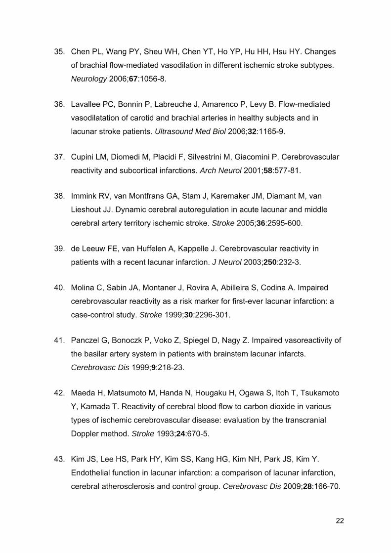
35. Chen PL, Wang PY, Sheu WH, Chen YT, Ho YP, Hu HH, Hsu HY. Changes
of brachial flow-mediated vasodilation in different ischemic stroke subtypes.
Neurology 2006;67:1056-8.
36. Lavallee PC, Bonnin P, Labreuche J, Amarenco P, Levy B. Flow-mediated
vasodilatation of carotid and brachial arteries in healthy subjects and in
lacunar stroke patients. Ultrasound Med Biol 2006;32:1165-9.
37. Cupini LM, Diomedi M, Placidi F, Silvestrini M, Giacomini P. Cerebrovascular
reactivity and subcortical infarctions. Arch Neurol 2001;58:577-81.
38. Immink RV, van Montfrans GA, Stam J, Karemaker JM, Diamant M, van
Lieshout JJ. Dynamic cerebral autoregulation in acute lacunar and middle
cerebral artery territory ischemic stroke. Stroke 2005;36:2595-600.
39. de Leeuw FE, van Huffelen A, Kappelle J. Cerebrovascular reactivity in
patients with a recent lacunar infarction. J Neurol 2003;250:232-3.
40. Molina C, Sabin JA, Montaner J, Rovira A, Abilleira S, Codina A. Impaired
cerebrovascular reactivity as a risk marker for first-ever lacunar infarction: a
case-control study. Stroke 1999;30:2296-301.
41. Panczel G, Bonoczk P, Voko Z, Spiegel D, Nagy Z. Impaired vasoreactivity of
the basilar artery system in patients with brainstem lacunar infarcts.
Cerebrovasc Dis 1999;9:218-23.
42. Maeda H, Matsumoto M, Handa N, Hougaku H, Ogawa S, Itoh T, Tsukamoto
Y, Kamada T. Reactivity of cerebral blood flow to carbon dioxide in various
types of ischemic cerebrovascular disease: evaluation by the transcranial
Doppler method. Stroke 1993;24:670-5.
43. Kim JS, Lee HS, Park HY, Kim SS, Kang HG, Kim NH, Park JS, Kim Y.
Endothelial function in lacunar infarction: a comparison of lacunar infarction,
cerebral atherosclerosis and control group. Cerebrovasc Dis 2009;28:166-70.
22
23
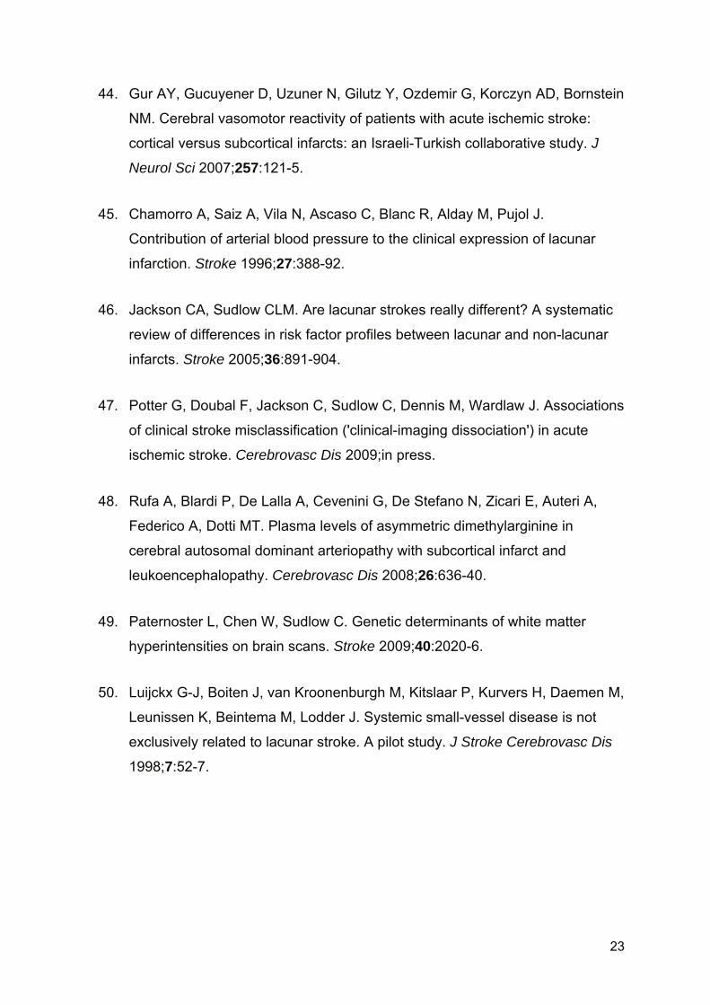
44. Gur AY, Gucuyener D, Uzuner N, Gilutz Y, Ozdemir G, Korczyn AD, Bornstein
NM. Cerebral vasomotor reactivity of patients with acute ischemic stroke:
cortical versus subcortical infarcts: an Israeli-Turkish collaborative study. J
Neurol Sci 2007;257:121-5.
45. Chamorro A, Saiz A, Vila N, Ascaso C, Blanc R, Alday M, Pujol J.
Contribution of arterial blood pressure to the clinical expression of lacunar
infarction. Stroke 1996;27:388-92.
46. Jackson CA, Sudlow CLM. Are lacunar strokes really different? A systematic
review of differences in risk factor profiles between lacunar and non-lacunar
infarcts. Stroke 2005;36:891-904.
47. Potter G, Doubal F, Jackson C, Sudlow C, Dennis M, Wardlaw J. Associations
of clinical stroke misclassification ('clinical-imaging dissociation') in acute
ischemic stroke. Cerebrovasc Dis 2009;in press.
48. Rufa A, Blardi P, De Lalla A, Cevenini G, De Stefano N, Zicari E, Auteri A,
Federico A, Dotti MT. Plasma levels of asymmetric dimethylarginine in
cerebral autosomal dominant arteriopathy with subcortical infarct and
leukoencephalopathy. Cerebrovasc Dis 2008;26:636-40.
49. Paternoster L, Chen W, Sudlow C. Genetic determinants of white matter
hyperintensities on brain scans. Stroke 2009;40:2020-6.
50. Luijckx G-J, Boiten J, van Kroonenburgh M, Kitslaar P, Kurvers H, Daemen M,
Leunissen K, Beintema M, Lodder J. Systemic small-vessel disease is not
exclusively related to lacunar stroke. A pilot study. J Stroke Cerebrovasc Dis
1998;7:52-7.
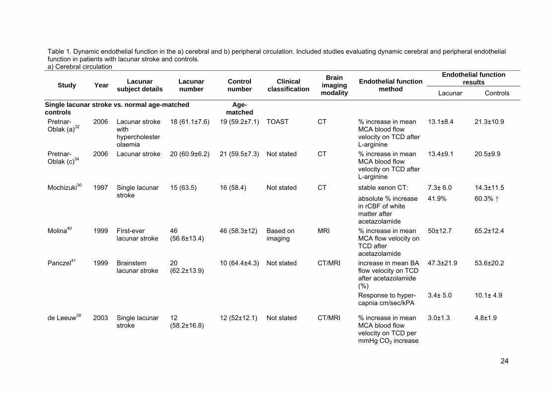
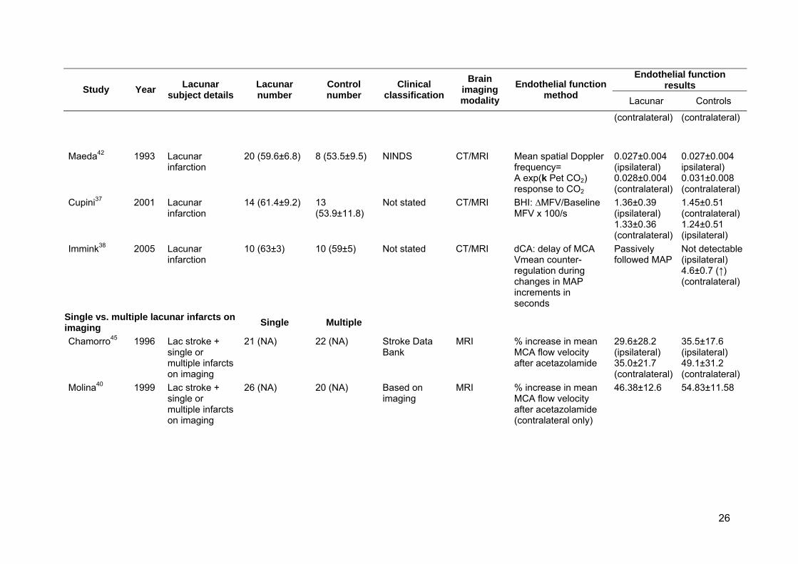
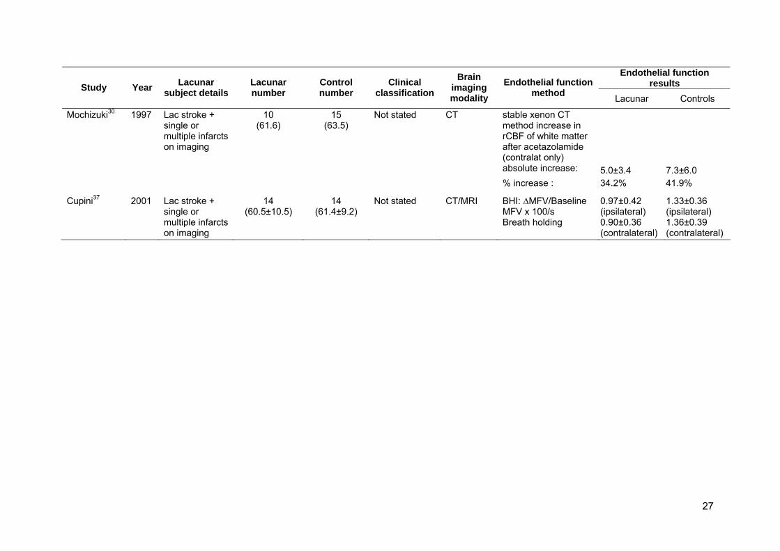
Table 1. Dynamic endothelial function in the a) cerebral and b) peripheral circulation. Included studies evaluating dynamic cerebral and peripheral endothelial function in patients with lacunar stroke and controls. a) Cerebral circulation
Endothelial function results Study Year Lacunar
subject detailsLacunar number
Control number
Clinical classification
Brain imaging modality
Endothelial function method Lacunar Controls
Single lacunar stroke vs. normal age-matched controls
Age-matched
Pretnar-Oblak (a)32
2006 Lacunar stroke with hypercholesterolaemia
18 (61.1±7.6) 19 (59.2±7.1) TOAST CT % increase in mean MCA blood flow velocity on TCD after L-arginine
13.1±8.4 21.3±10.9
Pretnar-Oblak (c)34
2006 Lacunar stroke 20 (60.9±6.2) 21 (59.5±7.3) Not stated CT % increase in mean MCA blood flow velocity on TCD after L-arginine
13.4±9.1 20.5±9.9
stable xenon CT: 7.3± 6.0 14.3±11.5 Mochizuki30 1997 Single lacunar stroke
15 (63.5) 16 (58.4) Not stated CT absolute % increase in rCBF of white matter after acetazolamide
41.9% 60.3% ↑
Molina40 1999 First-ever lacunar stroke
46 (56.6±13.4)
46 (58.3±12) Based on imaging
MRI % increase in mean MCA flow velocity on TCD after acetazolamide
50±12.7 65.2±12.4
increase in mean BA flow velocity on TCD after acetazolamide (%)
47.3±21.9 53.6±20.2 Panczel41 1999 Brainstem lacunar stroke
20 (62.2±13.9)
10 (64.4±4.3) Not stated CT/MRI
Response to hyper-capnia cm/sec/kPA
3.4± 5.0 10.1± 4.9
de Leeuw39 2003 Single lacunar stroke
12 (58.2±16.8)
12 (52±12.1) Not stated CT/MRI % increase in mean MCA blood flow velocity on TCD per mmHg CO2 increase
3.0±1.3 4.8±1.9
24
Endothelial function results Study Year Lacunar
subject detailsLacunar number
Control number
Clinical classification
Brain imaging modality
Endothelial function method Lacunar Controls
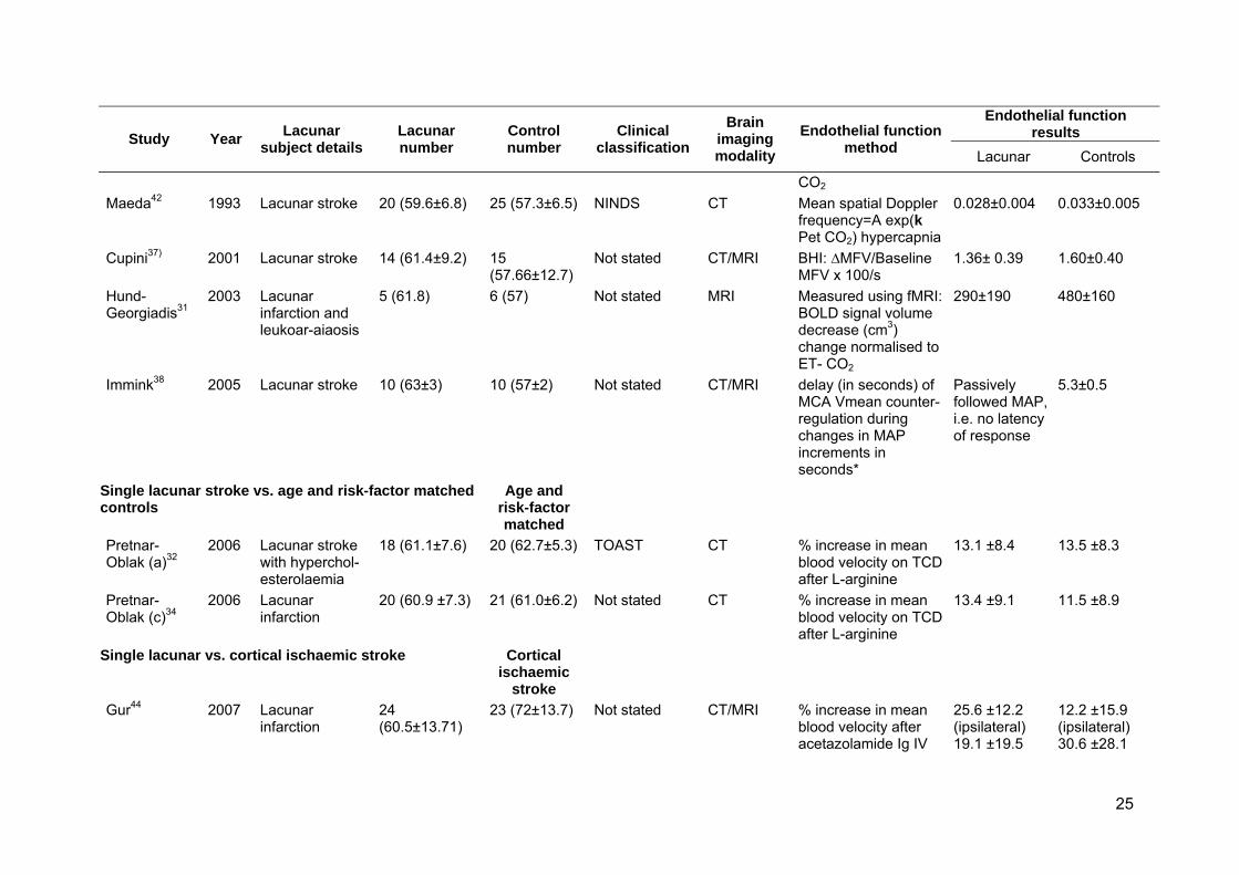
CO2 Maeda42 1993 Lacunar stroke 20 (59.6±6.8) 25 (57.3±6.5) NINDS CT Mean spatial Doppler
frequency=A exp(k Pet CO2) hypercapnia
0.028±0.004 0.033±0.005
Cupini37) 2001 Lacunar stroke 14 (61.4±9.2) 15 (57.66±12.7)
Not stated CT/MRI BHI: ∆MFV/Baseline MFV x 100/s
1.36± 0.39 1.60±0.40
Hund-Georgiadis31
2003 Lacunar infarction and leukoar-aiaosis
5 (61.8) 6 (57) Not stated MRI Measured using fMRI: BOLD signal volume decrease (cm3) change normalised to ET- CO2
290±190 480±160
Immink38 2005 Lacunar stroke 10 (63±3) 10 (57±2) Not stated CT/MRI delay (in seconds) of MCA Vmean counter-regulation during changes in MAP increments in seconds*
Passively followed MAP, i.e. no latency of response
5.3±0.5
Single lacunar stroke vs. age and risk-factor matched controls
Age and risk-factor matched
Pretnar-Oblak (a)32
2006 Lacunar stroke with hyperchol-esterolaemia
18 (61.1±7.6) 20 (62.7±5.3) TOAST CT % increase in mean blood velocity on TCD after L-arginine
13.1 ±8.4 13.5 ±8.3
Pretnar-Oblak (c)34
2006 Lacunar infarction
20 (60.9 ±7.3) 21 (61.0±6.2) Not stated CT % increase in mean blood velocity on TCD after L-arginine
13.4 ±9.1 11.5 ±8.9
Single lacunar vs. cortical ischaemic stroke Cortical ischaemic
stroke
Gur44 2007 Lacunar infarction
24 (60.5±13.71)
23 (72±13.7) Not stated CT/MRI % increase in mean blood velocity after acetazolamide Ig IV
25.6 ±12.2 (ipsilateral) 19.1 ±19.5
12.2 ±15.9 (ipsilateral) 30.6 ±28.1
25
Endothelial function results Study Year Lacunar
subject detailsLacunar number
Control number
Clinical classification
Brain imaging modality
Endothelial function method Lacunar Controls
(contralateral) (contralateral)
Maeda42 1993 Lacunar infarction
20 (59.6±6.8) 8 (53.5±9.5) NINDS CT/MRI Mean spatial Doppler frequency= A exp(k Pet CO2) response to CO2
0.027±0.004 (ipsilateral) 0.028±0.004 (contralateral)
0.027±0.004 ipsilateral) 0.031±0.008 (contralateral)
Cupini37 2001 Lacunar infarction
14 (61.4±9.2) 13 (53.9±11.8)
Not stated CT/MRI BHI: ∆MFV/Baseline MFV x 100/s
1.36±0.39 (ipsilateral) 1.33±0.36 (contralateral)
1.45±0.51 (contralateral) 1.24±0.51 (ipsilateral)
Immink38 2005 Lacunar infarction
10 (63±3) 10 (59±5) Not stated CT/MRI dCA: delay of MCA Vmean counter-regulation during changes in MAP increments in seconds
Passively followed MAP
Not detectable (ipsilateral) 4.6±0.7 (↑) (contralateral)
Single vs. multiple lacunar infarcts on imaging Single Multiple
Chamorro45 1996 Lac stroke + single or multiple infarcts on imaging
21 (NA) 22 (NA) Stroke Data Bank
MRI % increase in mean MCA flow velocity after acetazolamide
29.6±28.2 (ipsilateral) 35.0±21.7 (contralateral)
35.5±17.6 (ipsilateral) 49.1±31.2 (contralateral)
Molina40 1999 Lac stroke + single or multiple infarcts on imaging
26 (NA) 20 (NA) Based on imaging
MRI % increase in mean MCA flow velocity after acetazolamide (contralateral only)
46.38±12.6 54.83±11.58
26
Endothelial function results Study Year Lacunar
subject detailsLacunar number
Control number
Clinical classification
Brain imaging modality
Endothelial function method Lacunar Controls
stable xenon CT method increase in rCBF of white matter after acetazolamide (contralat only) absolute increase: 5.0±3.4 7.3±6.0
Mochizuki30 1997 Lac stroke + single or multiple infarcts on imaging
10 (61.6)
15 (63.5)
Not stated CT
% increase : 34.2% 41.9%
Cupini37 2001 Lac stroke + single or multiple infarcts on imaging
14 (60.5±10.5)
14 (61.4±9.2)
Not stated CT/MRI BHI: ∆MFV/Baseline MFV x 100/s Breath holding
0.97±0.42 (ipsilateral) 0.90±0.36 (contralateral)
1.33±0.36 (ipsilateral) 1.36±0.39 (contralateral)
27
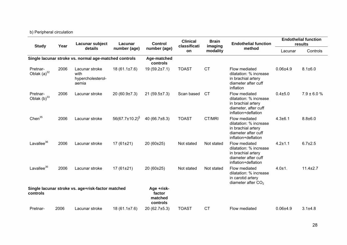
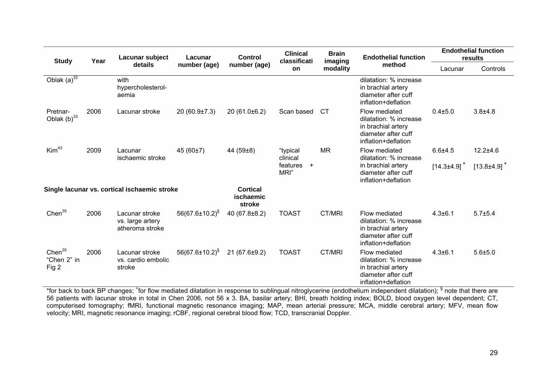
b) Peripheral circulation
Endothelial function results Study Year Lacunar subject
details Lacunar
number (age) Control
number (age) Clinical
classification
Brain imaging modality
Endothelial function method Lacunar Controls
Single lacunar stroke vs. normal age-matched controls Age-matched controls
Pretnar-Oblak (a)32
2006 Lacunar stroke with hypercholesterol-aemia
18 (61.1±7.6) 19 (59.2±7.1) TOAST CT Flow mediated dilatation: % increase in brachial artery diameter after cuff inflation
0.06±4.9 8.1±6.0
Pretnar-Oblak (b)33
2006 Lacunar stroke 20 (60.9±7.3) 21 (59.5±7.3) Scan based CT Flow mediated dilatation: % increase in brachial artery diameter, after cuff inflation+deflation
0.4±5.0 7.9 ± 6.0 %
Chen35 2006 Lacunar stroke 56(67.7±10.2)§ 40 (66.7±8.3) TOAST CT/MRI Flow mediated dilatation: % increase in brachial artery diameter after cuff inflation+deflation
4.3±6.1 8.8±6.0
Lavallee36 2006 Lacunar stroke 17 (61±21) 20 (60±25) Not stated Not stated Flow mediated dilatation: % increase in brachial artery diameter after cuff inflation+deflation
4.2±1.1 6.7±2.5
Lavallee36 2006 Lacunar stroke 17 (61±21) 20 (60±25) Not stated Not stated Flow mediated dilatation: % increase in carotid artery diameter after CO2
4.0±1. 11.4±2.7
Single lacunar stroke vs. age+risk-factor matched controls
Age +risk-factor
matched controls
Pretnar- 2006 Lacunar stroke 18 (61.1±7.6) 20 (62.7±5.3) TOAST CT Flow mediated 0.06±4.9 3.1±4.8
28
29
Endothelial function results Study Year Lacunar subject
details Lacunar
number (age) Control
number (age) Clinical
classification
Brain imaging modality
Endothelial function method Lacunar Controls
Oblak (a)32 with hypercholesterol-aemia
dilatation: % increase in brachial artery diameter after cuff inflation+deflation
Pretnar-Oblak (b)33
2006 Lacunar stroke 20 (60.9±7.3) 20 (61.0±6.2) Scan based CT Flow mediated dilatation: % increase in brachial artery diameter after cuff inflation+deflation
0.4±5.0 3.8±4.8
Kim43 2009 Lacunar ischaemic stroke
45 (60±7) 44 (59±8) “typical clinical features + MRI”
MR Flow mediated dilatation: % increase in brachial artery diameter after cuff inflation+deflation
6.6±4.5 [14.3±4.9] +
12.2±4.6 [13.8±4.9] +
Single lacunar vs. cortical ischaemic stroke Cortical ischaemic
stroke
Chen35 2006 Lacunar stroke vs. large artery atheroma stroke
56(67.6±10.2)§ 40 (67.8±8.2) TOAST CT/MRI Flow mediated dilatation: % increase in brachial artery diameter after cuff inflation+deflation
4.3±6.1 5.7±5.4
Chen35
“Chen 2” in Fig 2
2006 Lacunar stroke vs. cardio embolic stroke
56(67.6±10.2)§ 21 (67.6±9.2) TOAST CT/MRI Flow mediated dilatation: % increase in brachial artery diameter after cuff inflation+deflation
4.3±6.1 5.6±5.0
*for back to back BP changes; +for flow mediated dilatation in response to sublingual nitroglycerine (endothelium independent dilatation); § note that there are 56 patients with lacunar stroke in total in Chen 2006, not 56 x 3. BA, basilar artery; BHI, breath holding index; BOLD, blood oxygen level dependent; CT, computerised tomography; fMRI, functional magnetic resonance imaging; MAP, mean arterial pressure; MCA, middle cerebral artery; MFV, mean flow velocity; MRI, magnetic resonance imaging; rCBF, regional cerebral blood flow; TCD, transcranial Doppler.
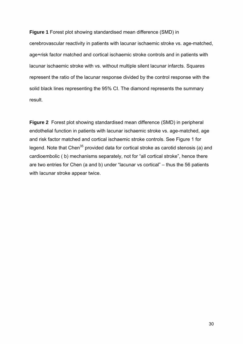
Figure 1 Forest plot showing standardised mean difference (SMD) in
cerebrovascular reactivity in patients with lacunar ischaemic stroke vs. age-matched,
age+risk factor matched and cortical ischaemic stroke controls and in patients with
lacunar ischaemic stroke with vs. without multiple silent lacunar infarcts. Squares
represent the ratio of the lacunar response divided by the control response with the
solid black lines representing the 95% CI. The diamond represents the summary
result.
Figure 2 Forest plot showing standardised mean difference (SMD) in peripheral
endothelial function in patients with lacunar ischaemic stroke vs. age-matched, age
and risk factor matched and cortical ischaemic stroke controls. See Figure 1 for
legend. Note that Chen35 provided data for cortical stroke as carotid stenosis (a) and
cardioembolic ( b) mechanisms separately, not for “all cortical stroke”, hence there
are two entries for Chen (a and b) under “lacunar vs cortical” – thus the 56 patients
with lacunar stroke appear twice.
30
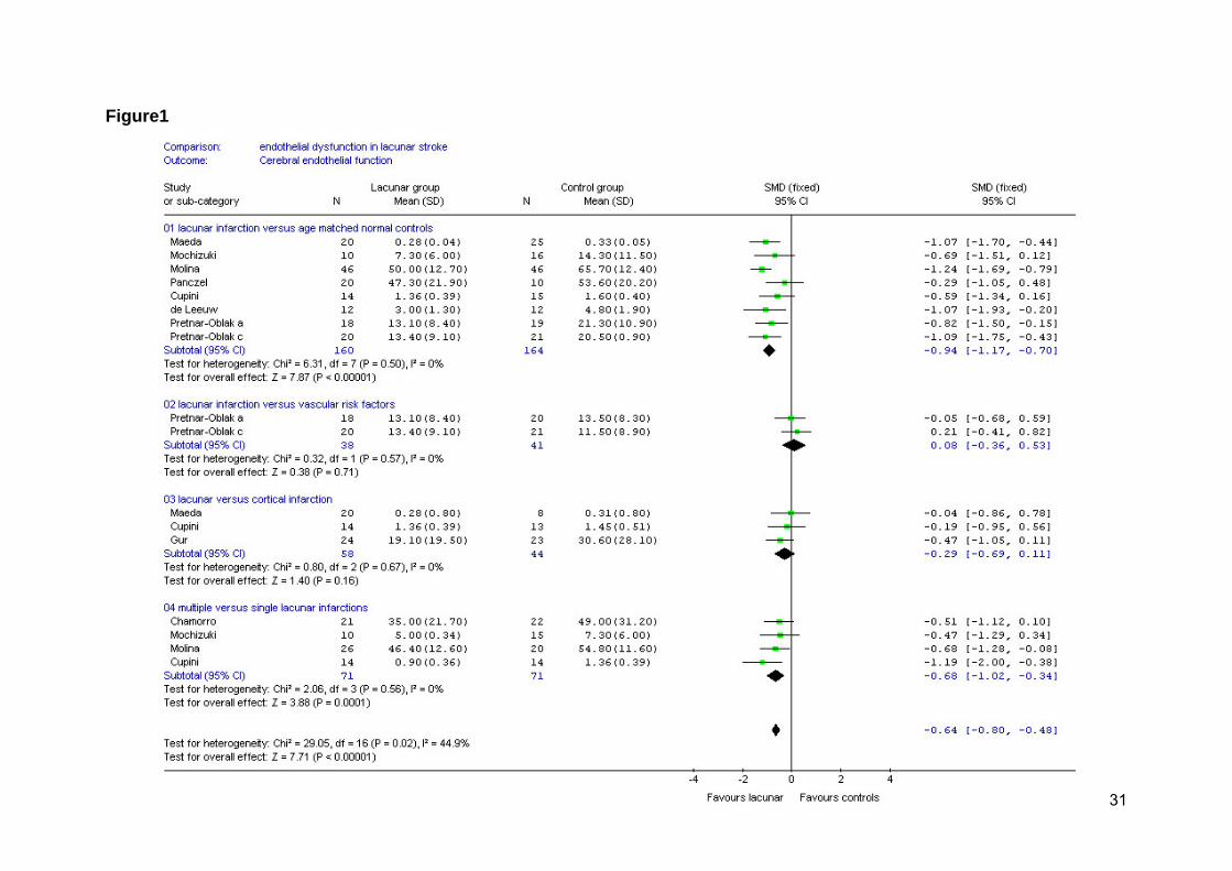
Figure1
31

32
Figure 2

Appendix 1: Search Strategy
1. brain ischemia/ or brain infarction/ or brain stem infarctions/ or cerebral infarction/
or hypoxia-ischemia, brain/ or stroke/
2. (isch?emi$ adj6 (stroke$ or apoplex$ or cerebral vasc$ or cerebrovasc$ or cva or
attack$)).tw.
3. ((brain or cerebr$ or cerebell$ or vertebrobasil$ or hemispher$ or intracran$ or
intracerebral or infratentorial or supratentorial or middle cerebr$ or mca$ or anterior
circulation) adj5 (isch?emi$ or infarct$ or thrombo$ or emboli$ or occlus$ or
hypoxi$)).tw.
4. 1 or 2 or 3
5. (lacun$ or small vessel$ or small infarct$ or microinfarct$ or subcortical lesion$ or
subcortical infarct$ or microvascular$ or microcirculation$).tw.
6. 4 and 5
7. blood-brain barrier/ or endothel$, vascular/ or tunica intima/ or microcirculation/
8. (endotheli$ adj5 (function$ or dysfunction$ or impairment$)).tw.
9. ((vascular or capillary) adj5 endotheli$).tw.
10. (endotheli$ adj5 (contraction or relaxation)).tw.
11. vascular tone/ or arterial stiffness.mp. [mp=title, original title, abstract, name of
substance word, subject heading word]
12. (vascul$ tone or neurovasc$ coupl$ or arterial stiff$ or vascul$ remodel$ or
cerebrovascular reactiv$ or cerebral autoregulation).tw.
13. (Flow mediated adj3 (dilat$ or vasodilat$)).tw.
14. exp Ultrasonography, Doppler, Transcranial/
15. pulse wave analysis.tw.
16. strain gauge plethysmography.tw.
33
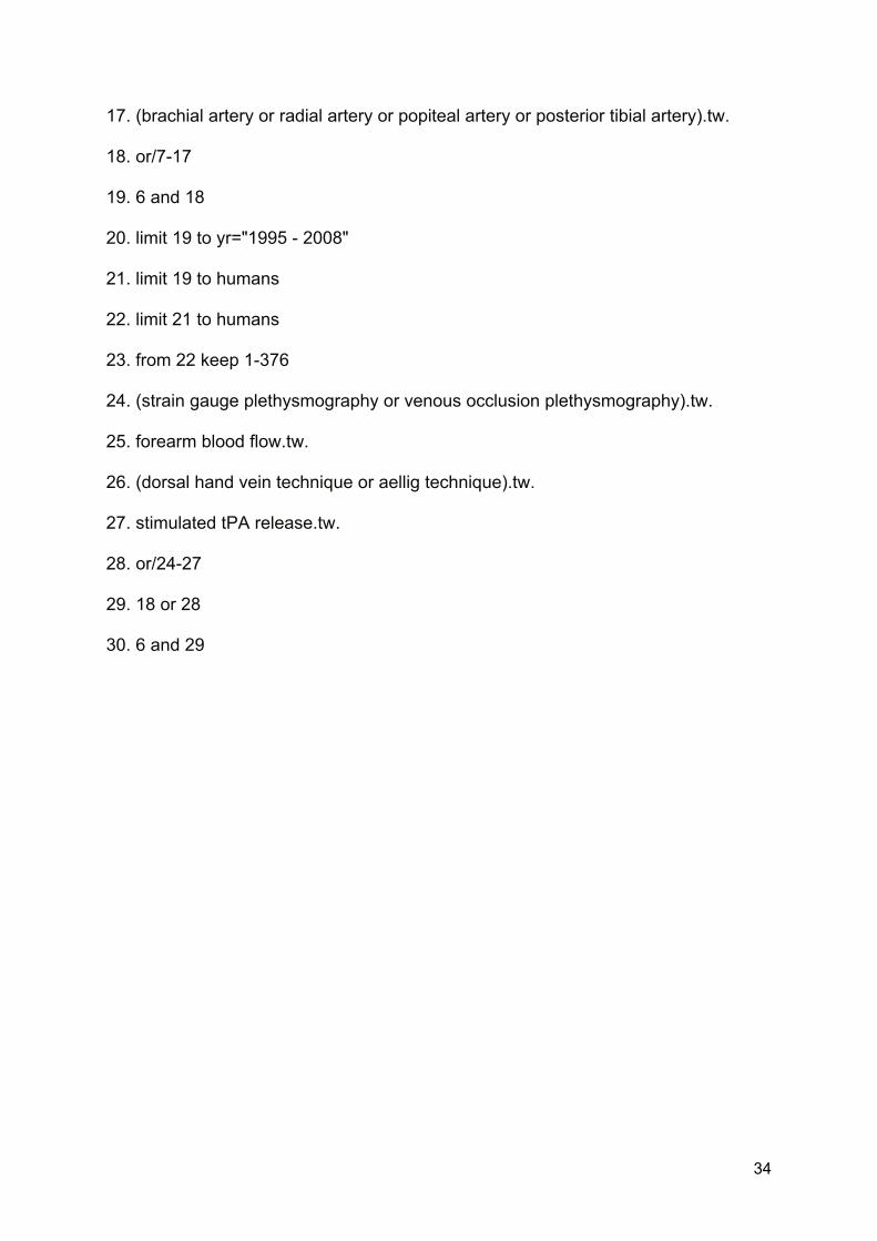
34
17. (brachial artery or radial artery or popiteal artery or posterior tibial artery).tw.
18. or/7-17
19. 6 and 18
20. limit 19 to yr="1995 - 2008"
21. limit 19 to humans
22. limit 21 to humans
23. from 22 keep 1-376
24. (strain gauge plethysmography or venous occlusion plethysmography).tw.
25. forearm blood flow.tw.
26. (dorsal hand vein technique or aellig technique).tw.
27. stimulated tPA release.tw.
28. or/24-27
29. 18 or 28
30. 6 and 29