Embed Size (px)
Citation preview

1
SUPPORTING INFORMATION
Synthesis and Functionalization of Dextran–Based Single–Chain Nanoparticles in Aqueous Media
Raquel Gracia,a Marco Marradi,a Unai Cossío,b Ana Benito,a Adrián Pérez-San Vicente,a Vanessa Gómez-Vallejo,b Hans-Jürgen Grande,a Jordi Llop,b Iraida Loinaza,*
aBiomaterials Unit, IK4-CIDETEC, Pº Miramón 196, 20014, Donostia-San Sebastián, Spain; bRadiochemistry and Nuclear Imaging Group, CIC biomaGUNE, Pº Miramón 182, 20014, Donostia-San Sebastián, Spain.
Materials and methods
Dextran from Leuconostoc spp. (DXT-40, Mr ~40 kDa), glycidyl methacrylate (GMA)
(97%), dimethyl sulfoxide (DMSO) (98%), 3-mercaptopropionic acid (≥99%), 4-(4,6-
dimethoxy-1,3,5-triazin-2-yl)-4-methylmorpholinium chloride (DMTMM·HCl) (96%)
and 2,2′-(ethylenedioxy)diethanethiol [3,6-dioxa-1,8-octane-dithiol (DODT)] (95%)
were purchased from Aldrich. Phosphate-buffered saline (PBS) was purchased from
Scharlau. 4-(Dimethylamino)pyridine (DMAP) was purchased from Acros-Organics.
2,2'-(7-(4-((2-Aminoethyl)amino)-1-carboxy-4-oxobutyl)-1,4,7-triazonane-1,4-
diyl)diaceticacid (NH2-NODA-GA) (98%) was purchased from CheMatech. Water
(H2O) used in the syntheses, unless otherwise stated, was deionized water from a MilliQ
A10 Gradient equipment (Millipore).
Dynamic Light Scattering (DLS): DLS analyses were conducted using a Zetasizer Nano
ZS, ZEN3600 Model (Malvern Instruments Ltd). All measurements were performed in
disposable sizing cuvettes at a laser wavelength of 633 nm and a scattering angle of 173°,
while the zeta-potential measurements were performed in disposable zeta potential cells
(pH 7.4, 25 ºC). Before the measurement, the samples were dispersed in saline solution
(0.9 wt% NaCl for size measurements and 1 mM NaCl for zeta-potential measurements)
at a concentration of 1 mg/mL. Each measurement was repeated for three runs per sample
at 25 °C.
Gel permeation chromatography (GPC): The weight-average molecular weight (Mw),
number-average molecular weight (Mn) and polydispersity index (PDI; Mw/Mn) were
measured at 40 ºC on an Agilent GPC-50 system equipped with 2x PL-Aquagel Mixed-
Electronic Supplementary Material (ESI) for Journal of Materials Chemistry B.This journal is © The Royal Society of Chemistry 2017

2
OH, Guard-Aquagel-OH columns and a differential refractive index (RI) detector. 0.3 M
NaNO3, 0.01 M NaH2PO4, pH 7 was used as eluent at a flow rate of 1 mL/min. The system
was calibrated using polyethylene oxide (PEO) standards.
Transmission electron microscopy (TEM): TEM analyses were performed in a TECNAI
G2 20 TWIN microscope (FEI, Eindhoven, The Netherlands), operating at an accelerating
voltage of 200 KeV in a bright-field image mode. One drop of the sample dispersion in
water (~3 μL, 0.035 mg/mL) was deposited on a carbon film supported on a copper grid
(300 mesh), hydrophilized by a glow discharge process just prior to use. After staining
for 20 seconds with a uranyl acetate aqueous solution (1% w/v), the sample was rotated
at high speed in order to dry at room temperature quickly by spinning process. Number-
average diameter was calculated by ImageJ platform analysis using a Gaussian curve
fitting after counting about 300 nanoparticles.
Nuclear magnetic resonance (1H NMR and DOSY NMR): NMR spectra were recorded on
a Bruker AVANCE III spectrometer at 500 MHz and 25 ºC. Chemical shifts (δ) are given
in ppm relative to the residual signal of the solvent. Splitting patterns: b, broad; s, singlet;
d, doublet; t, triplet; q, quartet; m, multiplet.
Diffusion Coefficient calculations: Taylor dispersion analysis (TDA) studies were
performed on a Viscosizer-TD using fused silica capillaries. The mobile phase was water
and the solutes were monitored by UV absorbance (UV wavelength filter 214 nm) at two
fixed windows. Diffusion coefficient experiments were run at 25 ºC and 140 mbar.
Fourier transform infrared (FTIR) spectra were registered at room temperature in a Jasco
FT/IR 4100 spectrophotometer, using a Gladi ATR accessory.
Synthesis of the dextran methacrylated precursor polymer [DXT-MA (DS ~52%)]
Dextran methacrylated polymer (DXT-MA) was synthesized following a slightly
modified published procedure [1S]. Dextran (DXT-40, 1g, 6.2 mmol) was dissolved in
30 mL of dimethyl sulfoxide (DMSO) under a nitrogen atmosphere, to this solution 200
mg of 4-(N,N-dimethylamino)pyridine (DMAP, 1.6 mmol) was added. Then, 1 mL of
1S van Dijk-Wolthuis, W. N. E.; Kettenes-van den Bosch, J. J.; van der Kerk-van Hoof, A.; Hennink, W. E. Macromolecules 1997, 30, 3411−3413.

3
glycidyl methacrylate (GMA, 7.5 mmol) was incorporated and the mixture was stirred at
room temperature during 4 days. The reaction was stopped by adding an equimolar
amount of concentrated HCl solution (37% v/v, 1.6 mmol, 0.132 mL) to neutralize
DMAP. The modified dextran solution was purified by dialysis against distilled water
(MWCO 3500 Da) at room temperature until reaching deionized water conductivity
values < 1µS (9 days, refreshing with 4 L of deionized water twice per day). Yield: 75%,
DS 52%.1H NMR (500 MHz, D2O) δ ppm: 6.35-6.10 (m, 1H, methacrylic-CH), 5.92-5.72
(m, 1H, methacrylic-CH), 5.54-4.86 (2.4H, including H-1 and H-2/3 MA-substituted),
4.20-3.33 (10.6H, m, rest of Glc), 1.98 (s, 3H, methacrylic-CH3).
Figure S1: 1H NMR (D2O, 500 MHz) of DXT-MA. For clarity reasons, only MA substitution at position 3 of glucose (Glc) has been depicted. DXT has been drawn as linear polysaccharide (α-1,6 glucosidic linkages) although it is known to be branched, as ramifications (α-1,3 glucosidic linkages) are also present.
The degree of substitution (DS, percent of modified hydroxyl groups per repeating unit)
was calculated by 1H NMR through integration of the MA proton signal (integration
reference 1.0) with respect to the signals at 3.3-4.2 ppm corresponding to the protons of
the glucose (Glc) moiety (6H for unsubstituted Glc and 5H for substituted Glc) except the
anomeric protons and the substituted positions (mainly position 3).

4
Preparation of dextran-based single-chain polymer nanoparticles (DXT-SCPN)
In a standard procedure, 0.38 mL of a previously prepared 0.15 M solution (2 mL,
MeOH/PBS, 1:1, v/v, pH= 9.5) of cross-linker DODT (0.06 mmol, 49 L) was added
dropwise using a syringe pump (0.05 mL/h) over a 0.02 M solution of DXT-MA
(DS=52%) (100 mg, 0.025 mmol, 13 mL PBS, pH= 9.5) during 8 h at room temperature
and under constant stirring. After addition, the reaction was maintained stirred at room
temperature for 12 h. Then, the disappearance of the -SH groups from the
homobifunctional cross-linker DODT was checked by Ellamn´s test. Further
characterization studies were carried out after purification of 5 mL sample from the
reaction mixture by dialysis against distilled water (MWCO 3500 Da) until reaching
deionized water conductivity values < 1S (5 days, refreshing with 4 L of deionized water
twice per day). Finally the resulting aqueous solution was freeze-dried to obtain
nanoparticles as a white solid. Yield >90%. 1H NMR (500 MHz, D2O) δ ppm: 6.34-6.12
(m, 1H, methacrylic-CH), 5.94-5.70 (m, 1H, methacrylic-CH), 5.55-4.85 (5.5H, including
H-1 and H-2/3 MA-substituted), 4.34-3.28 (28H, m, rest of Glc and 2xCH2O of cross-
linker), 3.06-2.53 (5H, m, CH(CH3)CH2S, CH2S of cross-linker), 1.98 (s, 3H,
methacrylic-CH3), 1.29 (s, 3H, cross-linker-CH3).Dh (DLS) = 13 ± 8 nm; PDI 0.2.
0
2
4
6
8
10
0.1 1 10 100 1000 10000
Inte
nsity
(Per
cent
)
Size (d.nm)
Size Distribution by Intensity
Record 21: SH 1-84 after reaction
Figure S2: DLS (PBS 10 mM, pH 7.4, 25 ºC) of DXT-SCPN.

5
Figure S3: 1H NMR (D2O, 500 MHz) of DXT-SCPN.
Functionalization of DXT-SCPN with 3-mercaptopropionic acid (DXT-SCPN-F)
One batch synthesis of the functionalized DXT-SCPN-F was achieved by adding slowly
2 mL of an aqueous solution of 3-mercapto propionic acid (61.4 L, 7.5 mol, pH= 9.5)
to the reaction flask in the previously reported synthesis of the DXT-SCPN. The reaction
was stirred for 24 h and the excess acid was removed by dialysis against distilled water
(MWCO 3500Da) until reaching deionized water conductivity values < 1S (5 days,
refreshing with 4L of deionized water twice per day). The resulting aqueous solution was
freeze-dried to obtain nanoparticles as a white solid. Yield >90%. 1H NMR (500 MHz,
D2O) δ ppm: 5.45-4.90 (6H, including H-1 and H-2/3 MA-substituted), 4.13-3.41 (31H,
m, rest of Glc and 2xCH2O of cross-linker), 3.02-2.71 (8H, m, 2x CH(CH3)CH2S, CH2S
of cross-linker and MPA), 2.70-2.49 (2H, m, CH2COOH of MPA), 1.29 (s, 5.4H, cross-
linker- and MPA-CH3) Mw (GPC) = 38KDa, Mw/Mn = 1.7; Dh (DLS) = 15 ± 4 nm; PDI
0.2, Zetapotential (pH = 7.2, 25 °C) = -20mV ± 5. TEM (uranyl acetate staining): 13 ± 3 nm.
6
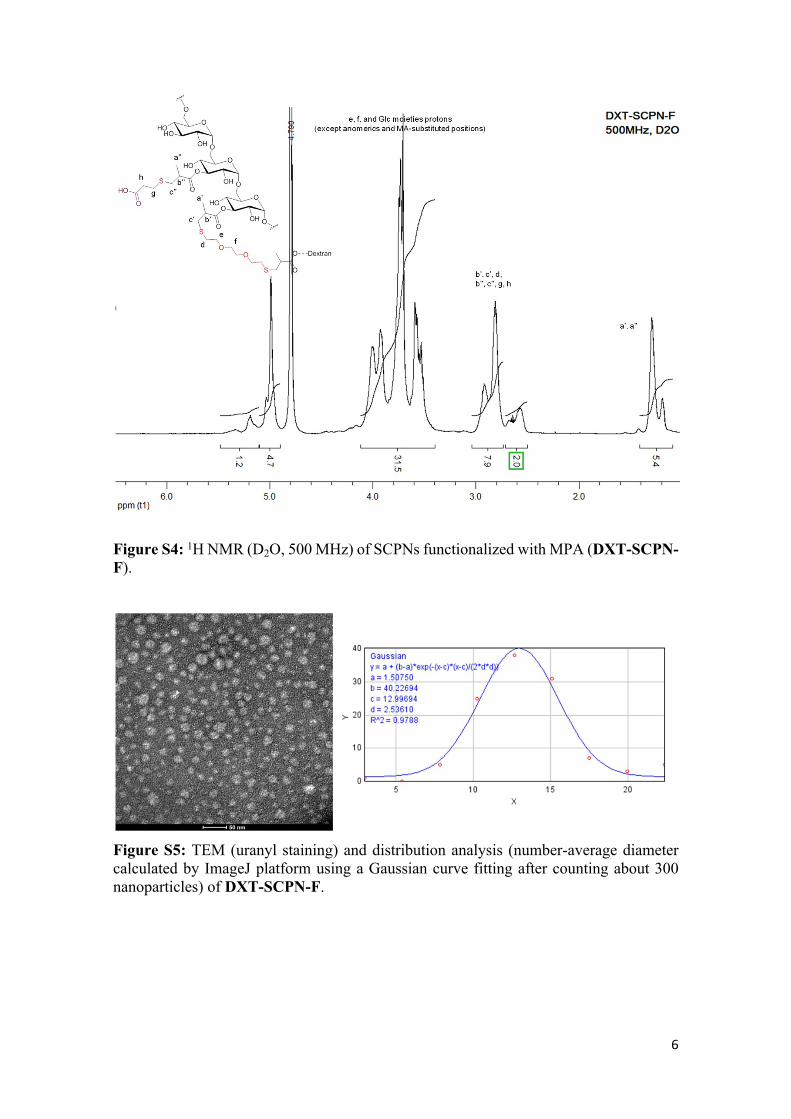
Figure S4: 1H NMR (D2O, 500 MHz) of SCPNs functionalized with MPA (DXT-SCPN-F).
Figure S5: TEM (uranyl staining) and distribution analysis (number-average diameter calculated by ImageJ platform using a Gaussian curve fitting after counting about 300 nanoparticles) of DXT-SCPN-F.
7
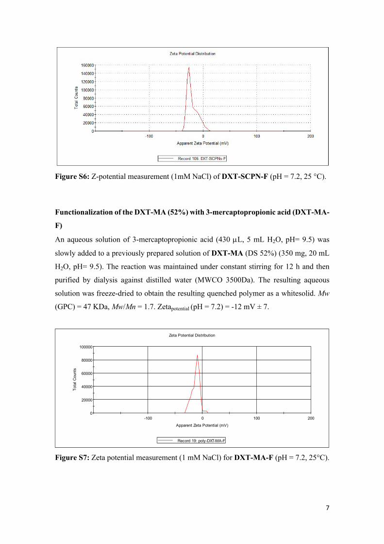
Figure S6: Z-potential measurement (1mM NaCl) of DXT-SCPN-F (pH = 7.2, 25 °C).
Functionalization of the DXT-MA (52%) with 3-mercaptopropionic acid (DXT-MA-
F)
An aqueous solution of 3-mercaptopropionic acid (430 L, 5 mL H2O, pH= 9.5) was
slowly added to a previously prepared solution of DXT-MA (DS 52%) (350 mg, 20 mL
H2O, pH= 9.5). The reaction was maintained under constant stirring for 12 h and then
purified by dialysis against distilled water (MWCO 3500Da). The resulting aqueous
solution was freeze-dried to obtain the resulting quenched polymer as a whitesolid. Mw
(GPC) = 47 KDa, Mw/Mn = 1.7. Zetapotential (pH = 7.2) = -12 mV ± 7.
0
20000
40000
60000
80000
100000
-100 0 100 200
Tota
l Cou
nts
Apparent Zeta Potential (mV)
Zeta Potential Distribution
Record 19: poly-DXT-MA-F
Figure S7: Zeta potential measurement (1 mM NaCl) for DXT-MA-F (pH = 7.2, 25°C).
8
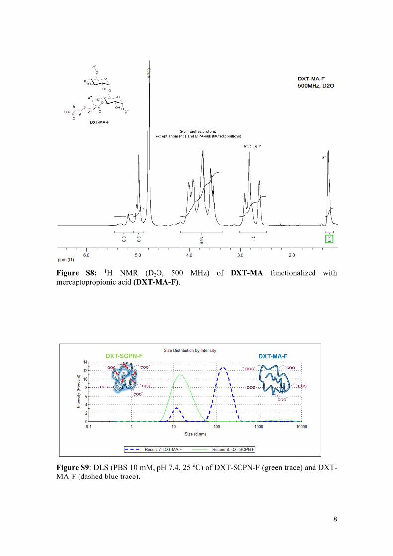
Figure S8: 1H NMR (D2O, 500 MHz) of DXT-MA functionalized with mercaptopropionic acid (DXT-MA-F).
Figure S9: DLS (PBS 10 mM, pH 7.4, 25 ºC) of DXT-SCPN-F (green trace) and DXT-MA-F (dashed blue trace).
9
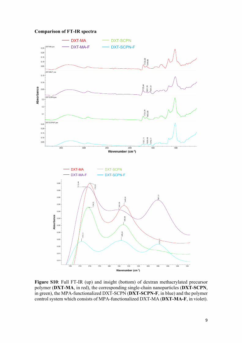
Comparison of FT-IR spectra
Figure S10: Full FT-IR (up) and insight (bottom) of dextran methacrylated precursor polymer (DXT-MA, in red), the corresponding single-chain nanoparticles (DXT-SCPN, in green), the MPA-functionalized DXT-SCPN (DXT-SCPN-F, in blue) and the polymer control system which consists of MPA-functionalized DXT-MA (DXT-MA-F, in violet).
10
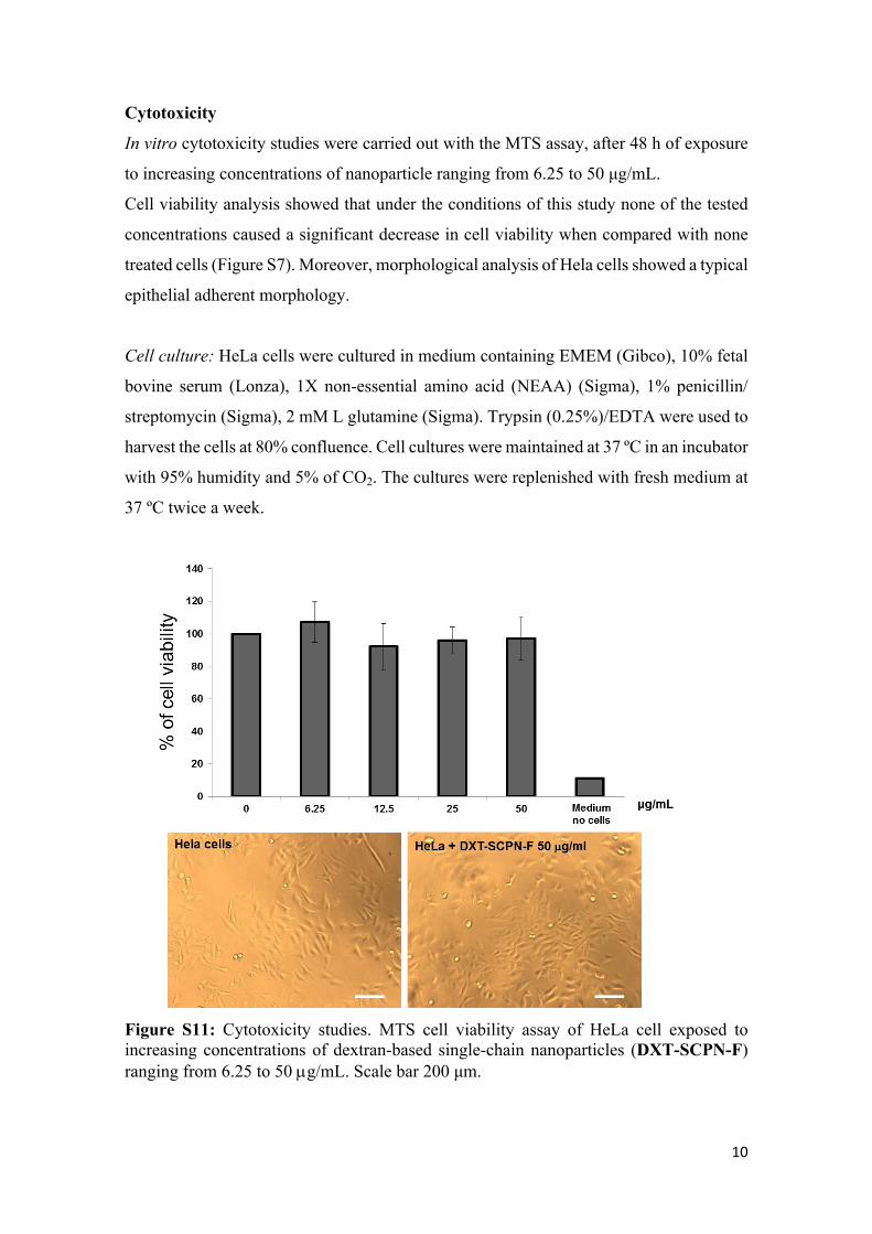
Cytotoxicity
In vitro cytotoxicity studies were carried out with the MTS assay, after 48 h of exposure
to increasing concentrations of nanoparticle ranging from 6.25 to 50 μg/mL.
Cell viability analysis showed that under the conditions of this study none of the tested
concentrations caused a significant decrease in cell viability when compared with none
treated cells (Figure S7). Moreover, morphological analysis of Hela cells showed a typical
epithelial adherent morphology.
Cell culture: HeLa cells were cultured in medium containing EMEM (Gibco), 10% fetal
bovine serum (Lonza), 1X non-essential amino acid (NEAA) (Sigma), 1% penicillin/
streptomycin (Sigma), 2 mM L glutamine (Sigma). Trypsin (0.25%)/EDTA were used to
harvest the cells at 80% confluence. Cell cultures were maintained at 37 ºC in an incubator
with 95% humidity and 5% of CO2. The cultures were replenished with fresh medium at
37 ºC twice a week.
Figure S11: Cytotoxicity studies. MTS cell viability assay of HeLa cell exposed to increasing concentrations of dextran-based single-chain nanoparticles (DXT-SCPN-F) ranging from 6.25 to 50 g/mL. Scale bar 200 μm.
11
MTS assay: HeLa cell growth was evaluated using Cell Titer 96 ® Aqueous One Solution
Cell proliferation Assay (Promega). HeLa cells were seeded at a density of 10000cl/cm2
in p96 well plates and allowed to grow for 24 h. After removing the medium, 100 µl of
HeLa medium containing various concentrations nanoparticles ranging from 6.25 to 50
µg/mL were added and further incubated for 48 h. At 48 h post nanoparticles incubation,
cells were cultured in a 37 ºC of humidified incubator for 2 h with 20µL of Cell Titer 96
® Aqueous One Solution Reagent containing tetrazolium compound [3-(4,5-
dimethylthiazol-2-yl)-5-(3-carboxymethoxyphenyl)-2-(4 sulfophenyl)-2H-tetrazolium)]
and an electron coupling reagent, phenazine ethosulfate (PES) per 100 µL of cultured
media. The absorbance per well was measured at 490 nm using a micro-plate reader
(Multiscan ascent, Thermo). All experiments were performed in triplicate.
Functionalization of the SCPNs with NODA and radiolabeling for in vivo
biodistribution studies
Functionalization: To a stirred solution of DXT-SCPN-F (20 mg) in DMSO (8 mL),
10mg of DMTMM·HCl (0.04 mmol) were added and the mixture was maintained under
constant stirring for 15 min. Then, a solution of NH2-NODA-GA (10mg, 0.02mmol) in
DMSO (1mL) was added to the reaction flask and the reaction was stirred at room
temperature for 12 h. The reaction was purified by dialysis against distilled water
(MWCO 1000 Da) until reaching deionized water conductivity values (< 1 S). The
amount of NODA (0.1mg NODA/1mg DXT-SCPN-F) was calculated by loading Zn(II)
which concentration was determined by colorimetric titration in acetate buffer (pH=4.5)
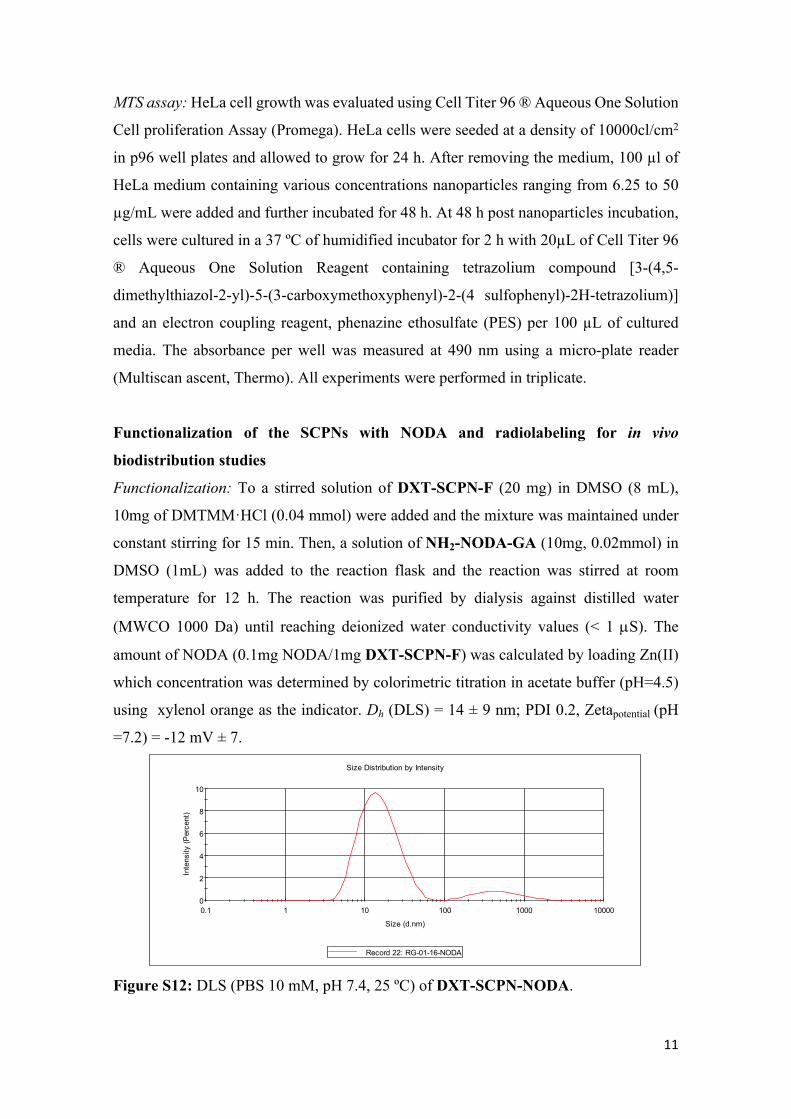
using xylenol orange as the indicator. Dh (DLS) = 14 ± 9 nm; PDI 0.2, Zetapotential (pH
=7.2) = -12 mV ± 7.
0
2
4
6
8
10
0.1 1 10 100 1000 10000
Inte
nsity
(Per
cent
)
Size (d.nm)
Size Distribution by Intensity
Record 22: RG-01-16-NODA
Figure S12: DLS (PBS 10 mM, pH 7.4, 25 ºC) of DXT-SCPN-NODA.
12
0
20000
40000
60000
80000
100000
120000
140000
160000
-100 0 100 200
Tota
l Cou
nts
Apparent Zeta Potential (mV)
Zeta Potential Distribution
Record 11: 3
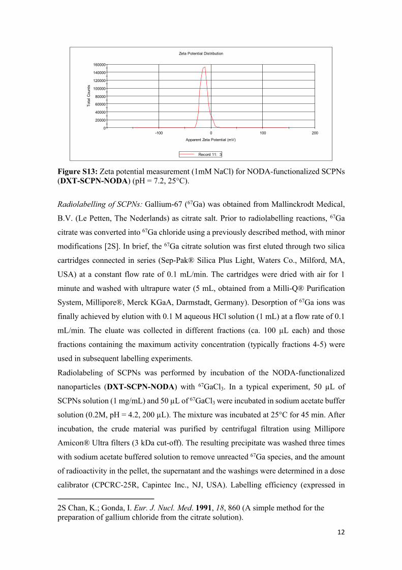
Figure S13: Zeta potential measurement (1mM NaCl) for NODA-functionalized SCPNs (DXT-SCPN-NODA) (pH = 7.2, 25°C).
Radiolabelling of SCPNs: Gallium-67 (67Ga) was obtained from Mallinckrodt Medical,
B.V. (Le Petten, The Nederlands) as citrate salt. Prior to radiolabelling reactions, 67Ga
citrate was converted into 67Ga chloride using a previously described method, with minor
modifications [2S]. In brief, the 67Ga citrate solution was first eluted through two silica
cartridges connected in series (Sep-Pak® Silica Plus Light, Waters Co., Milford, MA,
USA) at a constant flow rate of 0.1 mL/min. The cartridges were dried with air for 1
minute and washed with ultrapure water (5 mL, obtained from a Milli-Q® Purification
System, Millipore®, Merck KGaA, Darmstadt, Germany). Desorption of 67Ga ions was
finally achieved by elution with 0.1 M aqueous HCl solution (1 mL) at a flow rate of 0.1
mL/min. The eluate was collected in different fractions (ca. 100 µL each) and those
fractions containing the maximum activity concentration (typically fractions 4-5) were
used in subsequent labelling experiments.
Radiolabeling of SCPNs was performed by incubation of the NODA-functionalized
nanoparticles (DXT-SCPN-NODA) with 67GaCl3. In a typical experiment, 50 µL of
SCPNs solution (1 mg/mL) and 50 µL of 67GaCl3 were incubated in sodium acetate buffer
solution (0.2M, pH = 4.2, 200 µL). The mixture was incubated at 25°C for 45 min. After
incubation, the crude material was purified by centrifugal filtration using Millipore
Amicon® Ultra filters (3 kDa cut-off). The resulting precipitate was washed three times
with sodium acetate buffered solution to remove unreacted 67Ga species, and the amount
of radioactivity in the pellet, the supernatant and the washings were determined in a dose
calibrator (CPCRC-25R, Capintec Inc., NJ, USA). Labelling efficiency (expressed in
2S Chan, K.; Gonda, I. Eur. J. Nucl. Med. 1991, 18, 860 (A simple method for the preparation of gallium chloride from the citrate solution).

13
percentage) was calculated as the ratio between the amount of radioactivity in the filter
and the total amount of radioactivity in all fractions. Finally, the nanoparticles were
suspended in 0.2 M sodium acetate buffer solution (pH 4.2). Radiochemical yield was
calculated as the ratio between the amount of radioactivity in the resuspended fraction
and the starting amount of radioactivity.
Radiochemical stability of SCPNs: Radiolabelled nanoparticles prepared as described
above were incubated in sodium acetate buffered solution at 37 °C using a digital block
heater. At different time points (1, 3, 24, 48, 72 and 144 h) samples were withdrawn and
the amount of radioactivity was measured. The 67Ga-radiolabelled SCPNs were filtered,
washed twice with ultrapure water, and the amount of radioactivity in the filter and the
filtrate/washings was measured. The radiochemical stability was calculated as the
percentage of radioactivity in the pellet with respect to the total amount of radioactivity
(pellet + filtrate + washings).
Animal studies: Animals were maintained and handled in accordance with the Guidelines
for Accommodation and Care of Animals (European Convention for the Protection of
Vertebrate Animals Used for Experimental and Other Scientific Purposes). All animal
procedures were performed in accordance with the Spanish policy for animal protection
(RD53/2013), which meets the requirements of the European Union directive
2010/63/UE regarding their protection during experimental procedures. Experimental
procedures were approved by the Ethical Committee of CIC biomaGUNE and authorized
by the regional government.
Administration of the radiolabelled SCPNs: Six-to–eight weeks-old female Sprague
Dawley rats (Janvier, Le Genest-Saint-Isle, France) were used. Rats were anesthetized by
an intraperitoneal injection of a mixture of medetomidine, midazolam and fentanyl (0.6,
6 and 0.02 mg/Kg, respectively). Once animals (n = 4) were under sedation, 67Ga-
radiolabelled SCPNs were administered by intratracheal nebulization using a Penn-
Century MicroSprayer® Aerosolizer (FMJ-250 High Pressure Syringe Model, Penn-
Century. Inc. Wyndmoor, USA). A small animal Laryngoscope (Model LS-2, Penn-
Century. Inc.) was used for correct visualization of the epiglottis, ensuring a correct
positioning of the tip just above the carina. A pre-defined volume of radiolabelled SCPNs
(50 µL, established by using spacers in the syringe plunger) was administered (amount of

14
radioactivity around 1.85 MBq). Immediately after, rats were submitted to in vivo
imaging studies.
In vivo imaging studies: Immediately after administration of the radiolabeled SCPNs, and
without recovering from sedation, animals were positioned in an eXplore speCZT CT
preclinical imaging system (GE Healthcare, USA) to perform in vivo studies. Body
temperature was maintained with a homeothermic blanket control unit (Bruker BioSpin
GmbH, Karlsruhe, Germany) to prevent hypothermia, and SPECT scans were acquired
for 30 min. After the SPECT scan, a CT acquisition was performed to provide anatomical
information of each animal. The SPECT images were reconstructed using a ordered-
subset expectation maximization (OSEM) iterative algorithm (3 iterations/3 subsets,
128 x 128 x 32 array with a voxel size of 0.55 x 0.55 x 2.46 mm3), whereas for the CT a
cone beam filtered back-projection a Feldkamp algorithm (437 x 437 x 800 array with a
voxel size of 0.2 x 0.2 x 0.2 mm3) was used. After reconstruction, images were quantified
using πMOD analysis software (version 3.4, PMOD Technologies Ltd.). Volumes of
interest (VOIs) were manually drawn in the lungs on the CT images and translated to the
SPECT images. The relative concentration of radioactivity in the different VOIs was
finally determined.