Embed Size (px)
DESCRIPTION
بسم الله الرحمن الرحيم. ﴿ و قل رب زدنى علماً ﴾. صدق الله العظيم. Introduction to Radiotherapy. RAD 461. Dr. Ahmed Al Far, M.D. Professor of Radiation Oncology. What is the Target Volumes?. The Target Volumes. ◘ Before Planning: - PowerPoint PPT Presentation
Citation preview

بسم الله الرحمن الرحيمبسم الله الرحمن الرحيم
و قل رب زدنى و قل رب زدنى ﴿﴾علمًا�علمًا�
صدق الله العظيمصدق الله العظيم

Introduction to Radiotherapy
Dr. Ahmed Al Far, M.D.Professor of Radiation Oncology
RAD 461RAD 461

What is the Target VolumesWhat is the Target Volumes??
The Target Volumes
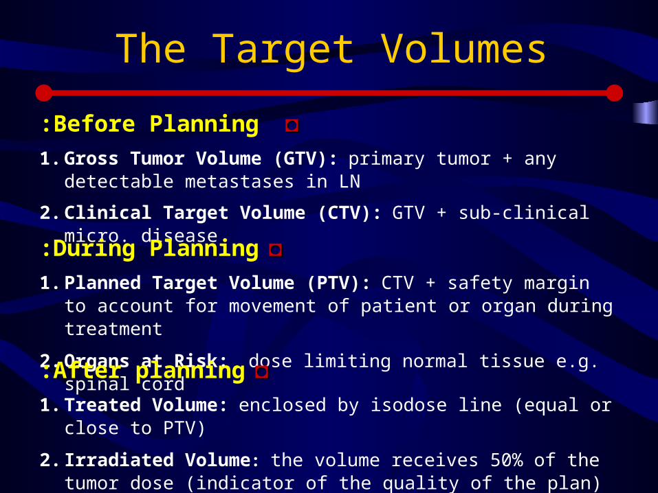
◘◘ Before Planning:
1. Gross Tumor Volume (GTV): primary tumor + any detectable metastases in LN
2. Clinical Target Volume (CTV): GTV + sub-clinical micro. disease
◘◘ During Planning:
1. Planned Target Volume (PTV): CTV + safety margin to account for movement of patient or organ during treatment
2. Organs at Risk: dose limiting normal tissue e.g. spinal cord
◘◘ After planning:
1. Treated Volume: enclosed by isodose line (equal or close to PTV)
2. Irradiated Volume: the volume receives 50% of the tumor dose (indicator of the quality of the plan)

The Target Volumes
Clinical target volume
Gross Tumor volume
Planning target volumeTreated volume
Irradiated volume
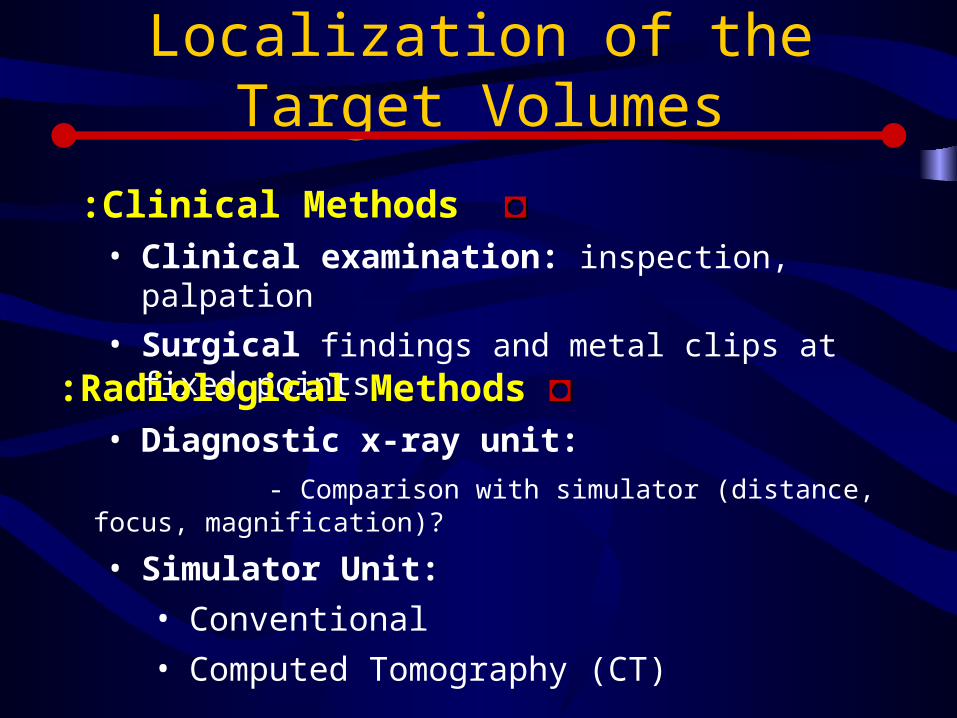
◘◘ Clinical Methods :• Clinical examination: inspection, palpation
• Surgical findings and metal clips at fixed points
◘◘ Radiological Methods:• Diagnostic x-ray unit:
- Comparison with simulator (distance, focus, magnification)?
• Simulator Unit:
• Conventional
• Computed Tomography (CT)
Localization of the Target Volumes

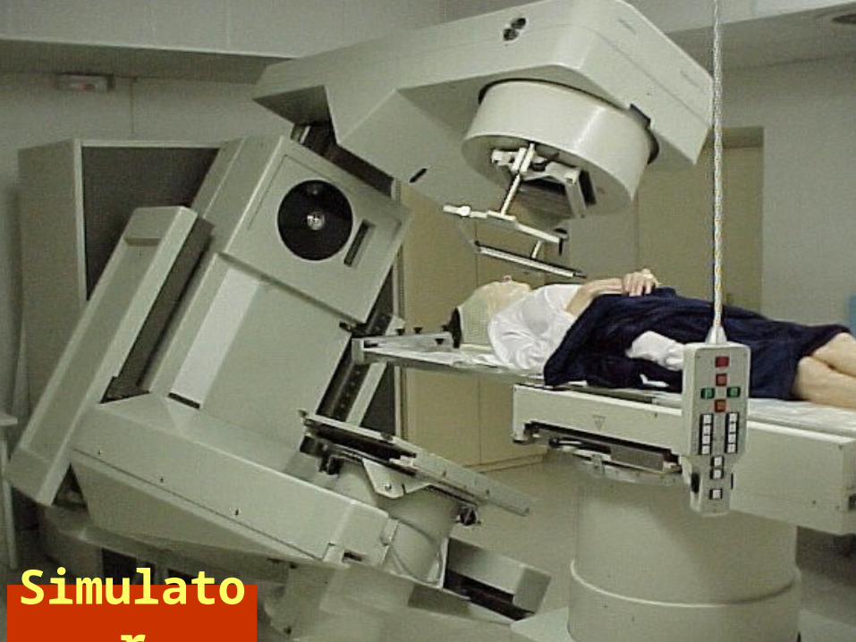
What is the Simulators?

Simulator
A diagnostic x-ray machine which:
accurately simulates the geometry, optical systems and mechanical movements of a radiotherapy machine.
Importance:
1. Localization: the target volume and critical normal tissue with respect to skin marks
2. Simulation: the planned treatment fields with respect to tumor and normal tissue

Simulator
ZX
Y

Simulator: Main components
• Gantry rotation around a horizontal axis
• Source axis distance (SAD) motion in radial direction
• Collimator rotation
• Image intensifier motion in the vertical, longitudinal and
radial directions
• Table movement ( rotation, vertical, longitudinal and
lateral)
• Movement of X-ray field collimators and the wires (ruler)
Simulator

Shielding Block Tray
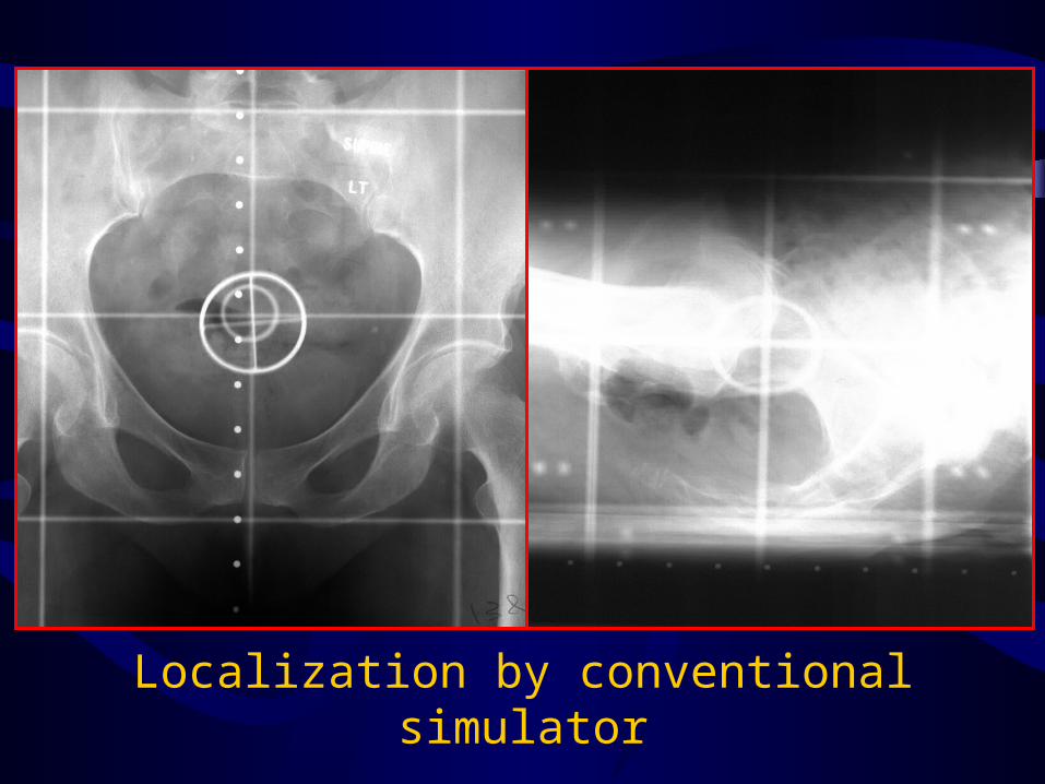
Localization by conventional simulator

Conventional & CT Simulators
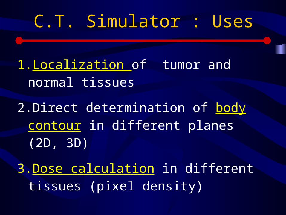
C.T. Simulator : Uses
1.Localization of tumor and normal tissues
2.Direct determination of body contour in different planes (2D, 3D)
3.Dose calculation in different tissues (pixel density)
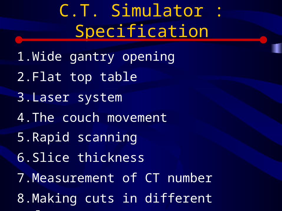
C.T. Simulator : Specification
1.Wide gantry opening
2.Flat top table
3.Laser system
4.The couch movement
5.Rapid scanning
6.Slice thickness
7.Measurement of CT number
8.Making cuts in different planes

Conventional & CT Simulators
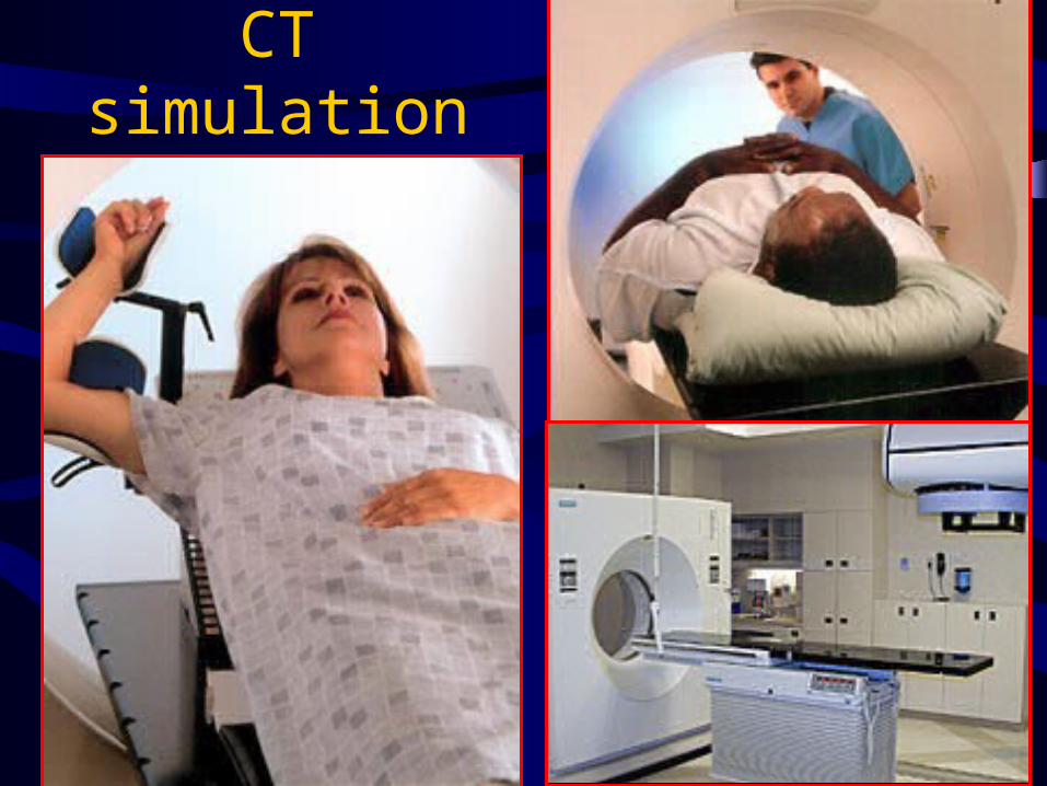
CT simulation

The CT scans are linked with a specialized treatment planning computer providing detailed information which
will assist with optimum treatment .
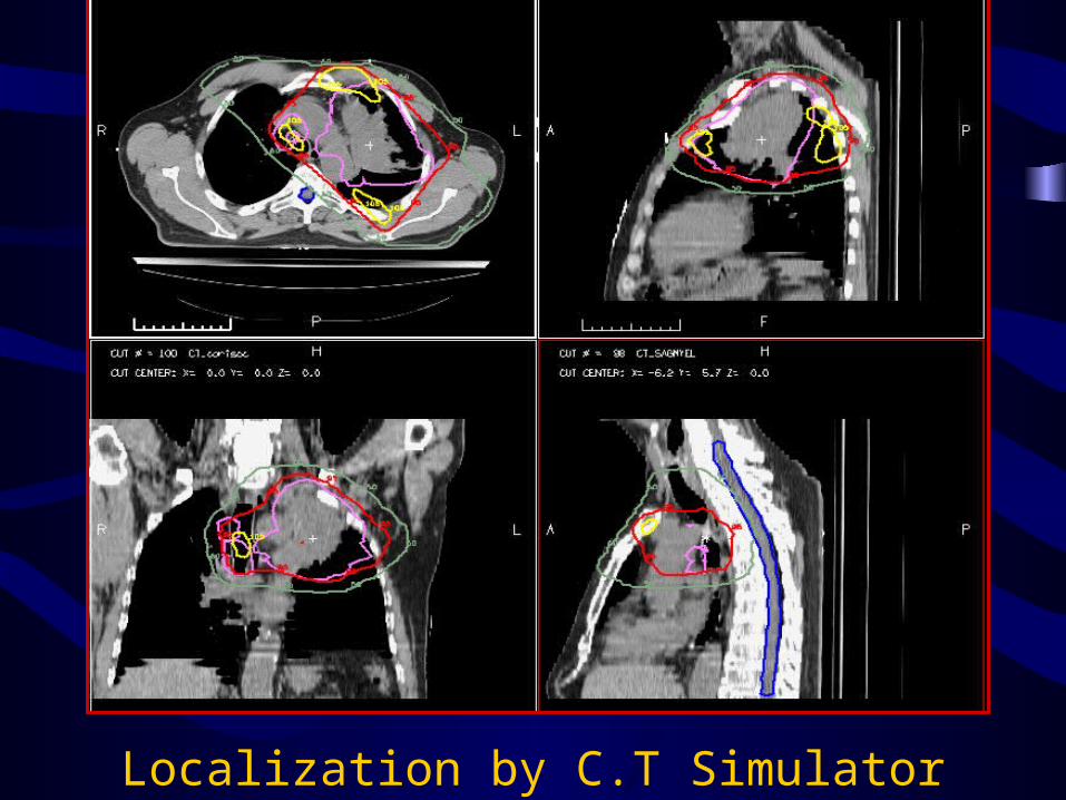
Localization by C.T Simulator