Embed Size (px)
Citation preview
© 2015 Pearson Education, Inc.
Muscle Tissue
• Function is to contract, or shorten, to produce movement
• Three types:1. Skeletal muscle2. Cardiac muscle3. Smooth muscle
Muscle and Nervous Tissue
Pages 97-101

Three types of muscle tissue
• Skeletal• Cardiac• Smooth
© 2015 Pearson Education, Inc.
Skeletal Muscle Tissue
• Voluntarily (consciously) controlled• Attached to the bones or skin• Produces gross body movements or facial
expressions• Characteristics of skeletal muscle cells– Striations (stripes)– Multinucleate (more than one nucleus)– Long, cylindrical shape
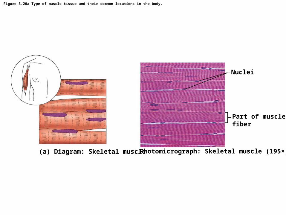
Figure 3.20a Type of muscle tissue and their common locations in the body.
Nuclei
Part of musclefiber
Photomicrograph: Skeletal muscle (195×)(a) Diagram: Skeletal muscle
© 2015 Pearson Education, Inc.
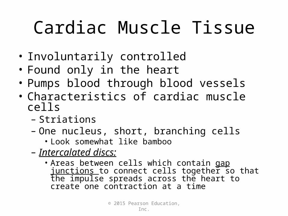
Cardiac Muscle Tissue• Involuntarily controlled• Found only in the heart• Pumps blood through blood vessels• Characteristics of cardiac muscle cells– Striations– One nucleus, short, branching cells
• Look somewhat like bamboo– Intercalated discs:
• Areas between cells which contain gap junctions to connect cells together so that the impulse spreads across the heart to create one contraction at a time
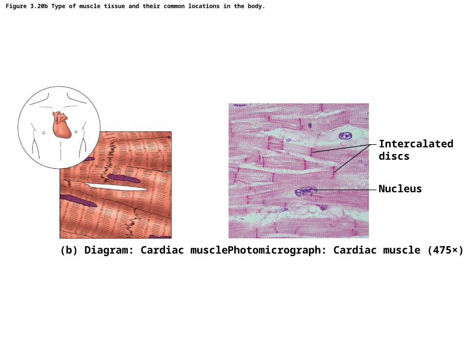
Figure 3.20b Type of muscle tissue and their common locations in the body.
Intercalateddiscs
Nucleus
Photomicrograph: Cardiac muscle (475×)(b) Diagram: Cardiac muscle
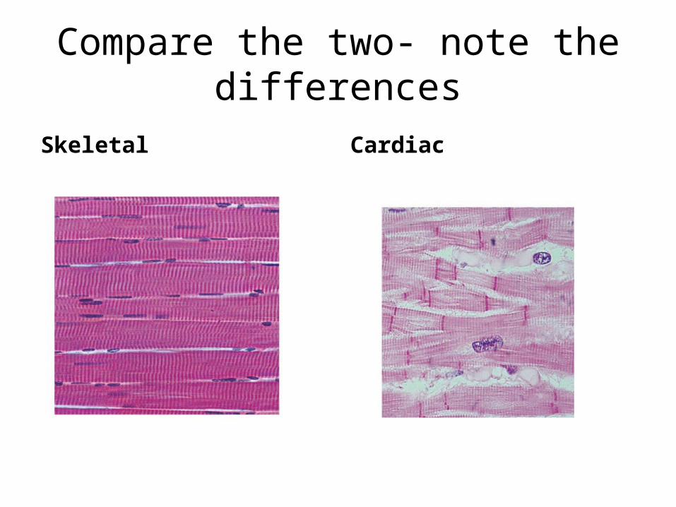
Compare the two- note the differences
Skeletal Cardiac
© 2015 Pearson Education, Inc.
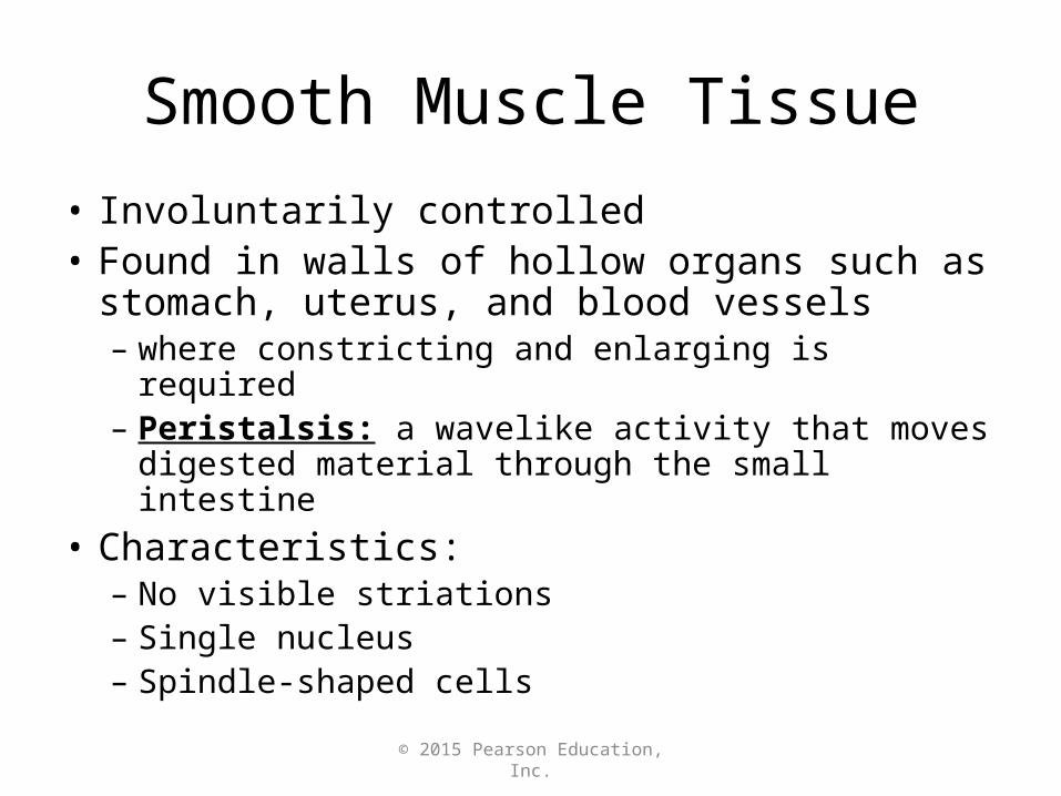
Smooth Muscle Tissue
• Involuntarily controlled• Found in walls of hollow organs such as stomach,
uterus, and blood vessels– where constricting and enlarging is required– Peristalsis: a wavelike activity that moves digested
material through the small intestine• Characteristics:– No visible striations– Single nucleus– Spindle-shaped cells
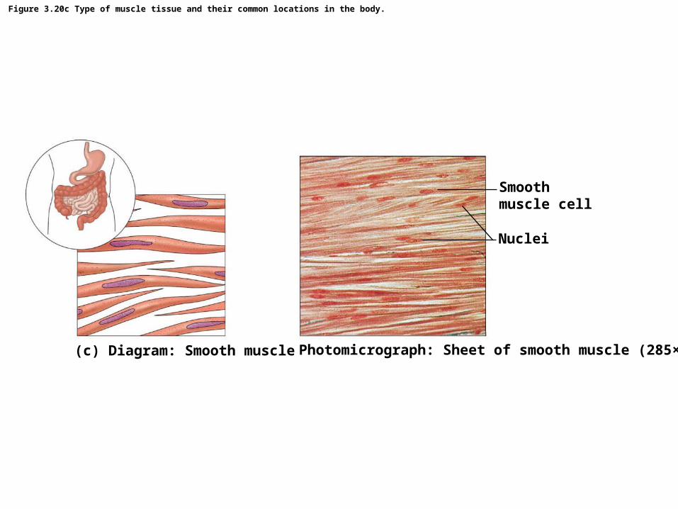
Figure 3.20c Type of muscle tissue and their common locations in the body.
Smoothmuscle cell
Nuclei
Photomicrograph: Sheet of smooth muscle (285×)(c) Diagram: Smooth muscle

© 2015 Pearson Education, Inc.
Nervous Tissue
• Two types of cells:– Neurons– nerve support cells called neuroglia• these insulate, protect, and support neurons
• Function: receive and conduct electrochemical impulses to and from body parts– Irritability– Conductivity

Figure 3.21 Nervous tissue.
Brain
Spinalcord
Nuclei ofsupportingcells
Cell bodyof neuron
Neuronprocesses
Nuclei ofsupportingcells
Neuronprocesses
Cell bodyof neuron
Diagram: Nervous tissue
Photomicrograph: Neurons (320×)

Figure 3.22 Summary of the major functions and body locations of the four tissue types: epithelial, connective, muscle, and nervous tissues.
Nervous tissue: Internal communication• Brain, spinal cord, and nerves
Muscle tissue: Contracts to cause movement
Epithelial tissue: Forms boundaries betweendifferent environments, protects, secretes, absorbs,filters
Connective tissue: Supports, protects, bindsother tissues together
• Muscles attached to bones (skeletal)• Muscles of heart (cardiac)• Muscles of walls of hollow organs (smooth)
• Lining of GI tract organs and other hollow organs• Skin surface (epidermis)
• Bones• Tendons• Fat and other soft padding tissue
© 2015 Pearson Education, Inc.
Tissue Repair (Wound Healing)
• Occurs in two ways:1. Regeneration• Replacement of destroyed tissue by the same kind of
cells; cells divide to make new ones
2. Fibrosis• Repair by dense (fibrous) connective tissue (scar tissue)

© 2015 Pearson Education, Inc.
Tissue Repair (Wound Healing)
• The type of healing is determined by:1. Type of tissue damaged2. Severity of the injury
© 2015 Pearson Education, Inc.
Events in Tissue Repair
• Inflammation– Capillaries become very permeable– Clotting proteins migrate into the area from the
bloodstream– A clot (visible fibers from blood) walls off the injured
area• Granulation tissue forms– Growth of new capillaries– Phagocytes dispose of blood clot and fibroblasts– Rebuild collagen fibers

© 2015 Pearson Education, Inc.
Events in Tissue Repair
• Regeneration of surface epithelium– Scab detaches– Whether scar is visible or invisible depends on
severity of wound• Scar tissue is constructed of many collagen fibers

© 2015 Pearson Education, Inc.
Regeneration of Tissues
• regenerate easily– Epithelial tissue (skin and mucous membranes)– Fibrous connective tissues and bone
• regenerate poorly– Skeletal muscle– Dense connective tissues like tendons, ligaments
• replaced largely with scar tissue– Cardiac muscle– Nervous tissue within the brain and spinal cord

© 2015 Pearson Education, Inc.
Development Aspects of Cells and Tissues
• Growth via cell division continues through puberty• Cell exposed to friction replace lost cells throughout
life (like epithelial cells)• Connective tissue forms repair (scar) tissue– Scar tissue is a form of connective tissue constructed of
many collagen fibers
• Cells that lose ability to divide: (become amitotic)– muscle tissue (by the end of puberty)– Nervous tissue (shortly after birth)