Embed Size (px)
Citation preview

© 2014 Pearson Education, Inc.
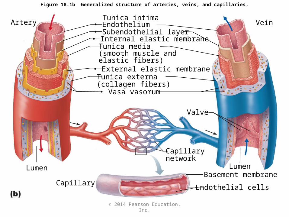
Figure 18.1a Generalized structure of arteries, veins, and capillaries.
Artery
Vein
© 2014 Pearson Education, Inc.
Tunica intima
• Internal elastic membraneTunica media(smooth muscle and elastic fibers)• External elastic membrane
Tunica externa(collagen fibers)
Lumen
Artery
Capillarynetwork
Lumen
Vein
Endothelial cellsCapillary
Basement membrane
• Vasa vasorum
Figure 18.1b Generalized structure of arteries, veins, and capillaries.
Valve
• Endothelium• Subendothelial layer
© 2014 Pearson Education, Inc.
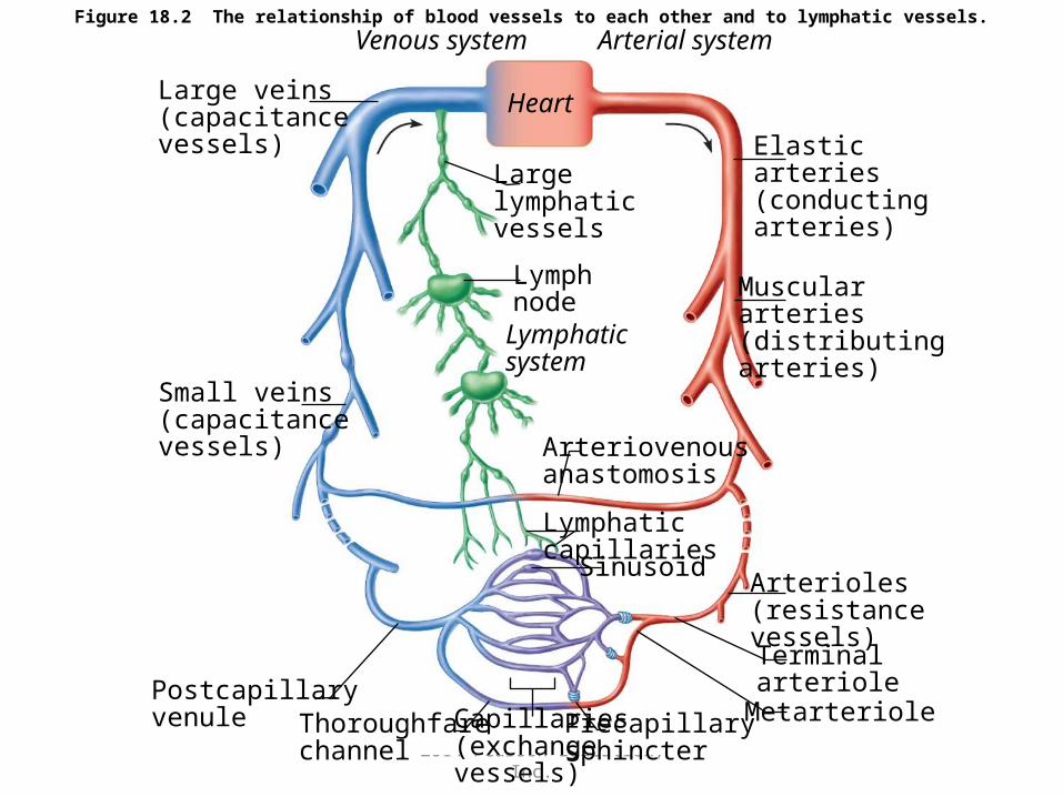
Figure 18.2 The relationship of blood vessels to each other and to lymphatic vessels.Venous system Arterial system
Large veins(capacitancevessels)
Largelymphaticvessels
Elasticarteries(conductingarteries)
Musculararteries(distributingarteries)
LymphnodeLymphaticsystem
Small veins(capacitancevessels) Arteriovenous
anastomosis
Lymphaticcapillaries
Sinusoid Arterioles(resistancevessels)Terminalarteriole
MetarteriolePrecapillarysphincter
Capillaries(exchangevessels)
Thoroughfarechannel
Postcapillaryvenule
Heart
© 2014 Pearson Education, Inc.
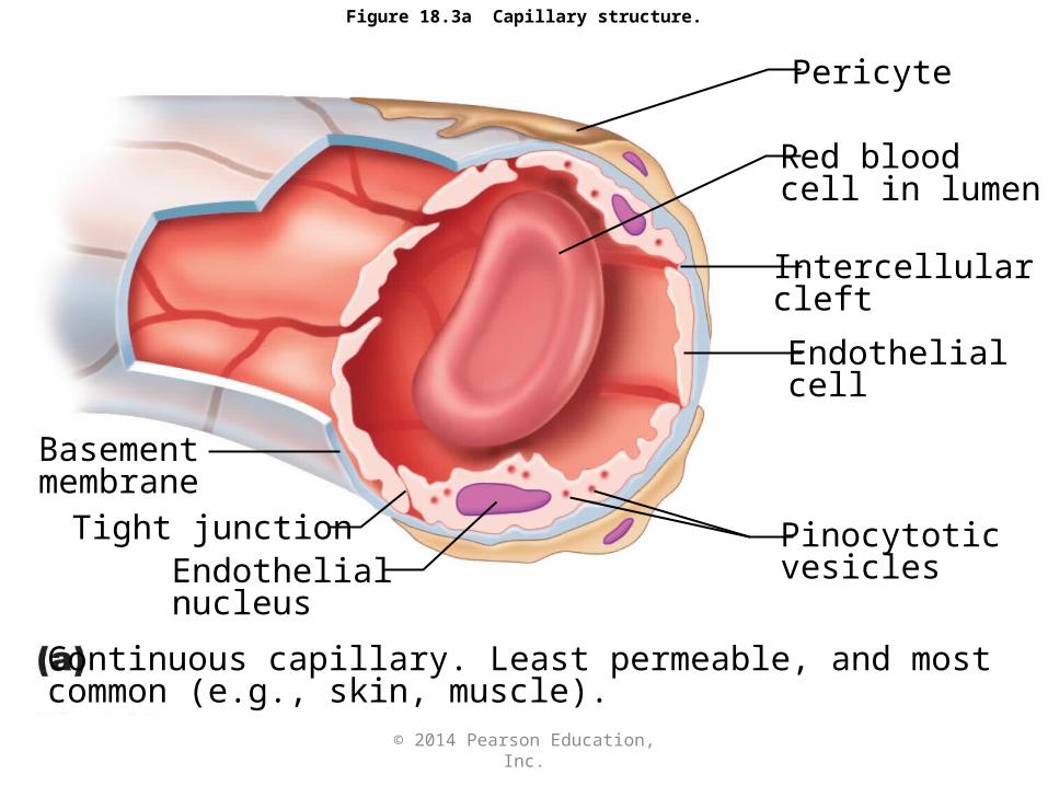
Figure 18.3a Capillary structure.
Pericyte
Red bloodcell in lumen
Intercellularcleft
Endothelialcell
Basementmembrane
Tight junctionEndothelialnucleus
Pinocytoticvesicles
Continuous capillary. Least permeable, and most common (e.g., skin, muscle).

© 2014 Pearson Education, Inc.
Figure 18.3b Capillary structure.
Pinocytoticvesicles
Red bloodcell in lumen
Fenestrations(pores)
Intercellularcleft
Endothelialcell
EndothelialnucleusBasement membrane
Tight junction
Fenestrated capillary. Large fenestrations (pores) increase permeability. Occurs in areas of active absorption or filtration (e.g., kidney, small intestine).
© 2014 Pearson Education, Inc.
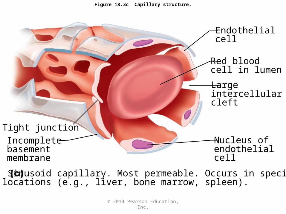
Figure 18.3c Capillary structure.
Endothelialcell
Red bloodcell in lumen
Largeintercellularcleft
Nucleus ofendothelialcell
Incompletebasementmembrane
Sinusoid capillary. Most permeable. Occurs in speciallocations (e.g., liver, bone marrow, spleen).
Tight junction

© 2014 Pearson Education, Inc.
Chapter Opener 18
© 2014 Pearson Education, Inc.
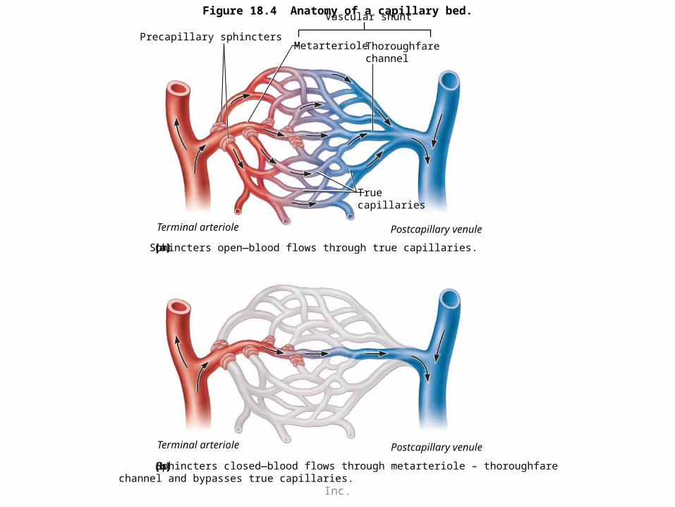
Figure 18.4 Anatomy of a capillary bed.Vascular shunt
Precapillary sphinctersMetarteriole Thoroughfare
channel
Terminal arteriole
Truecapillaries
Postcapillary venule
Sphincters open—blood flows through true capillaries.
Terminal arteriole Postcapillary venule
Sphincters closed—blood flows through metarteriole – thoroughfare channel and bypasses true capillaries.
© 2014 Pearson Education, Inc.
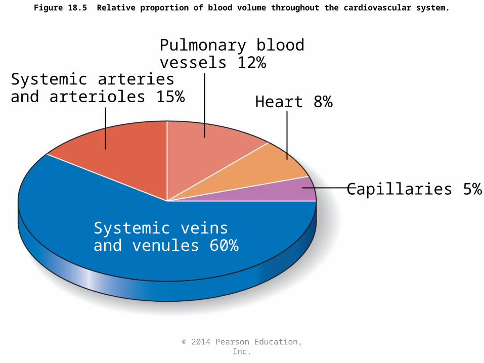
Figure 18.5 Relative proportion of blood volume throughout the cardiovascular system.
Pulmonary bloodvessels 12%
Systemic arteriesand arterioles 15% Heart 8%
Capillaries 5%
Systemic veinsand venules 60%
© 2014 Pearson Education, Inc.
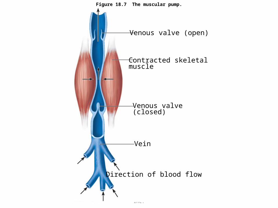
Figure 18.7 The muscular pump.
Venous valve (open)
Contracted skeletalmuscle
Venous valve(closed)
Vein
Direction of blood flow


![1 a veins arteries capillaries · veins arteries capillaries First _____ next _____ last _____ [1 mark] b Choose from the list of organs in the box to answer the question. From which](https://img.pdfslide.us/doc/110x75/5f1fa4f65f10160d415d4180/1-a-veins-arteries-capillaries-veins-arteries-capillaries-first-next-.jpg)