Embed Size (px)
Citation preview
© 2012 Pearson Education, Inc.
PowerPoint® Lecture Slides Prepared by Patty Bostwick-Taylor,Florence-Darlington Technical College
C H A P T E R 6
The Muscular System
© 2012 Pearson Education, Inc.
The Muscular System
•Muscles are responsible for all types of body movement
•Three basic muscle types are found in the body
•Skeletal muscle
•Cardiac muscle
•Smooth muscle
© 2012 Pearson Education, Inc.
Characteristics of Muscles
•Skeletal and smooth muscle cells are elongated (muscle cell = muscle fiber)
•Contraction and shortening of muscles is due to the movement of microfilaments
•All muscles share some terminology
•Prefixes myo and mys refer to “muscle”
•Prefix sarco refers to “flesh”
© 2012 Pearson Education, Inc. Table 6.1
© 2012 Pearson Education, Inc.
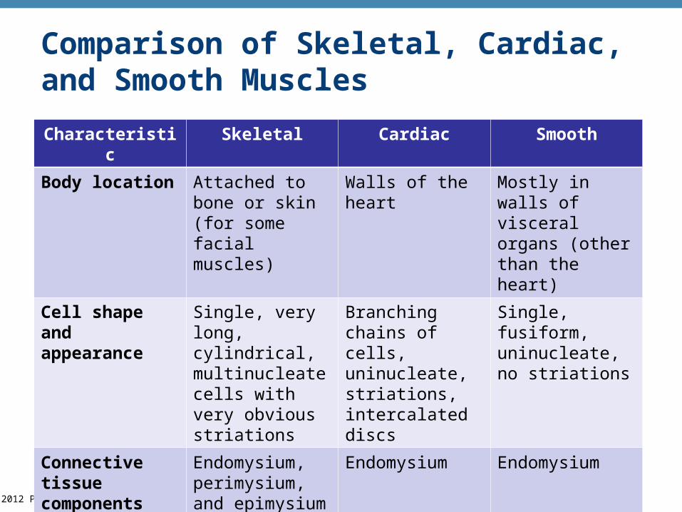
Comparison of Skeletal, Cardiac, and Smooth Muscles
Characteristic Skeletal Cardiac Smooth
Body location Attached to bone or skin (for some facial muscles)
Walls of the heart Mostly in walls of visceral organs (other than the heart)
Cell shape and appearance
Single, very long, cylindrical, multinucleate cells with very obvious striations
Branching chains of cells, uninucleate, striations, intercalated discs
Single, fusiform, uninucleate, no striations
Connective tissue components
Endomysium, perimysium, and epimysium
Endomysium Endomysium
© 2012 Pearson Education, Inc.
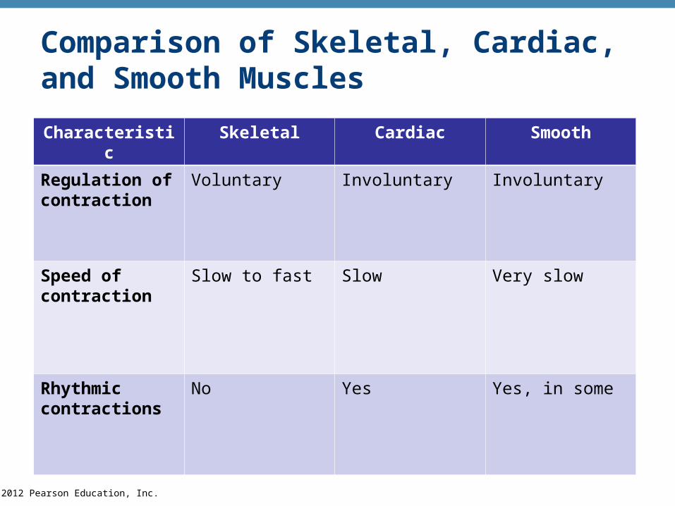
Comparison of Skeletal, Cardiac, and Smooth Muscles
Characteristic Skeletal Cardiac Smooth
Regulation of contraction
Voluntary Involuntary Involuntary
Speed of contraction
Slow to fast Slow Very slow
Rhythmic contractions
No Yes Yes, in some
© 2012 Pearson Education, Inc.
Skeletal Muscle Characteristics
•Most are attached by tendons to bones
•Cells are multinucleate
•Striated—have visible banding
•Voluntary—subject to conscious control
© 2012 Pearson Education, Inc.
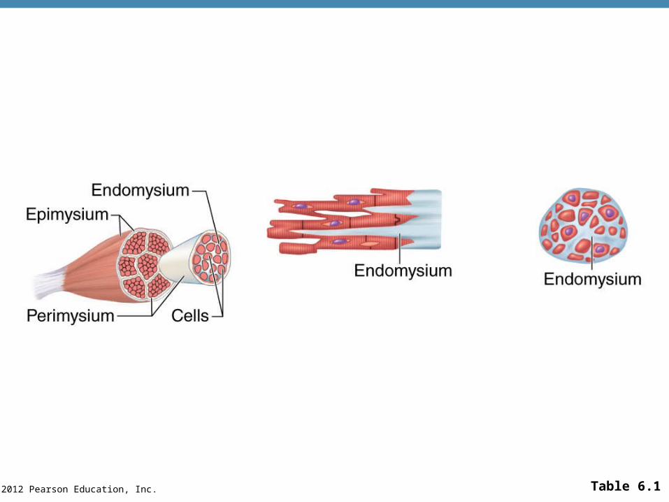
Connective Tissue Wrappings of Skeletal Muscle
•Cells are surrounded and bundled by connective tissue
•Endomysium—encloses a single muscle fiber
•Perimysium—wraps around a fascicle (bundle) of muscle fibers
•Epimysium—covers the entire skeletal muscle
•Fascia—on the outside of the epimysium
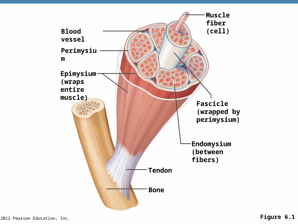
© 2012 Pearson Education, Inc. Figure 6.1
Blood vessel
Perimysium
Epimysium(wraps entiremuscle)
Musclefiber(cell)
Fascicle(wrapped byperimysium)
Endomysium(betweenfibers)
Tendon
Bone
© 2012 Pearson Education, Inc.
Skeletal Muscle Attachments
•Epimysium blends into a connective tissue attachment
•Tendons—cord-like structures
•Mostly collagen fibers
•Often cross a joint due to toughness and small size
•Aponeuroses—sheet-like structures
•Attach muscles indirectly to bones, cartilages, or connective tissue coverings
© 2012 Pearson Education, Inc.
Skeletal Muscle Attachments
•Sites of muscle attachment
•Bones
•Cartilages
•Connective tissue coverings
© 2012 Pearson Education, Inc.
Smooth Muscle Characteristics
•Lacks striations
•Spindle-shaped cells
•Single nucleus
•Involuntary—no conscious control
•Found mainly in the walls of hollow organs
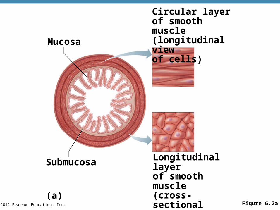
© 2012 Pearson Education, Inc. Figure 6.2a
Circular layerof smooth muscle(longitudinal viewof cells)Mucosa
Longitudinal layerof smooth muscle(cross-sectionalview of cells)
Submucosa
(a)
© 2012 Pearson Education, Inc.
Cardiac Muscle Characteristics
•Striations
•Usually has a single nucleus
•Branching cells
•Joined to another muscle cell at an intercalated disc
•Involuntary
•Found only in the walls of the heart
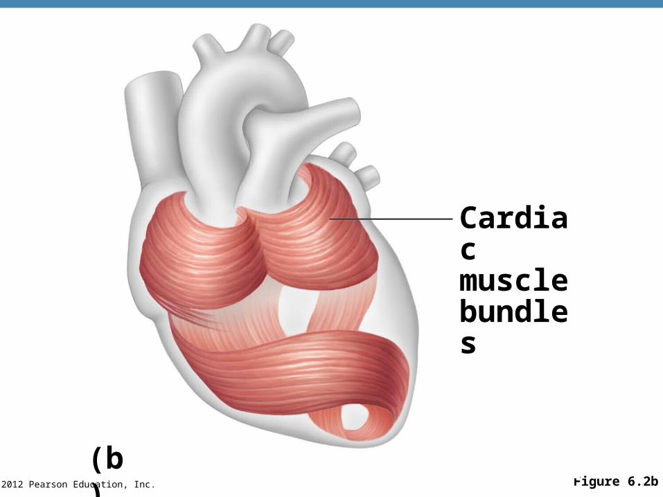
© 2012 Pearson Education, Inc. Figure 6.2b
Cardiacmusclebundles
(b)
© 2012 Pearson Education, Inc.
Skeletal Muscle Functions
•Produce movement
•Maintain posture
•Stabilize joints
•Generate heat
© 2012 Pearson Education, Inc.
Microscopic Anatomy of Skeletal Muscle
•Sarcolemma—specialized plasma membrane
•Myofibrils—long organelles inside muscle cell
•Sarcoplasmic reticulum—specialized smooth endoplasmic reticulum
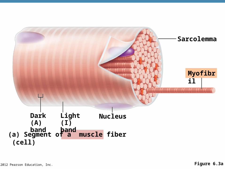
© 2012 Pearson Education, Inc. Figure 6.3a
Sarcolemma
Myofibril
Dark(A) band
Light(I) band
Nucleus
(a) Segment of a muscle fiber (cell)
© 2012 Pearson Education, Inc.
Microscopic Anatomy of Skeletal Muscle
•Myofibrils are aligned to give distinct bands
•I band = light band
•Contains only thin filaments
•A band = dark band
•Contains the entire length of the thick filaments
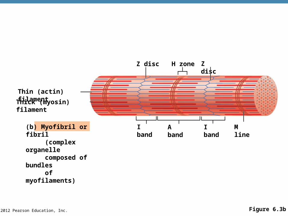
© 2012 Pearson Education, Inc. Figure 6.3b
(b) Myofibril or fibril (complex organelle composed of bundles of myofilaments)
Z disc H zone Z disc
I band A band I band M line
Thin (actin) filament
Thick (myosin) filament
© 2012 Pearson Education, Inc.
Microscopic Anatomy of Skeletal Muscle
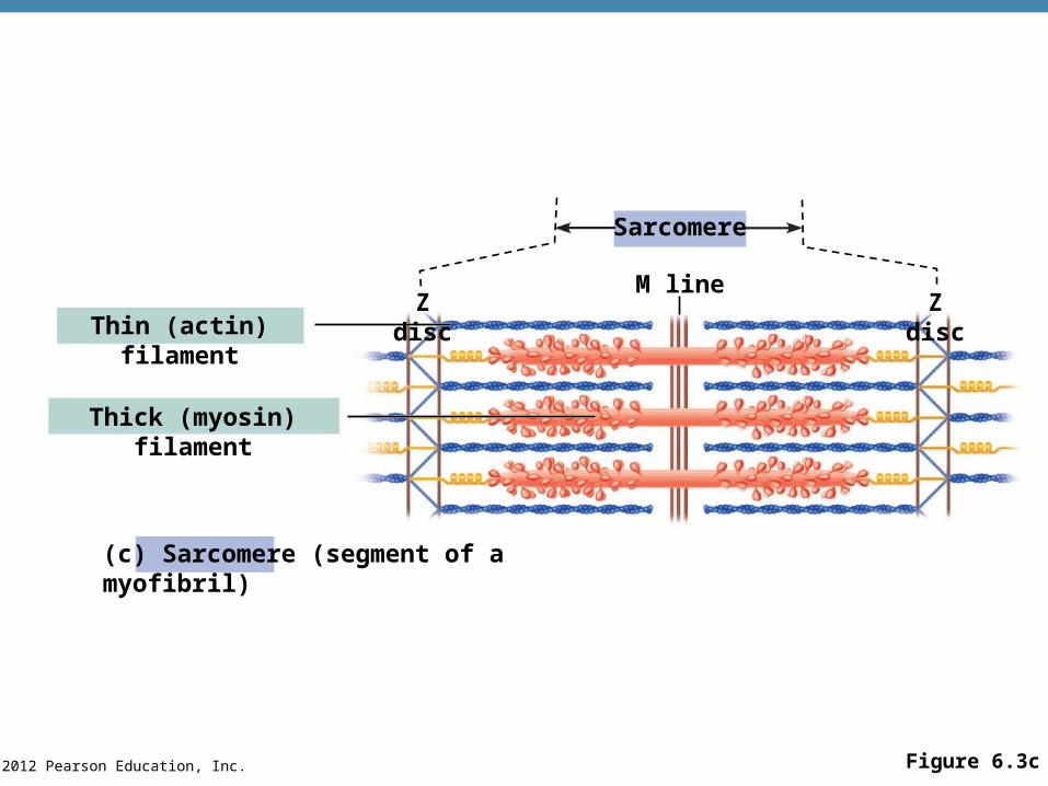
•Sarcomere—contractile unit of a muscle fiber
•Organization of the sarcomere
•Myofilaments
•Thick filaments = myosin filaments
•Thin filaments = actin filaments
© 2012 Pearson Education, Inc.
Microscopic Anatomy of Skeletal Muscle
•Thick filaments = myosin filaments•Composed of the protein myosin•Has ATPase enzymes•Myosin filaments have heads (extensions, or cross bridges)
•Myosin and actin overlap somewhat•Thin filaments = actin filaments
•Composed of the protein actin•Anchored to the Z disc
© 2012 Pearson Education, Inc. Figure 6.3c
Z disc
Sarcomere
M lineZ disc
Thin (actin) filament
Thick (myosin) filament
(c) Sarcomere (segment of a myofibril)
© 2012 Pearson Education, Inc.
Microscopic Anatomy of Skeletal Muscle
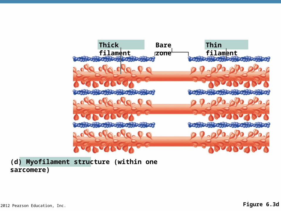
•At rest, within the A band there is a zone that lacks actin filaments
•Called either the H zone or bare zone
•Sarcoplasmic reticulum (SR)
•Stores and releases calcium
•Surrounds the myofibril
© 2012 Pearson Education, Inc. Figure 6.3d
Thick filament Bare zone Thin filament
(d) Myofilament structure (within one sarcomere)
© 2012 Pearson Education, Inc.
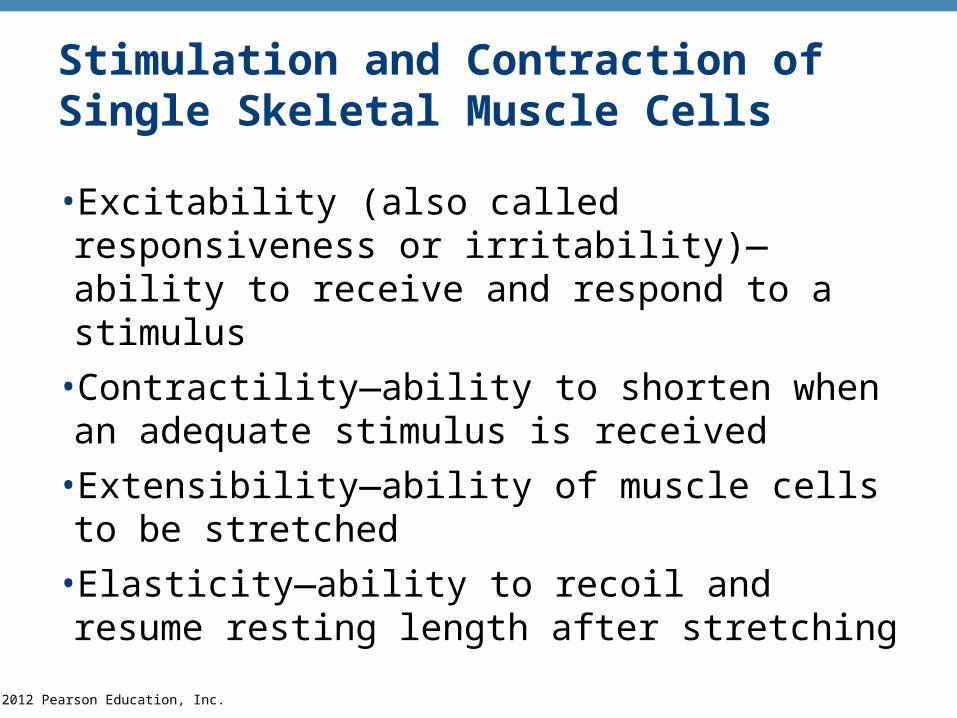
Stimulation and Contraction of Single Skeletal Muscle Cells
•Excitability (also called responsiveness or irritability)—ability to receive and respond to a stimulus
•Contractility—ability to shorten when an adequate stimulus is received
•Extensibility—ability of muscle cells to be stretched
•Elasticity—ability to recoil and resume resting length after stretching
© 2012 Pearson Education, Inc.
The Nerve Stimulus and Action Potential
•Skeletal muscles must be stimulated by a motor neuron (nerve cell) to contract
•Motor unit—one motor neuron and all the skeletal muscle cells stimulated by that neuron
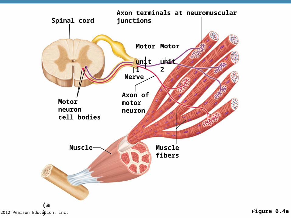
© 2012 Pearson Education, Inc. Figure 6.4a(a)
Spinal cord
Motor unit 1
Motor unit 2
Axon terminals at neuromuscular junctions
Nerve
Axon ofmotorneuron
Motor neuroncell bodies
Muscle Muscle fibers
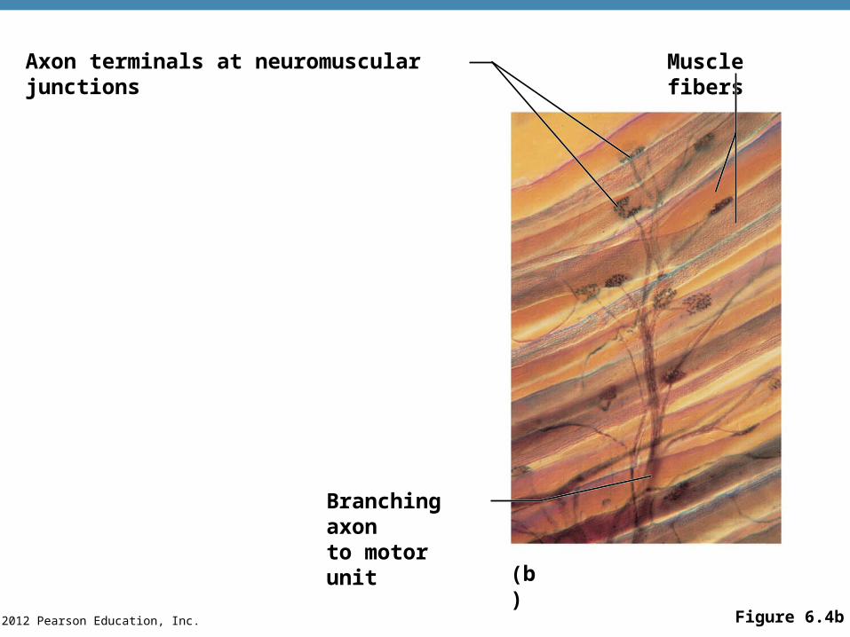
© 2012 Pearson Education, Inc. Figure 6.4b
Axon terminals at neuromuscular junctions Muscle fibers
Branching axonto motor unit
(b)
© 2012 Pearson Education, Inc.
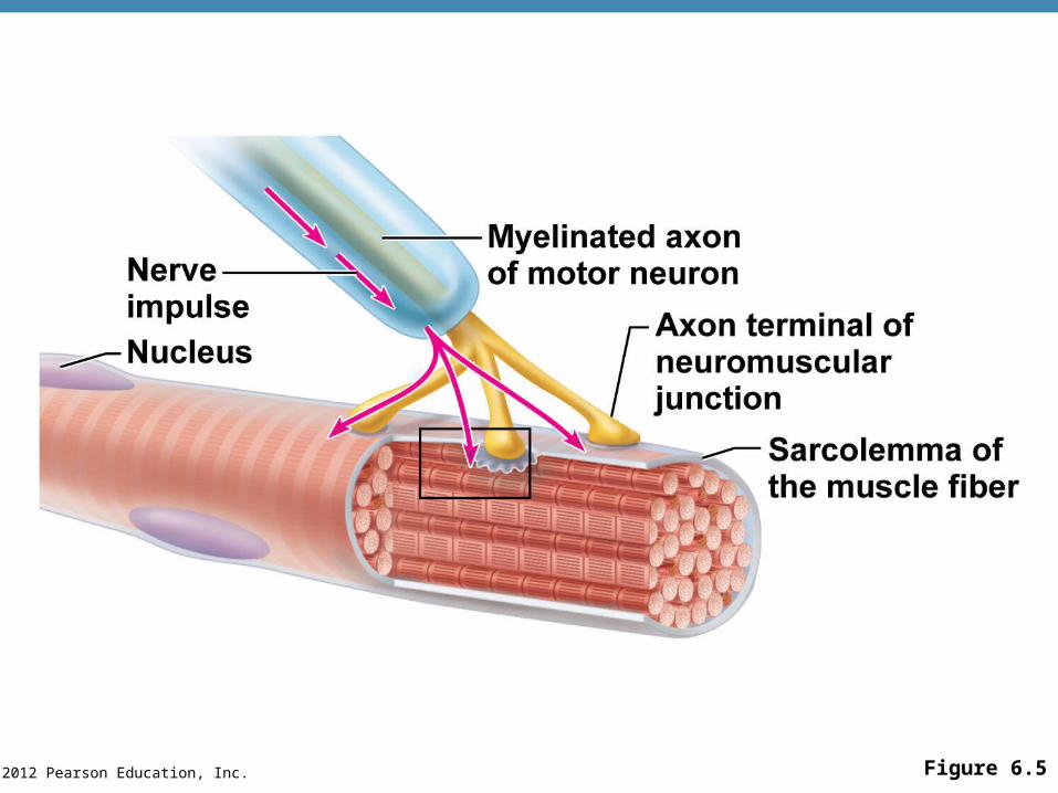
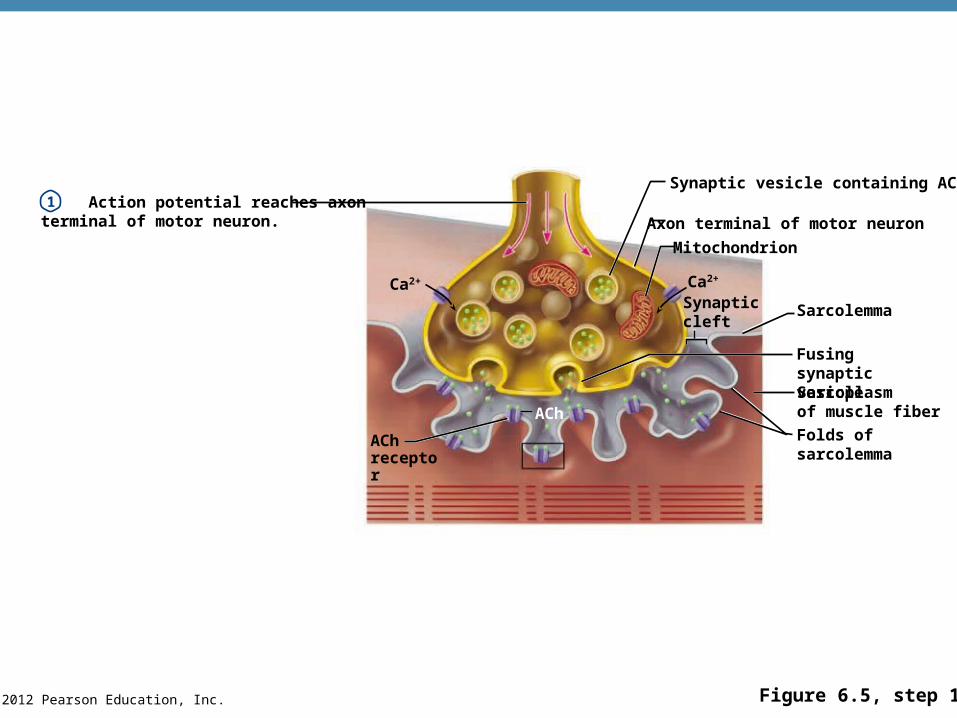
•Neuromuscular junction
•Association site of axon terminal of the motor neuron and muscle
The Nerve Stimulus and Action Potential
PLAYPLAY A&P Flix™: Events at the Neuromuscular Junction
© 2012 Pearson Education, Inc. Figure 6.5
© 2012 Pearson Education, Inc.
The Nerve Stimulus and Action Potential
•Synaptic cleft
•Gap between nerve and muscle
•Nerve and muscle do not make contact
•Area between nerve and muscle is filled with interstitial fluid
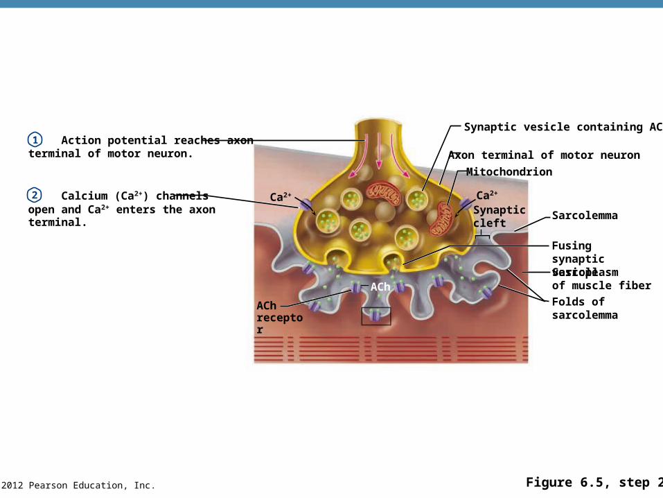
•Action potential reaches the axon terminal of the motor neuron
•Calcium channels open and calcium ions enter the axon terminal
© 2012 Pearson Education, Inc.
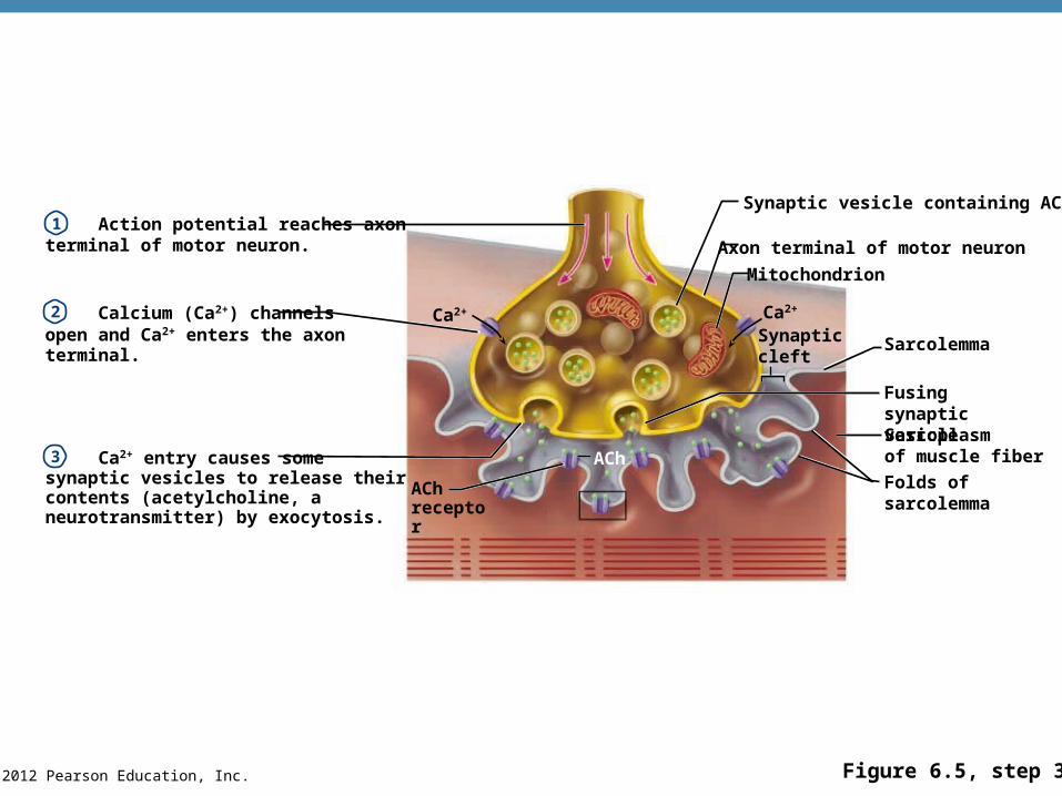
Transmission of Nerve Impulse to Muscle
•Calcium ion entry causes some synaptic vesicles to release their contents (acetylcholine, a neurotransmitter) by exocytosis
•Neurotransmitter—chemical released by nerve upon arrival of nerve impulse in the axon terminal
•The neurotransmitter for skeletal muscle is acetylcholine (ACh)
© 2012 Pearson Education, Inc.
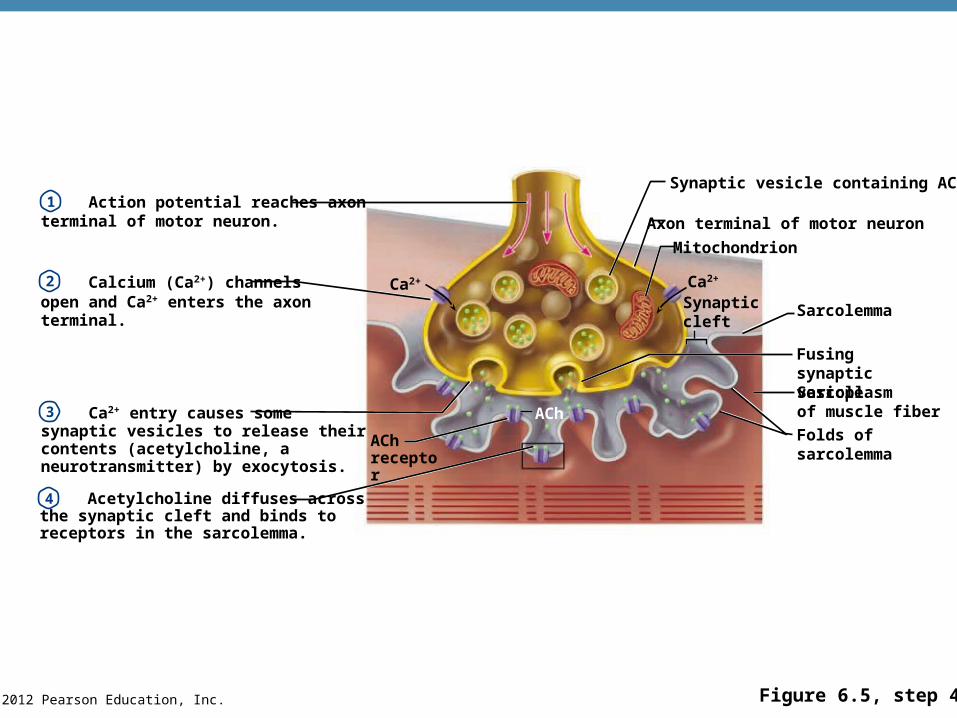
Transmission of Nerve Impulse to Muscle
•Acetylcholine attaches to receptors on the sarcolemma of the muscle cell
•In response to the binding of ACh to a receptor, the sarcolemma becomes permeable to sodium (Na+)
•Sodium rushes into the cell generating an action potential and potassium leaves the cell
•Once started, muscle contraction cannot be stopped
© 2012 Pearson Education, Inc. Figure 6.5, step 1
Synaptic vesicle containing ACh
Axon terminal of motor neuron
Mitochondrion
Ca2+
Synapticcleft
Sarcolemma
Fusing synapticvesicleSarcoplasmof muscle fiber
Folds ofsarcolemma
Ca2+
AChreceptor
ACh
Action potential reaches axonterminal of motor neuron.1
© 2012 Pearson Education, Inc. Figure 6.5, step 2
Synaptic vesicle containing ACh
Axon terminal of motor neuron
Mitochondrion
Ca2+
Fusing synapticvesicleSarcoplasmof muscle fiber
Folds ofsarcolemma
Ca2+
AChreceptor
ACh
SarcolemmaSynapticcleft
Action potential reaches axonterminal of motor neuron.
Calcium (Ca2+) channelsopen and Ca2+ enters the axon terminal.
1
2
© 2012 Pearson Education, Inc. Figure 6.5, step 3
Synaptic vesicle containing ACh
Axon terminal of motor neuron
Mitochondrion
Ca2+
Fusing synapticvesicleSarcoplasmof muscle fiber
Folds ofsarcolemma
Ca2+
AChreceptor
ACh
SarcolemmaSynapticcleft
Action potential reaches axonterminal of motor neuron.
Calcium (Ca2+) channelsopen and Ca2+ enters the axon terminal.
Ca2+ entry causes somesynaptic vesicles to release theircontents (acetylcholine, a neurotransmitter) by exocytosis.
1
2
3
© 2012 Pearson Education, Inc. Figure 6.5, step 4
Action potential reaches axonterminal of motor neuron.
Calcium (Ca2+) channelsopen and Ca2+ enters the axon terminal.
Ca2+ entry causes somesynaptic vesicles to release theircontents (acetylcholine, a neurotransmitter) by exocytosis.
Acetylcholine diffuses acrossthe synaptic cleft and binds toreceptors in the sarcolemma.
Synaptic vesicle containing ACh
Axon terminal of motor neuron
Mitochondrion
Ca2+
Fusing synapticvesicleSarcoplasmof muscle fiber
Folds ofsarcolemma
Ca2+
AChreceptor
ACh
1
2
3
4
SarcolemmaSynapticcleft
© 2012 Pearson Education, Inc. Figure 6.5, step 5
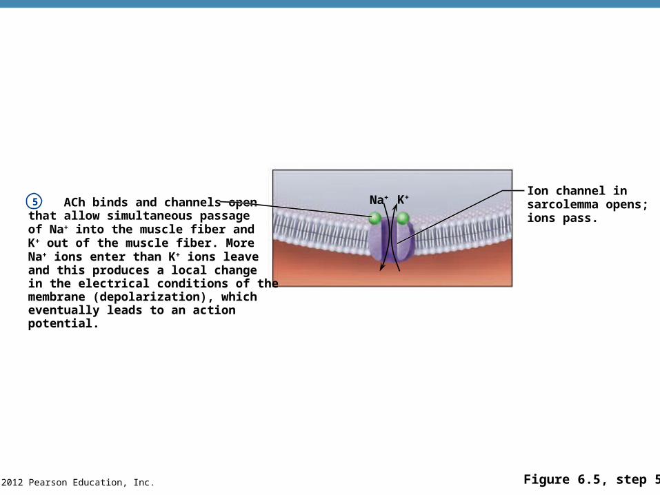
ACh binds and channels openthat allow simultaneous passageof Na+ into the muscle fiber and K+ out of the muscle fiber. MoreNa+ ions enter than K+ ions leaveand this produces a local changein the electrical conditions of themembrane (depolarization), whicheventually leads to an actionpotential.
5Ion channel insarcolemma opens;ions pass.
Na+ K+
© 2012 Pearson Education, Inc. Figure 6.5, step 6
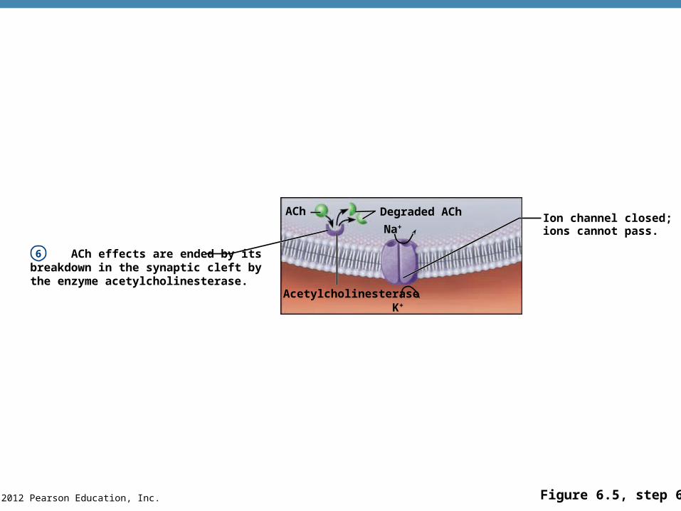
ACh effects are ended by itsbreakdown in the synaptic cleft bythe enzyme acetylcholinesterase.
6
Ion channel closed;ions cannot pass.
Acetylcholinesterase
Na+
Degraded AChACh
K+
© 2012 Pearson Education, Inc. Figure 6.6a-b
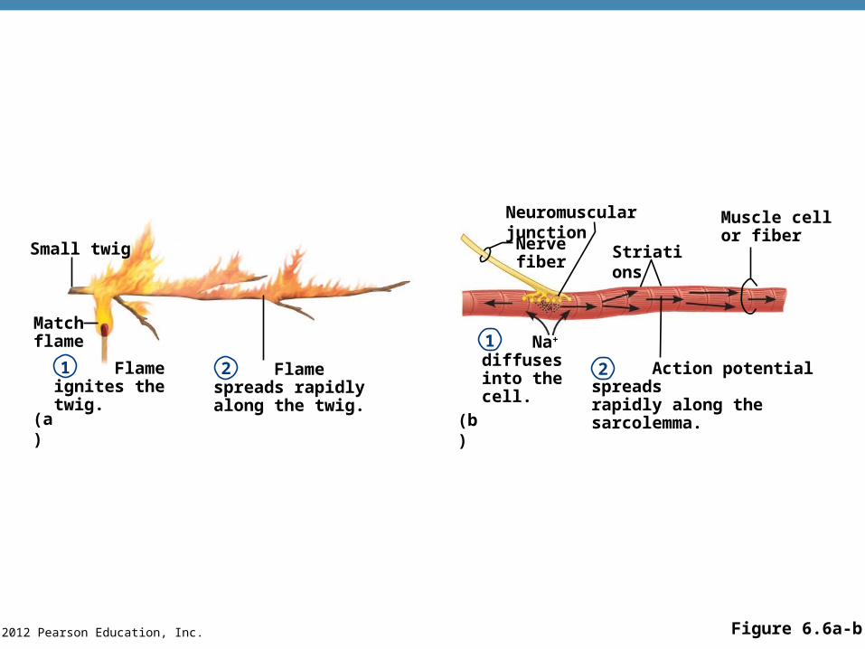
Small twig
Matchflame
Flame ignites the twig.
Flame spreads rapidly along the twig.
Neuromuscular junction
Nervefiber
Striations
Muscle cellor fiber
Na+ diffusesinto the cell. Action potential spreads
rapidly along the sarcolemma.
(b)
1 2
1
2
(a)
© 2012 Pearson Education, Inc.
The Sliding Filament Theory of Muscle Contraction
•Activation by nerve causes myosin heads (cross bridges) to attach to binding sites on the thin filament
•Myosin heads then bind to the next site of the thin filament and pull them toward the center of the sarcomere
•This continued action causes a sliding of the myosin along the actin
•The result is that the muscle is shortened (contracted)
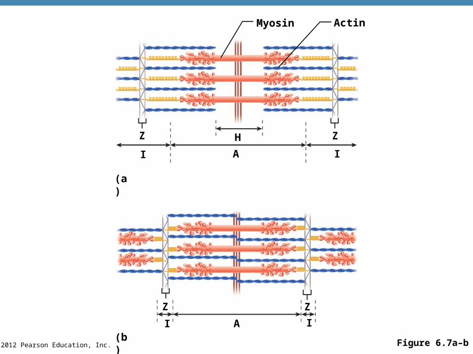
© 2012 Pearson Education, Inc. Figure 6.7a–b
Myosin Actin
Z H
I
Z
A I
(a)
(b)
Z
I A I
Z
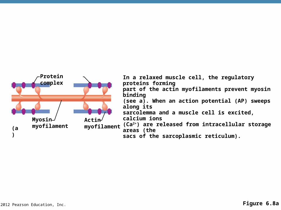
© 2012 Pearson Education, Inc. Figure 6.8a
Protein complex
Myosinmyofilament
Actinmyofilament(a)
In a relaxed muscle cell, the regulatory proteins formingpart of the actin myofilaments prevent myosin binding(see a). When an action potential (AP) sweeps along itssarcolemma and a muscle cell is excited, calcium ions(Ca2+) are released from intracellular storage areas (thesacs of the sarcoplasmic reticulum).
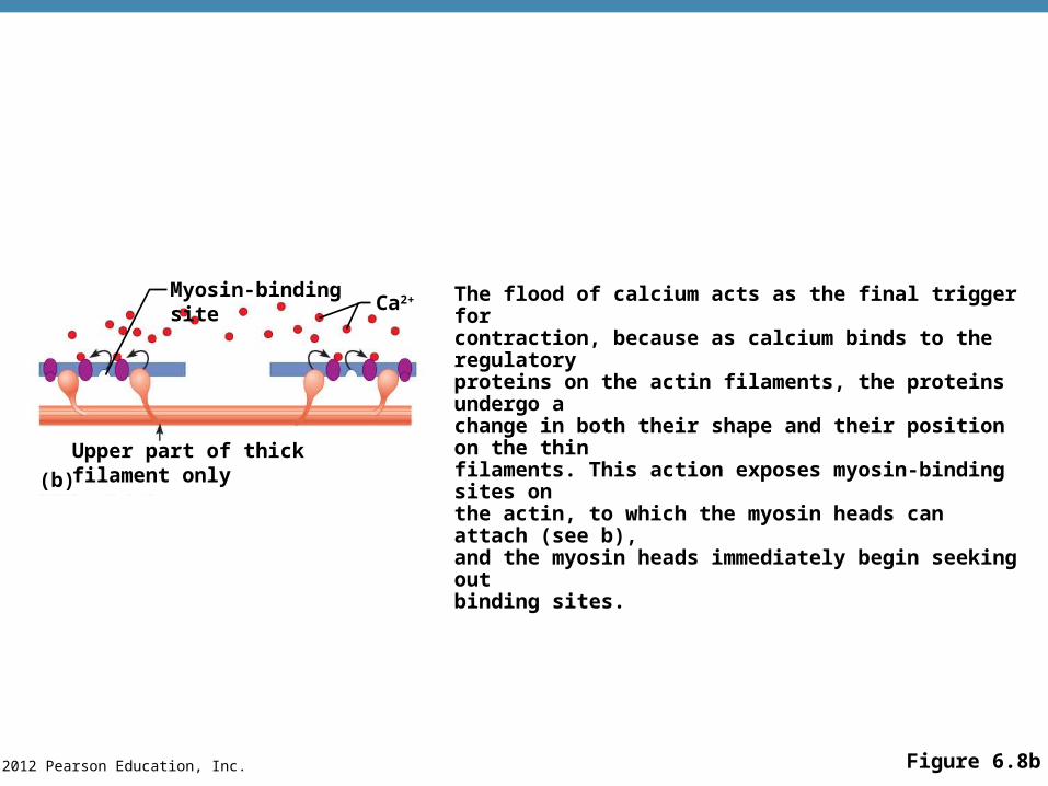
© 2012 Pearson Education, Inc. Figure 6.8b
Myosin-binding siteCa2+
Upper part of thick filament only
(b)
The flood of calcium acts as the final trigger forcontraction, because as calcium binds to the regulatoryproteins on the actin filaments, the proteins undergo a change in both their shape and their position on the thinfilaments. This action exposes myosin-binding sites onthe actin, to which the myosin heads can attach (see b),and the myosin heads immediately begin seeking out binding sites.
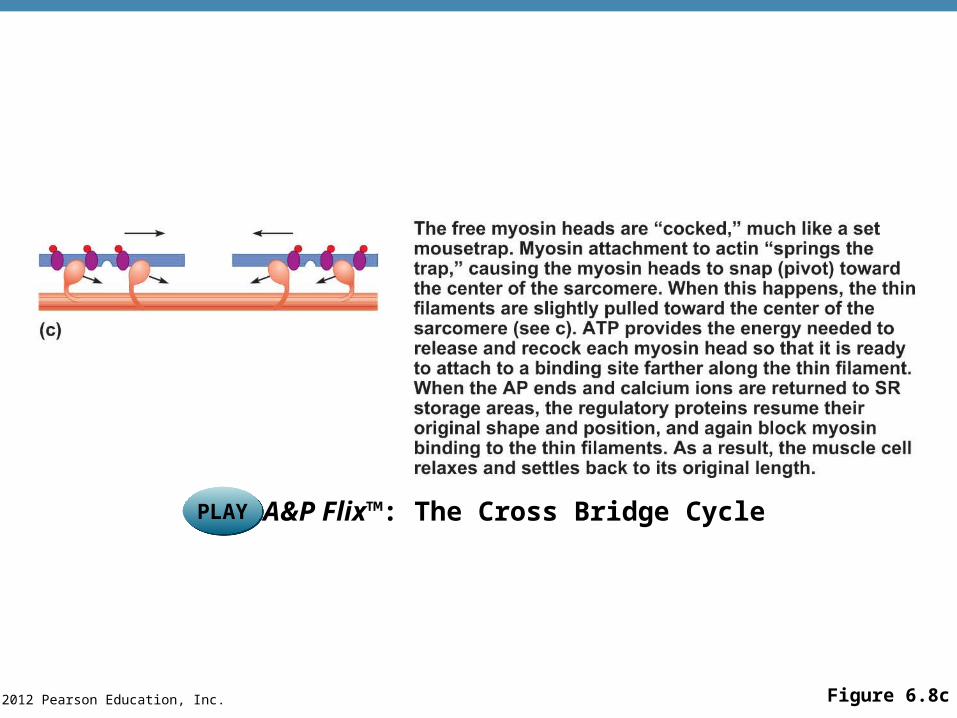
© 2012 Pearson Education, Inc. Figure 6.8c
PLAYPLAY A&P Flix™: The Cross Bridge Cycle
© 2012 Pearson Education, Inc.
Contraction of Skeletal Muscle
•Muscle fiber contraction is “all or none”
•Within a skeletal muscle, not all fibers may be stimulated during the same interval
•Different combinations of muscle fiber contractions may give differing responses
•Graded responses—different degrees of skeletal muscle shortening
© 2012 Pearson Education, Inc.
Contraction of Skeletal Muscle
•Graded responses can be produced by changing:
•The frequency of muscle stimulation
•The number of muscle cells being stimulated at one time
© 2012 Pearson Education, Inc.
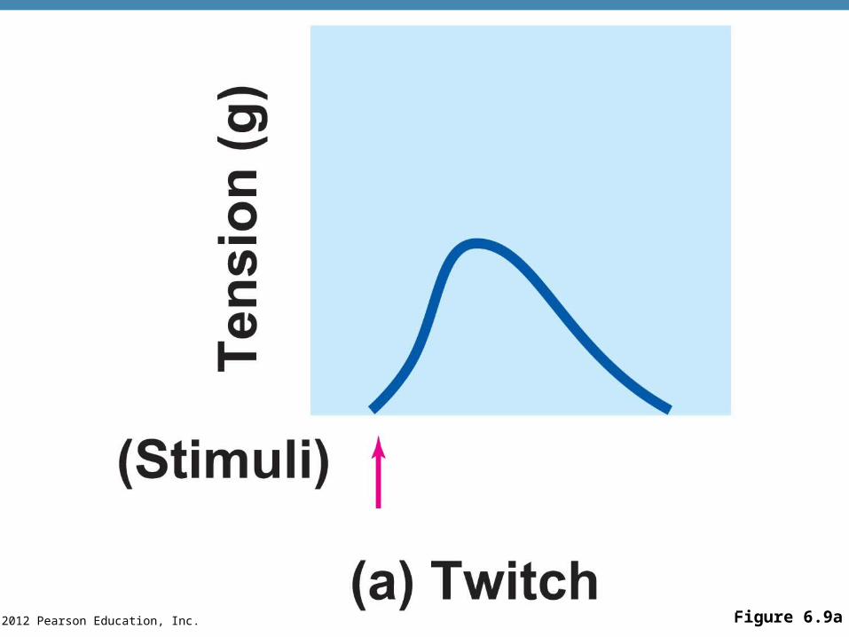
Types of Graded Responses
•Twitch
•Single, brief contraction
•Not a normal muscle function
© 2012 Pearson Education, Inc. Figure 6.9a
© 2012 Pearson Education, Inc.
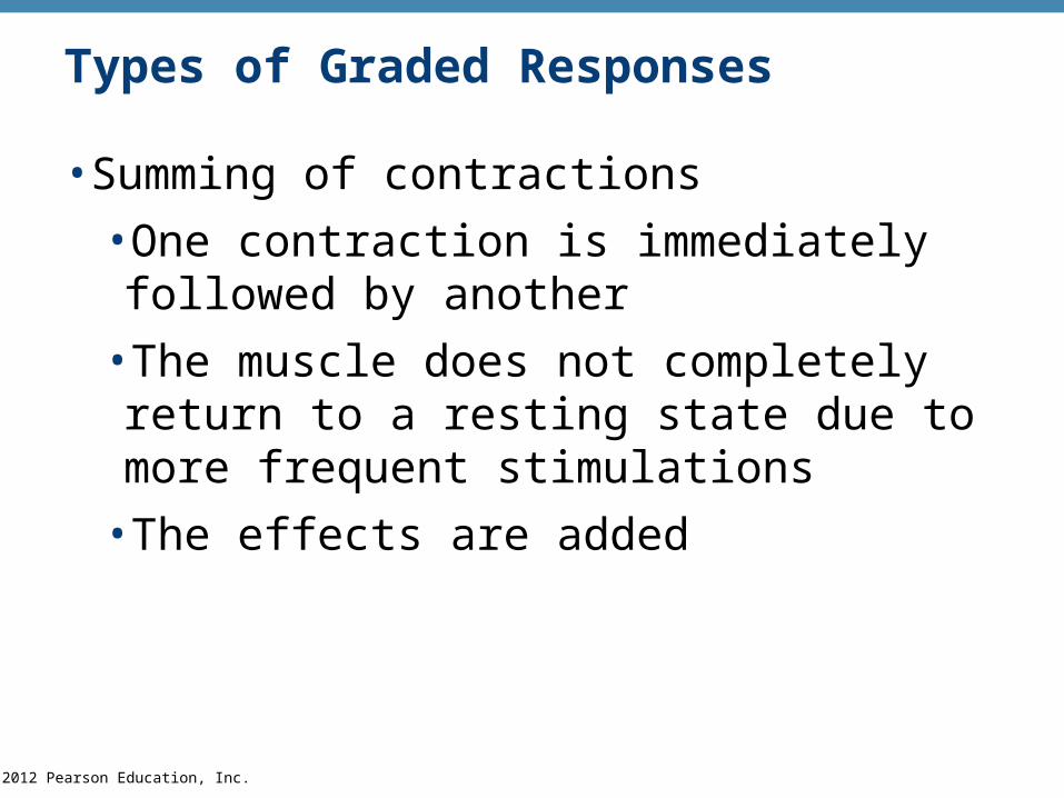
Types of Graded Responses
•Summing of contractions
•One contraction is immediately followed by another
•The muscle does not completely return to a resting state due to more frequent stimulations
•The effects are added
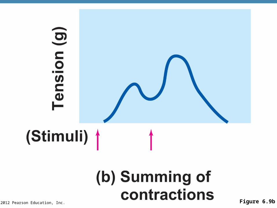
© 2012 Pearson Education, Inc. Figure 6.9b
© 2012 Pearson Education, Inc.
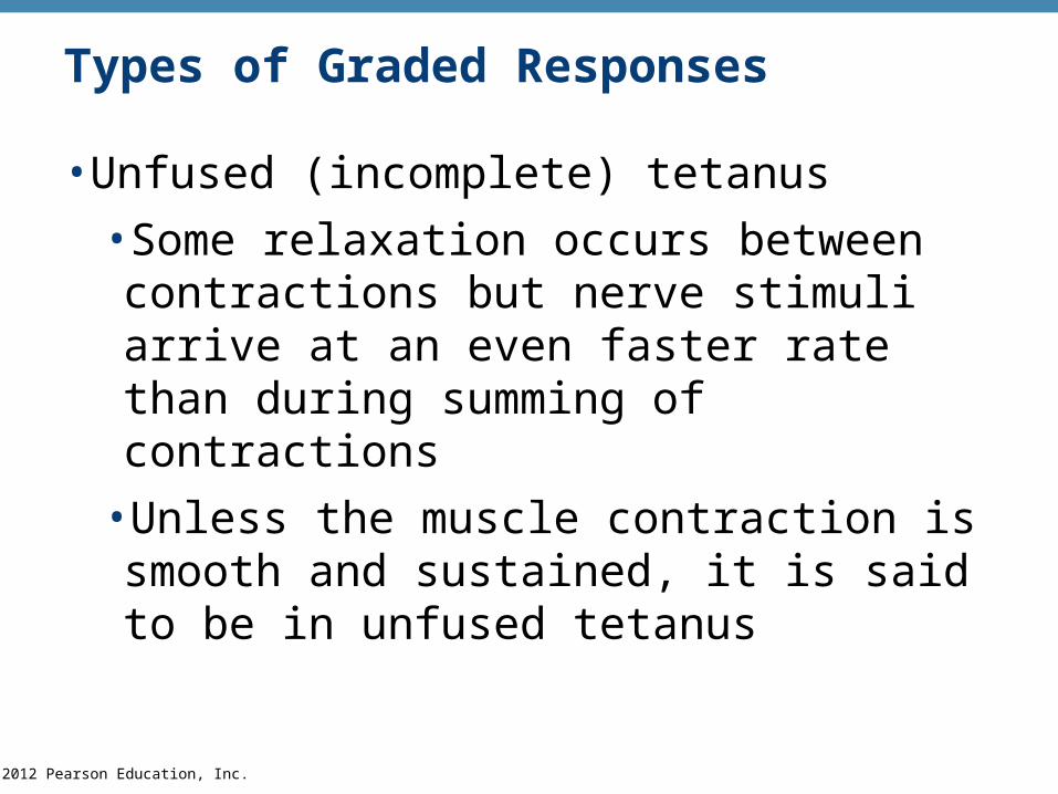
Types of Graded Responses
•Unfused (incomplete) tetanus
•Some relaxation occurs between contractions but nerve stimuli arrive at an even faster rate than during summing of contractions
•Unless the muscle contraction is smooth and sustained, it is said to be in unfused tetanus
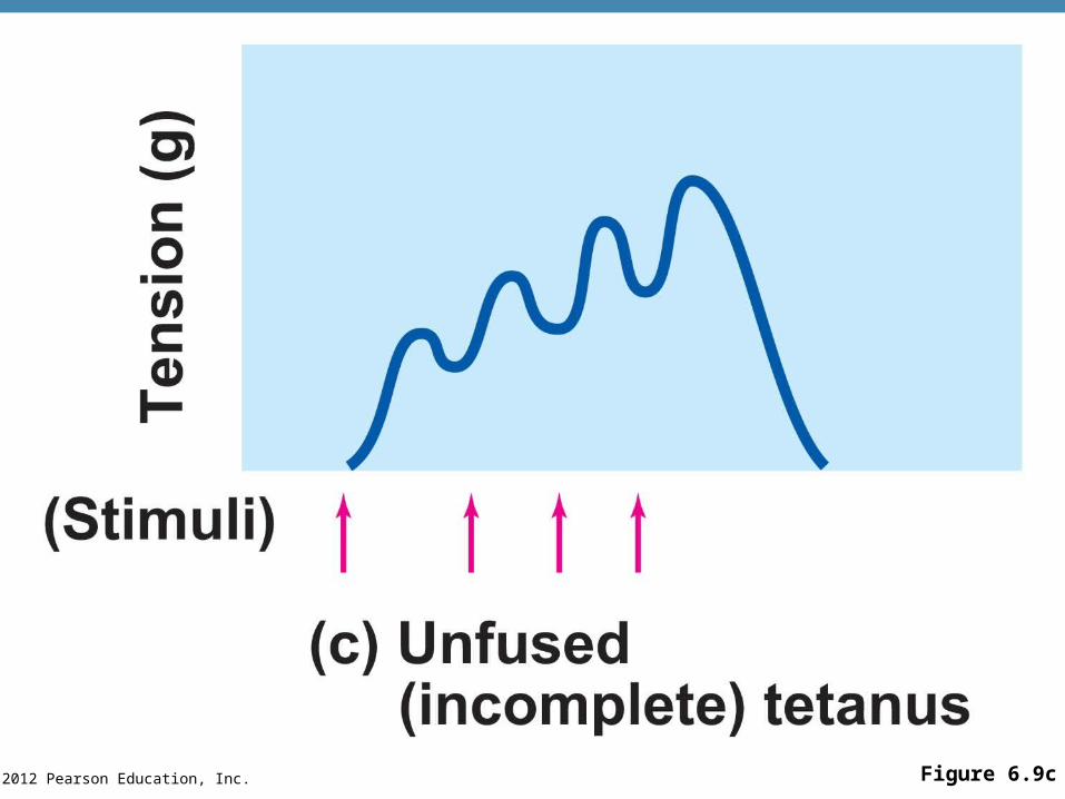
© 2012 Pearson Education, Inc. Figure 6.9c
© 2012 Pearson Education, Inc.
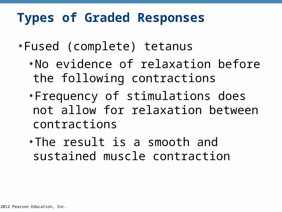
Types of Graded Responses
•Fused (complete) tetanus
•No evidence of relaxation before the following contractions
•Frequency of stimulations does not allow for relaxation between contractions
•The result is a smooth and sustained muscle contraction
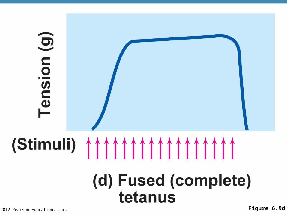
© 2012 Pearson Education, Inc. Figure 6.9d
© 2012 Pearson Education, Inc.
Muscle Response to Strong Stimuli
•Muscle force depends upon the number of fibers stimulated
•More fibers contracting results in greater muscle tension
•Muscles can continue to contract unless they run out of energy
© 2012 Pearson Education, Inc.
Energy for Muscle Contraction
•Initially, muscles use stored ATP for energy
•ATP bonds are broken to release energy
•Only 4–6 seconds worth of ATP is stored by muscles
•After this initial time, other pathways must be utilized to produce ATP
© 2012 Pearson Education, Inc.
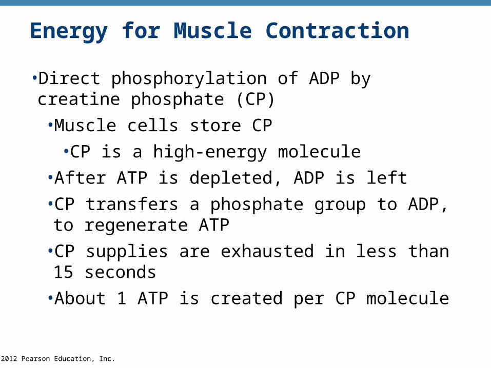
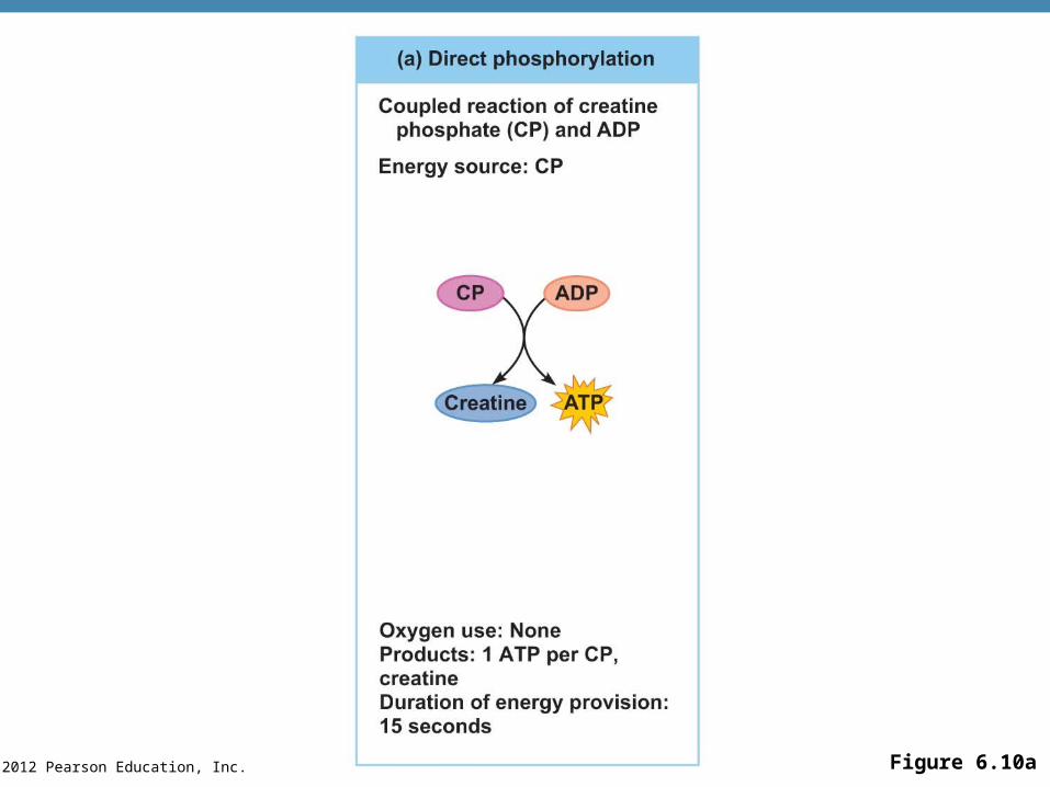
Energy for Muscle Contraction
•Direct phosphorylation of ADP by creatine phosphate (CP)
•Muscle cells store CP
•CP is a high-energy molecule
•After ATP is depleted, ADP is left
•CP transfers a phosphate group to ADP, to regenerate ATP
•CP supplies are exhausted in less than 15 seconds
•About 1 ATP is created per CP molecule
© 2012 Pearson Education, Inc. Figure 6.10a
© 2012 Pearson Education, Inc.
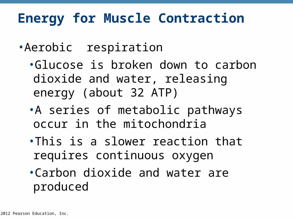
Energy for Muscle Contraction
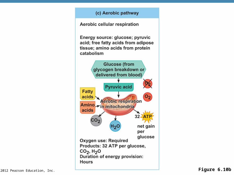
•Aerobic respiration
•Glucose is broken down to carbon dioxide and water, releasing energy (about 32 ATP)
•A series of metabolic pathways occur in the mitochondria
•This is a slower reaction that requires continuous oxygen
•Carbon dioxide and water are produced
© 2012 Pearson Education, Inc. Figure 6.10c
© 2012 Pearson Education, Inc.
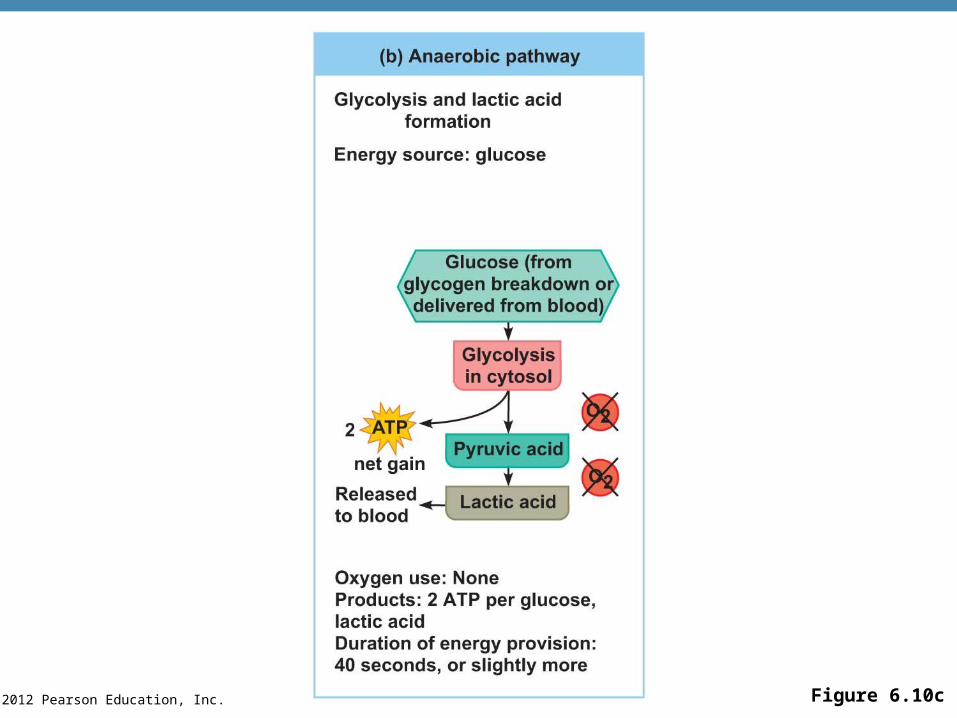
Energy for Muscle Contraction
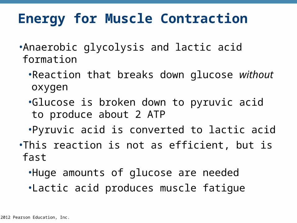
•Anaerobic glycolysis and lactic acid formation
•Reaction that breaks down glucose without oxygen
•Glucose is broken down to pyruvic acid to produce about 2 ATP
•Pyruvic acid is converted to lactic acid
•This reaction is not as efficient, but is fast
•Huge amounts of glucose are needed
•Lactic acid produces muscle fatigue
© 2012 Pearson Education, Inc. Figure 6.10b
© 2012 Pearson Education, Inc.
Muscle Fatigue and Oxygen Deficit
•When a muscle is fatigued, it is unable to contract even with a stimulus
•Common cause for muscle fatigue is oxygen debt
•Oxygen must be “repaid” to tissue to remove oxygen deficit
•Oxygen is required to get rid of accumulated lactic acid
• Increasing acidity (from lactic acid) and lack of ATP causes the muscle to contract less
© 2012 Pearson Education, Inc.
Types of Muscle Contractions
• Isotonic contractions
•Myofilaments are able to slide past each other during contractions
•The muscle shortens and movement occurs
•Example: bending the knee; rotating the arm
• Isometric contractions
•Tension in the muscles increases
•The muscle is unable to shorten or produce movement
•Example: push against a wall with bent elbows
© 2012 Pearson Education, Inc.
Muscle Tone
•Some fibers are contracted even in a relaxed muscle
•Different fibers contract at different times to provide muscle tone and to be constantly ready
© 2012 Pearson Education, Inc.
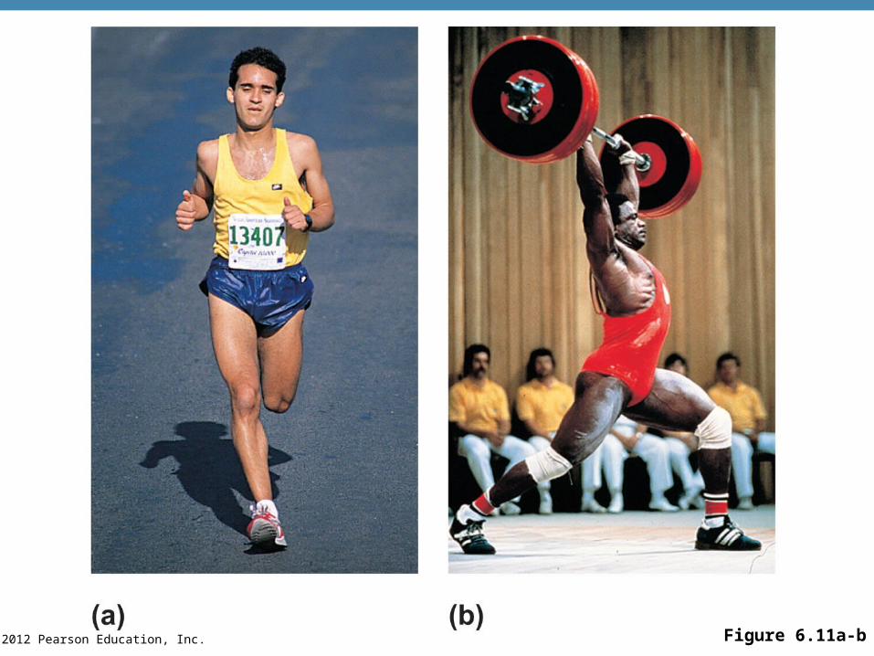
Effect of Exercise on Muscles
•Exercise increases muscle size, strength, and endurance
•Aerobic (endurance) exercise (biking, jogging) results in stronger, more flexible muscles with greater resistance to fatigue
•Makes body metabolism more efficient
•Improves digestion, coordination
•Resistance (isometric) exercise (weight lifting) increases muscle size and strength
© 2012 Pearson Education, Inc. Figure 6.11a-b
© 2012 Pearson Education, Inc.
Five Golden Rules of Skeletal Muscle Activity
1. With a few exceptions, all skeletal muscles cross at least one joint.
2. Typically, the bulk of a skeletal muscle lies proximal to the joint crossed.
3. All skeletal muscles have at least two attachments: the origin and the insertion.
4. Skeletal muscles can only pull; they never push.
5. During contraction, a skeletal muscle insertion moves toward the origin.
© 2012 Pearson Education, Inc.
Muscles and Body Movements
•Movement is attained due to a muscle moving an attached bone
•Muscles are attached to at least two points
•Origin
•Attachment to a moveable bone
•Insertion
•Attachment to an immovable bone
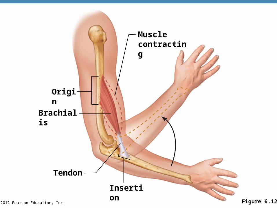
© 2012 Pearson Education, Inc. Figure 6.12
Tendon
Insertion
Brachialis
Origin
Musclecontracting
© 2012 Pearson Education, Inc.
Types of Body Movements
•Flexion
•Decreases the angle of the joint
•Brings two bones closer together
•Typical of bending hinge joints like knee and elbow or ball-and-socket joints like the hip
•Extension
•Opposite of flexion
• Increases angle between two bones
•Typical of straightening the elbow or knee
•Extension beyond 180° is hypertension
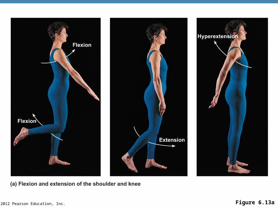
© 2012 Pearson Education, Inc. Figure 6.13a
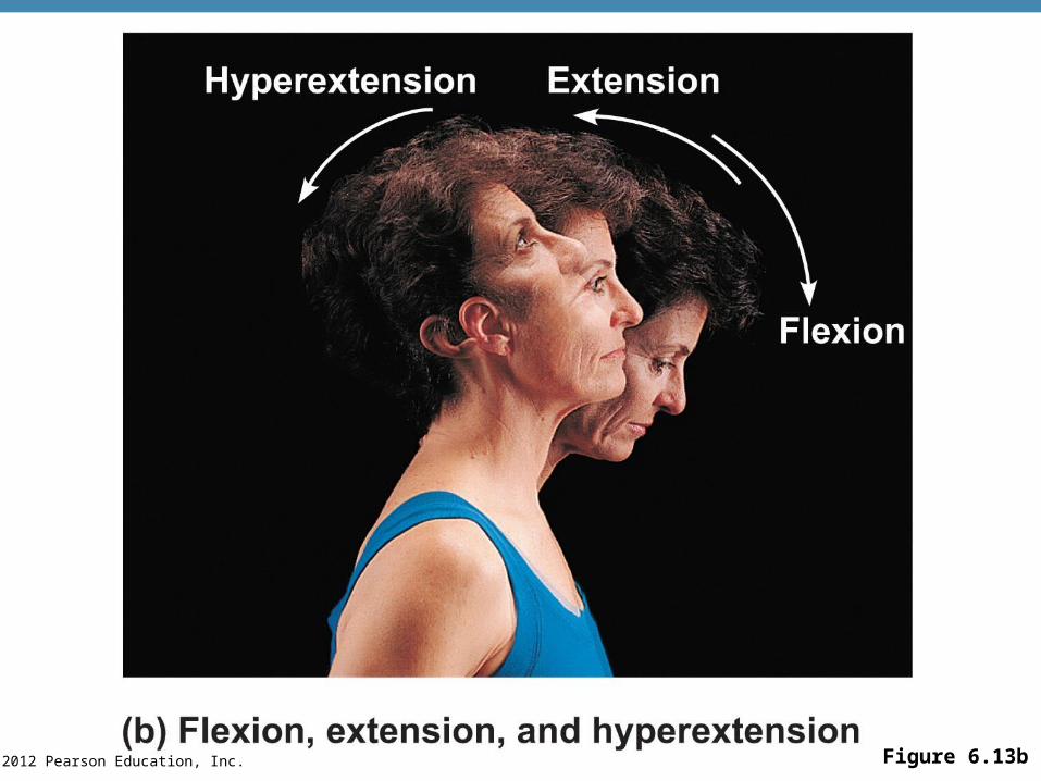
© 2012 Pearson Education, Inc. Figure 6.13b
© 2012 Pearson Education, Inc.
Types of Body Movements
•Rotation
•Movement of a bone around its longitudinal axis
•Common in ball-and-socket joints
•Example is when you move atlas around the dens of axis (shake your head “no”)
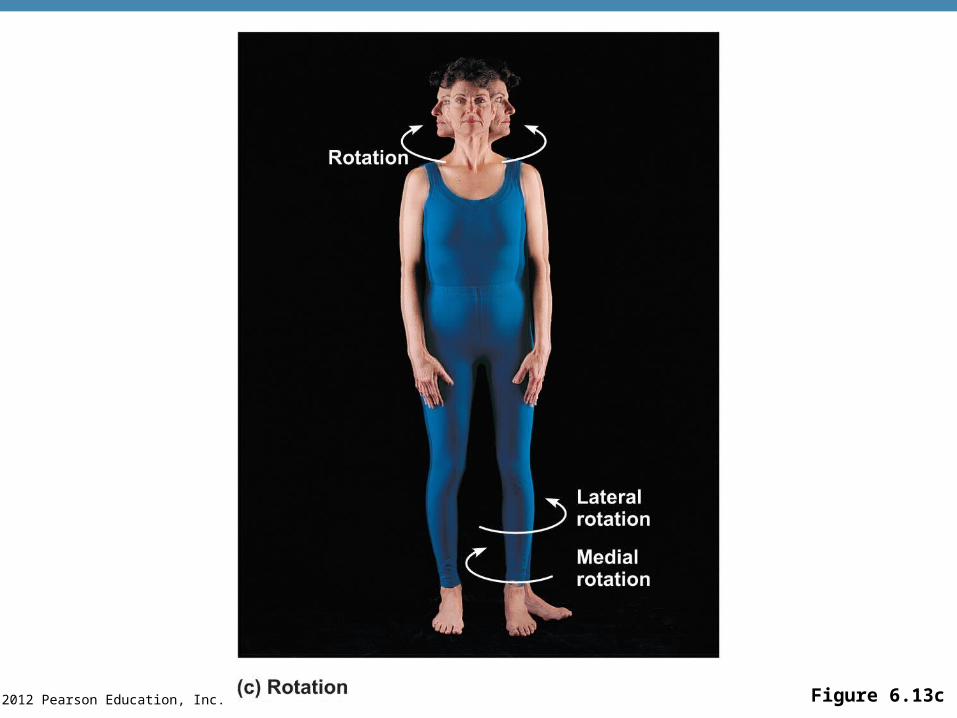
© 2012 Pearson Education, Inc. Figure 6.13c
© 2012 Pearson Education, Inc.
Types of Body Movements
•Abduction
•Movement of a limb away from the midline
•Adduction
•Opposite of abduction
•Movement of a limb toward the midline
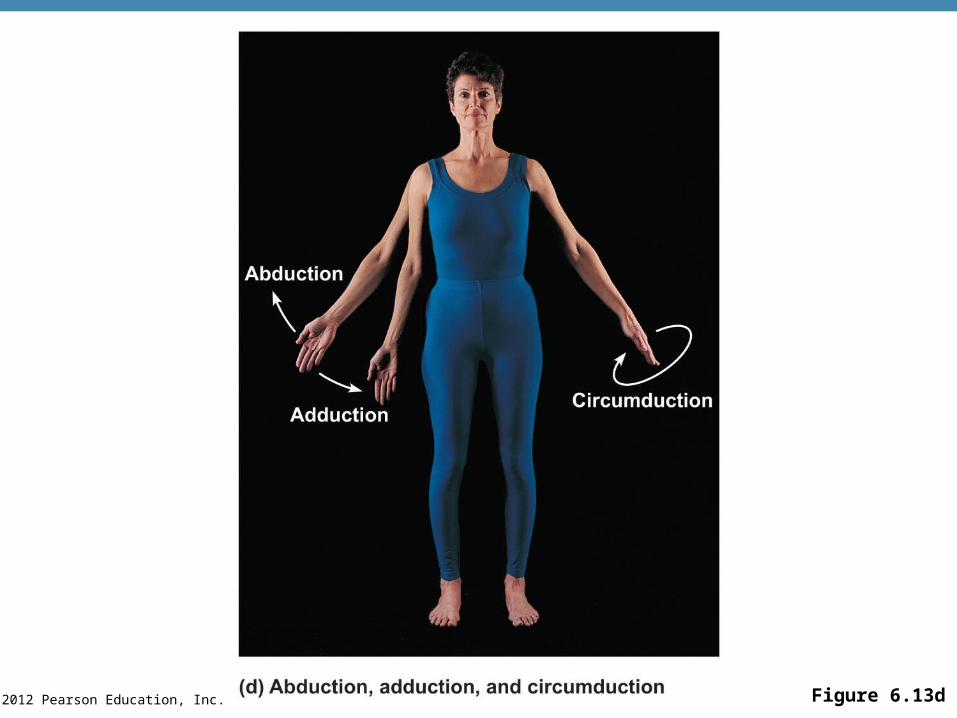
© 2012 Pearson Education, Inc. Figure 6.13d
© 2012 Pearson Education, Inc.
Types of Body Movements
•Circumduction
•Combination of flexion, extension, abduction, and adduction
•Common in ball-and-socket joints

© 2012 Pearson Education, Inc. Figure 6.13d
© 2012 Pearson Education, Inc.
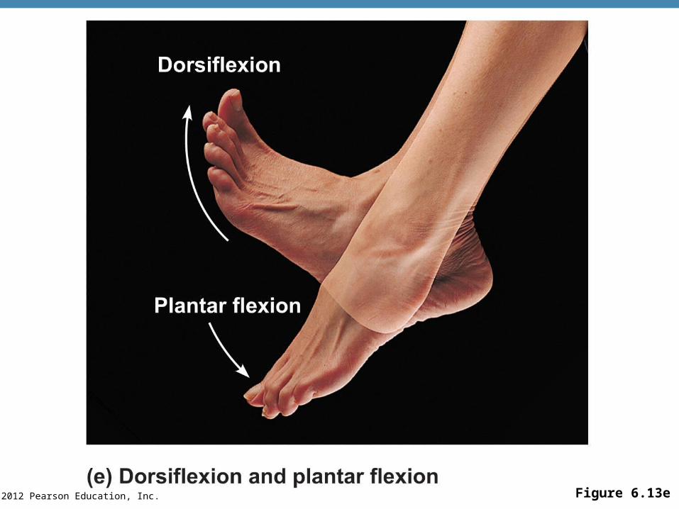
Special Movements
•Dorsiflexion
•Lifting the foot so that the superior surface approaches the shin (toward the dorsum)
•Plantar flexion
•Depressing the foot (pointing the toes)
•“Planting” the foot toward the sole
© 2012 Pearson Education, Inc. Figure 6.13e
© 2012 Pearson Education, Inc.
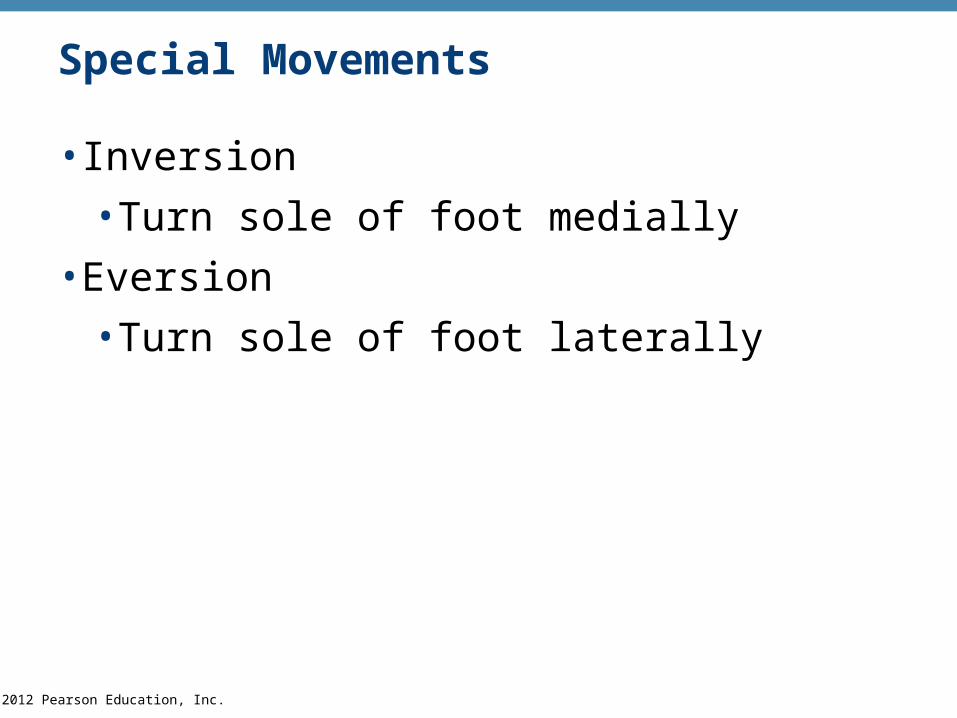
Special Movements
•Inversion
•Turn sole of foot medially
•Eversion
•Turn sole of foot laterally
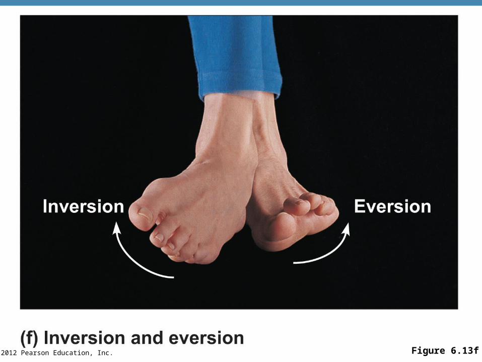
© 2012 Pearson Education, Inc. Figure 6.13f
© 2012 Pearson Education, Inc.
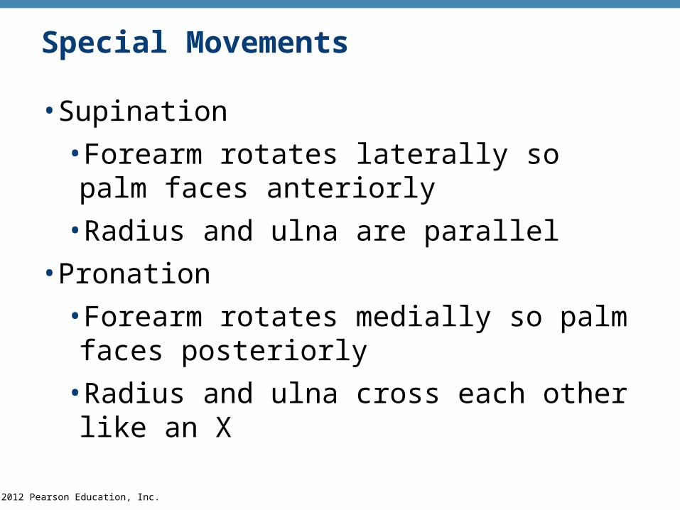
Special Movements
•Supination
•Forearm rotates laterally so palm faces anteriorly
•Radius and ulna are parallel
•Pronation
•Forearm rotates medially so palm faces posteriorly
•Radius and ulna cross each other like an X
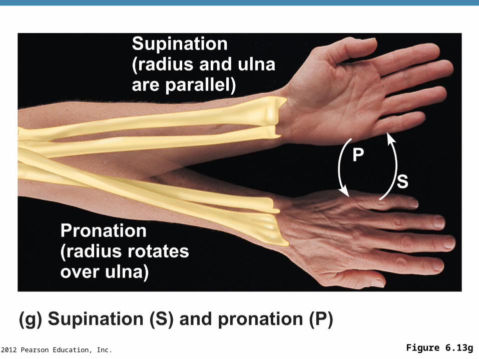
© 2012 Pearson Education, Inc. Figure 6.13g
© 2012 Pearson Education, Inc.
Special Movements
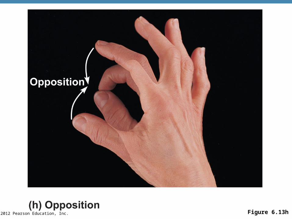
•Opposition
•Move thumb to touch the tips of other fingers on the same hand
© 2012 Pearson Education, Inc. Figure 6.13h
© 2012 Pearson Education, Inc.
Types of Muscles
•Prime mover—muscle with the major responsibility for a certain movement
•Antagonist—muscle that opposes or reverses a prime mover
•Synergist—muscle that aids a prime mover in a movement and helps prevent rotation
•Fixator—stabilizes the origin of a prime mover
© 2012 Pearson Education, Inc. Figure 6.14a
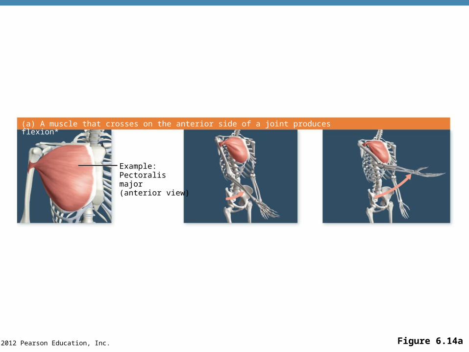
Example:Pectoralis major(anterior view)
(a) A muscle that crosses on the anterior side of a joint produces flexion*
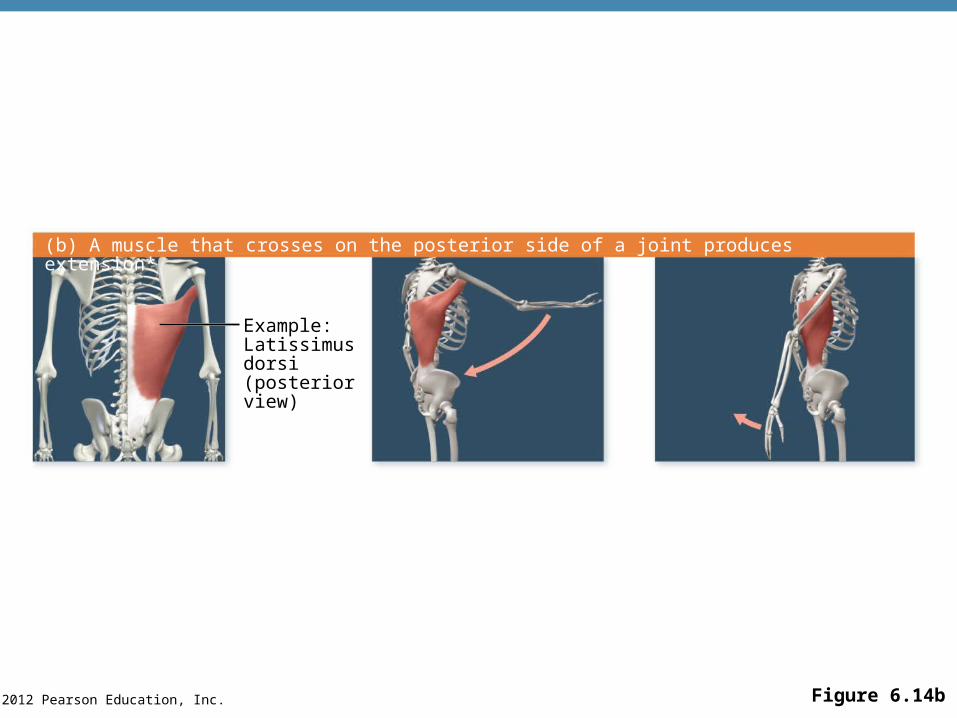
© 2012 Pearson Education, Inc. Figure 6.14b
Example:Latissimusdorsi(posteriorview)
(b) A muscle that crosses on the posterior side of a joint produces extension*
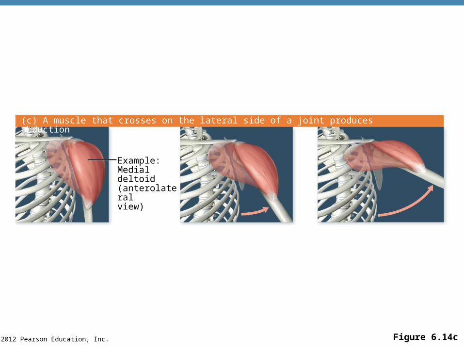
© 2012 Pearson Education, Inc. Figure 6.14c
Example:Medial deltoid(anterolateralview)
(c) A muscle that crosses on the lateral side of a joint produces abduction
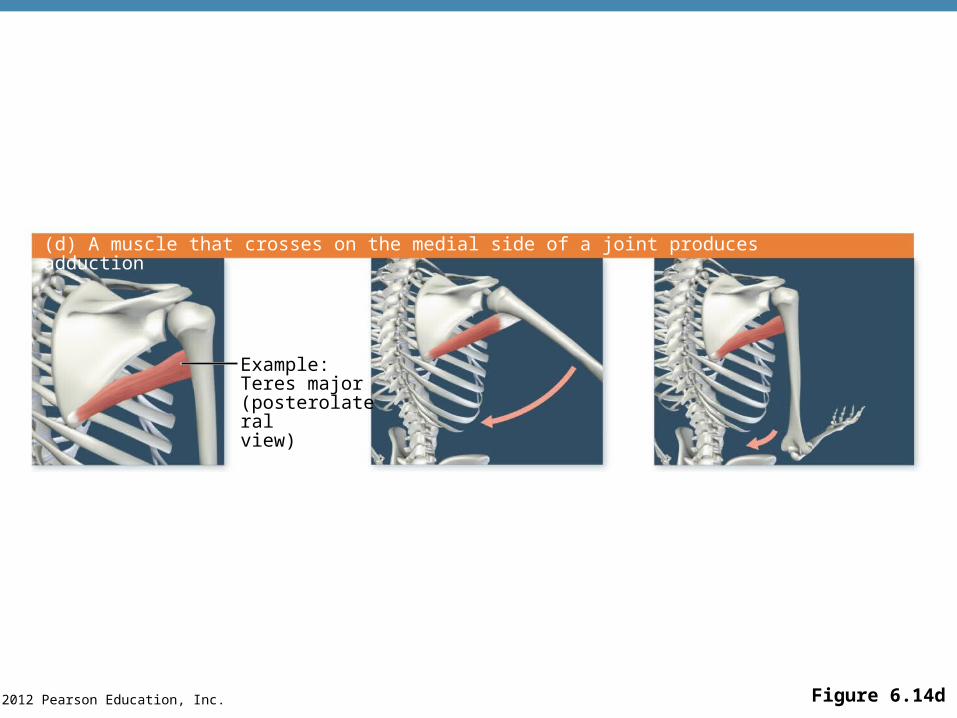
© 2012 Pearson Education, Inc. Figure 6.14d
Example:Teres major(posterolateralview)
(d) A muscle that crosses on the medial side of a joint produces adduction
© 2012 Pearson Education, Inc.
Naming Skeletal Muscles
•By direction of muscle fibers
•Example: Rectus (straight)
•By relative size of the muscle
•Example: Maximus (largest)
© 2012 Pearson Education, Inc.
Naming Skeletal Muscles
•By location of the muscle
•Example: Temporalis (temporal bone)
•By number of origins
•Example: Triceps (three heads)
© 2012 Pearson Education, Inc.
Naming Skeletal Muscles
•By location of the muscle’s origin and insertion
•Example: Sterno (on the sternum)
•By shape of the muscle
•Example: Deltoid (triangular)
•By action of the muscle
•Example: Flexor and extensor (flexes or extends a bone)
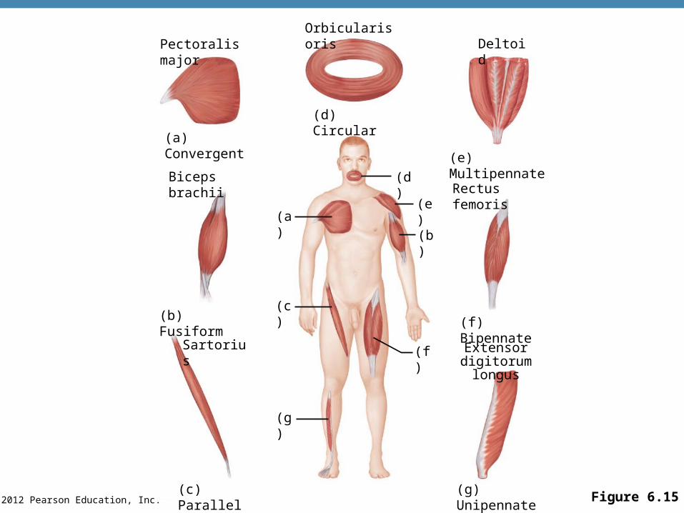
© 2012 Pearson Education, Inc. Figure 6.15(g) Unipennate
(f) Bipennate
Orbicularis orisDeltoid
(d) Circular
(a) Convergent
Biceps brachii
(a)
(c)(b) Fusiform
Sartorius
(g)
(c) Parallel
(d)
(e) Multipennate
(e)
(b)
(f) Extensor digitorum longus
Rectus femoris
Pectoralis major
© 2012 Pearson Education, Inc.
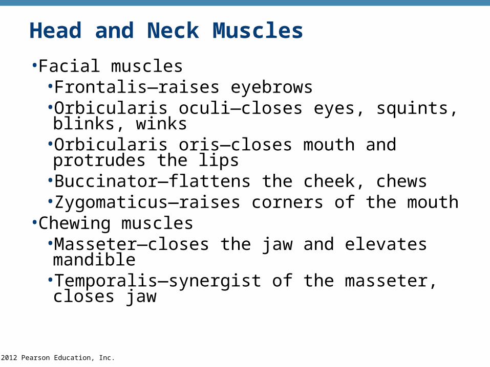
Head and Neck Muscles
•Facial muscles•Frontalis—raises eyebrows•Orbicularis oculi—closes eyes, squints, blinks, winks
•Orbicularis oris—closes mouth and protrudes the lips
•Buccinator—flattens the cheek, chews•Zygomaticus—raises corners of the mouth
•Chewing muscles•Masseter—closes the jaw and elevates mandible•Temporalis—synergist of the masseter, closes jaw
© 2012 Pearson Education, Inc.
Head and Neck Muscles
•Neck muscles
•Platysma—pulls the corners of the mouth inferiorly
•Sternocleidomastoid—flexes the neck, rotates the head
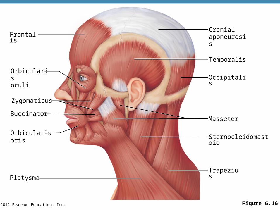
© 2012 Pearson Education, Inc. Figure 6.16
Cranialaponeurosis
Orbicularisoris
Orbicularisoculi
Frontalis
Zygomaticus
Buccinator
Platysma
Temporalis
Occipitalis
Masseter
Sternocleidomastoid
Trapezius
© 2012 Pearson Education, Inc.
Muscles of Trunk, Shoulder, Arm
•Anterior muscles
•Pectoralis major—adducts and flexes the humerus
•Intercostal muscles
•External intercostals—raise rib cage during inhalation
•Internal intercostals—depress the rib cage to move air out of the lungs when you exhale forcibly

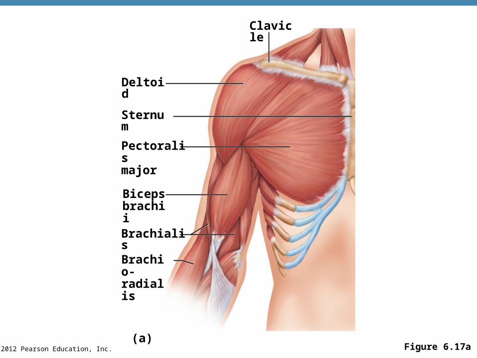
© 2012 Pearson Education, Inc. Figure 6.17a
Deltoid
Sternum
Pectoralismajor
Bicepsbrachii
Brachialis
Brachio-radialis
Clavicle
(a)
© 2012 Pearson Education, Inc.
Muscles of Trunk, Shoulder, Arm
•Muscles of the abdominal girdle
•Rectus abdominis—flexes vertebral column and compresses abdominal contents (defecation, childbirth, forced breathing)
•External oblique—flex vertebral column; rotate trunk and bend it laterally
• Internal oblique—flex vertebral column; rotate trunk and bend it laterally
•Transversus abdominis—compresses abdominal contents
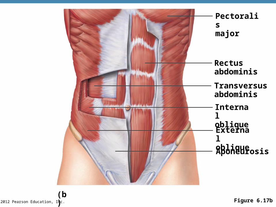
© 2012 Pearson Education, Inc. Figure 6.17b
Pectoralismajor
Rectusabdominis
Transversusabdominis
Internaloblique
Externaloblique
Aponeurosis
(b)
© 2012 Pearson Education, Inc.
Muscles of Trunk, Shoulder, Arm
•Posterior muscles
•Trapezius—elevates, depresses, adducts, and stabilizes the scapula
•Latissimus dorsi—extends and adducts the humerus
•Erector spinae—back extension
•Quadratus lumborum—flexes the spine laterally
•Deltoid—arm abduction
© 2012 Pearson Education, Inc.
Muscles of Trunk, Shoulder, Arm
•Muscles that arise from the shoulder girdle and cross the shoulder joint to insert into the humerus include:
•Pectoralis major
•Latissimus dorsi
•Deltoid
PLAYPLAY A&P Flix™: Movement at the glenohumeral joint: An overview.
PLAYPLAY A&P Flix™: Muscles that cross the glenohumeral joint.
PLAYPLAY A&P Flix™: Muscles of the pectoral girdle.
PLAYPLAYA&P Flix™: Muscles that act on the shoulder joint and humerus: An overview.

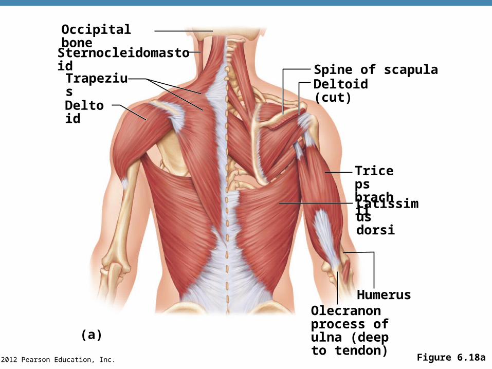
© 2012 Pearson Education, Inc. Figure 6.18a
Olecranonprocess ofulna (deepto tendon)
Tricepsbrachii
Latissimusdorsi
Occipital bone
Sternocleidomastoid
Trapezius
Deltoid
(a)
Spine of scapulaDeltoid (cut)
Humerus
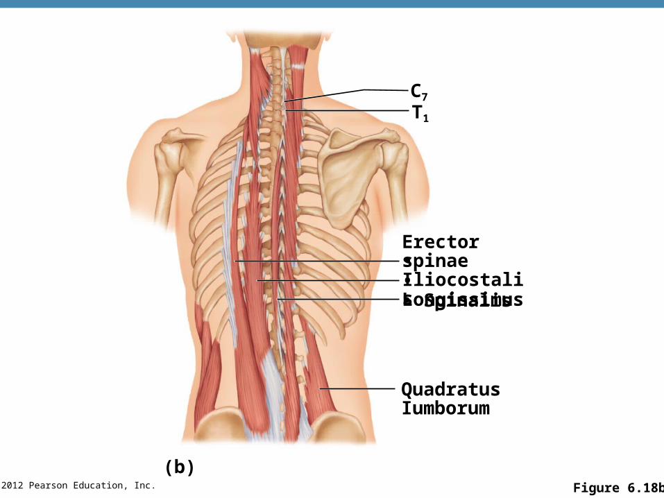
© 2012 Pearson Education, Inc. Figure 6.18b
C7
T1
Erector spinae• Iliocostalis• Longissimus• Spinalis
QuadratusIumborum
(b)
© 2012 Pearson Education, Inc.
Muscles of the Upper Limb
•Biceps brachii—supinates forearm, flexes elbow
•Brachialis—elbow flexion
•Brachioradialis—weak muscle; elbow flexion
•Triceps brachii—elbow extension (antagonist to biceps brachii)
PLAYPLAY A&P Flix™: Movement at the elbow joint.
PLAYPLAY A&P Flix™: Muscles of the elbow joint.
PLAYPLAY A&P Flix™: The elbow joint and forearm: An overview.
© 2012 Pearson Education, Inc. Figure 6.17a
Deltoid
Sternum
Pectoralismajor
Bicepsbrachii
Brachialis
Brachio-radialis
Clavicle
(a)
© 2012 Pearson Education, Inc. Figure 6.18a
Olecranonprocess ofulna (deepto tendon)
Tricepsbrachii
Latissimusdorsi
Occipital bone
Sternocleidomastoid
Trapezius
Deltoid
(a)
Spine of scapulaDeltoid (cut)
Humerus
© 2012 Pearson Education, Inc.
Muscles of the Upper Limb
•Muscles of the forearm, which insert on the hand bones and cause their movement include:
•Flexor carpi—wrist flexion
•Flexor digitorum—finger flexion
•Extensor carpi—wrist extension
•Extensor digitorum—finger extension
PLAYPLAY A&P Flix™: Movements of the wrist and fingers (b).
PLAYPLAY A&P Flix™: Movements of the wrist and fingers (a).
PLAYPLAYA&P Flix™: Muscles that act on the wrist and fingers: An overview.
© 2012 Pearson Education, Inc.
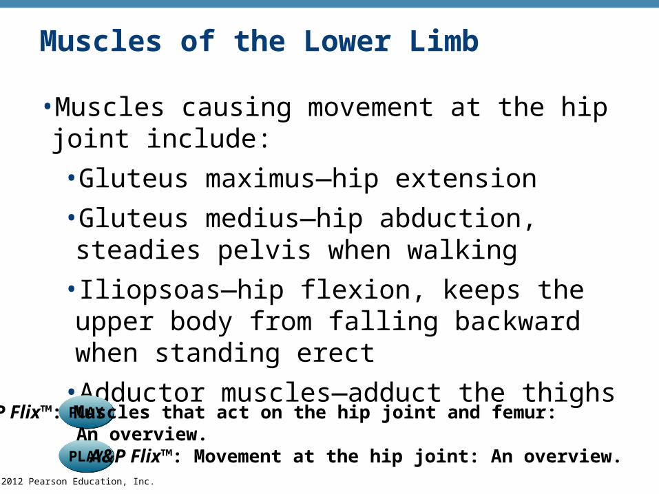
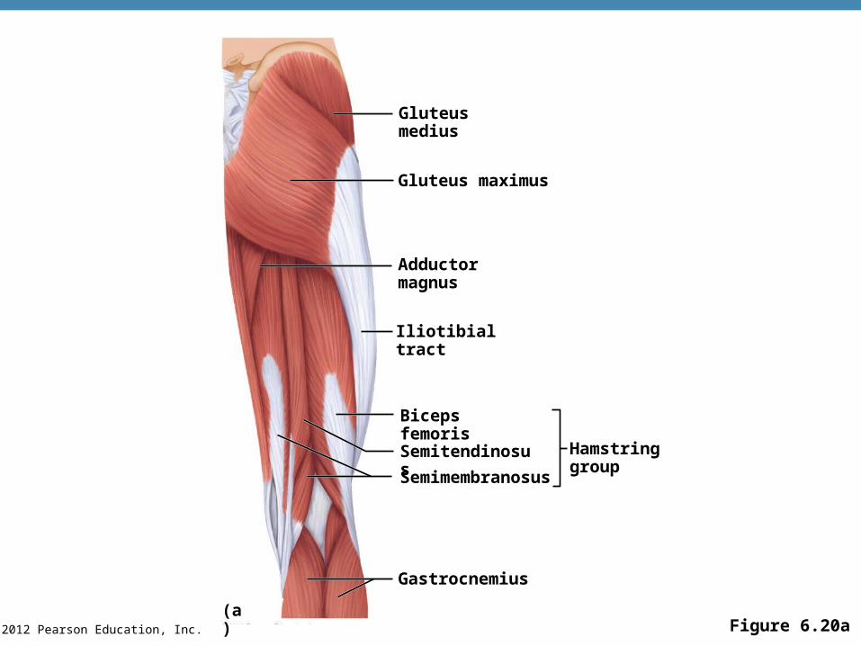
Muscles of the Lower Limb
•Muscles causing movement at the hip joint include:
•Gluteus maximus—hip extension
•Gluteus medius—hip abduction, steadies pelvis when walking
•Iliopsoas—hip flexion, keeps the upper body from falling backward when standing erect
•Adductor muscles—adduct the thighs
PLAYPLAY A&P Flix™: Movement at the hip joint: An overview.
PLAYPLAYA&P Flix™: Muscles that act on the hip joint and femur: An overview.

© 2012 Pearson Education, Inc. Figure 6.20a
Gluteus medius
Gluteus maximus
Adductormagnus
Iliotibial tract
Biceps femoris
Semitendinosus
Semimembranosus
Gastrocnemius
(a)
Hamstring group
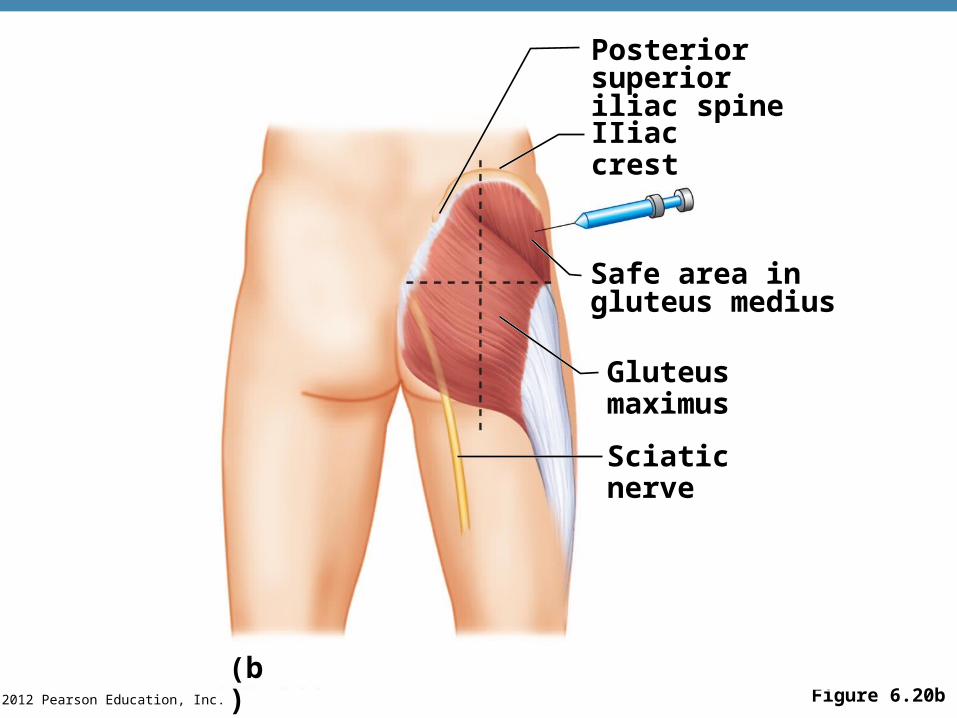
© 2012 Pearson Education, Inc. Figure 6.20b
Sciatic nerve
Gluteus maximus
Safe area ingluteus medius
IIiac crest
Posterior superioriliac spine
(b)
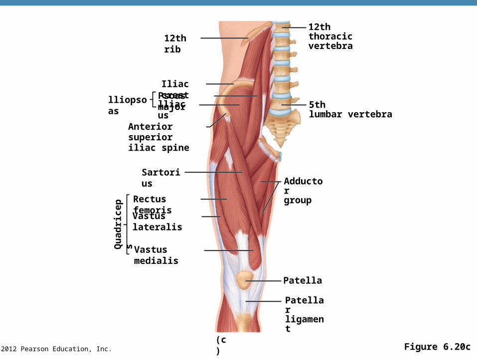
© 2012 Pearson Education, Inc. Figure 6.20c
12th rib
Iliac crest
lliopsoas Psoas majorlliacus
Anterior superioriliac spine
Sartorius
Rectus femoris
Vastus lateralis
Vastus medialisQu
adri
cep
s
Patellarligament
(c)
Patella
Adductorgroup
5thlumbar vertebra
12ththoracic vertebra
© 2012 Pearson Education, Inc.
Muscles of the Lower Limb
•Muscles causing movement at the knee joint
•Hamstring group—thigh extension and knee flexion
•Biceps femoris
•Semimembranosus
•Semitendinosus
© 2012 Pearson Education, Inc. Figure 6.20a
Gluteus medius
Gluteus maximus
Adductormagnus
Iliotibial tract
Biceps femoris
Semitendinosus
Semimembranosus
Gastrocnemius
(a)
Hamstring group
© 2012 Pearson Education, Inc.
Muscles of the Lower Limb
•Muscles causing movement at the knee joint
•Sartorius—flexes the thigh
•Quadriceps group—extends the knee
•Rectus femoris
•Vastus muscles (three)
PLAYPLAY A&P Flix™: Muscles that cross the knee joint: An overview.

© 2012 Pearson Education, Inc. Figure 6.20c
12th rib
Iliac crest
lliopsoas Psoas majorlliacus
Anterior superioriliac spine
Sartorius
Rectus femoris
Vastus lateralis
Vastus medialisQu
adri
cep
s
Patellarligament
(c)
Patella
Adductorgroup
5thlumbar vertebra
12ththoracic vertebra

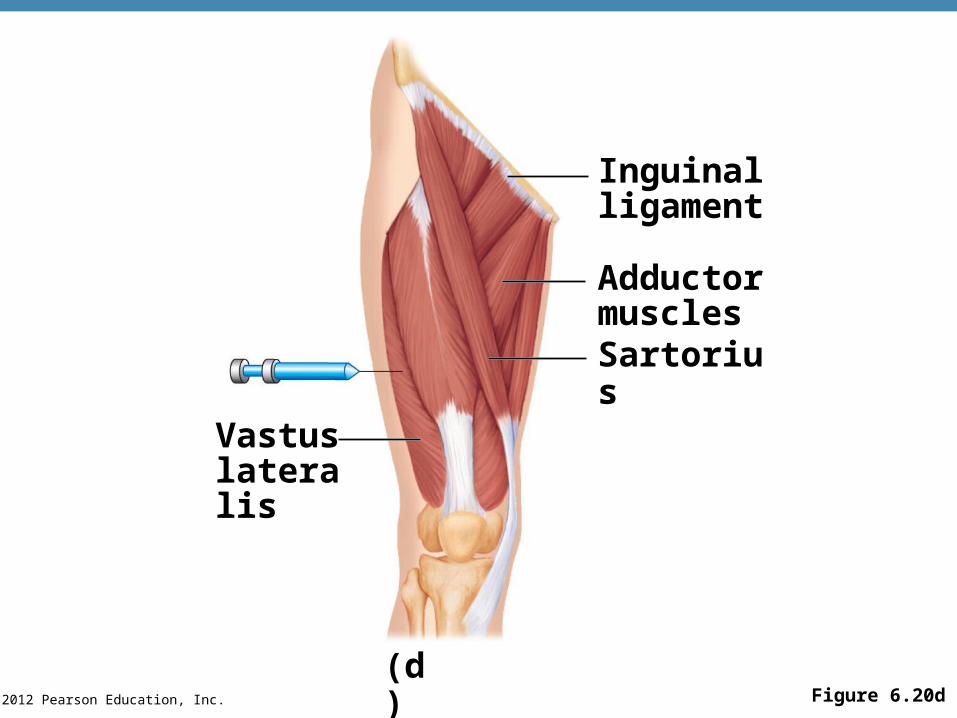
© 2012 Pearson Education, Inc. Figure 6.20d
Vastuslateralis
(d)
Sartorius
Adductormuscles
Inguinalligament
© 2012 Pearson Education, Inc.
Muscles of the Lower Limb
•Muscles causing movement at ankle and foot
•Tibialis anterior—dorsiflexion, foot inversion
•Extensor digitorum longus—toe extension and dorsiflexion of the foot
•Fibularis muscles—plantar flexion, foot eversion
•Soleus—plantar flexion
PLAYPLAY A&P Flix™: Movements of the ankle and foot.
PLAYPLAY A&P Flix™: Posterior muscles that act on the ankle and foot.
PLAYPLAYA&P Flix™: Muscles that act on the ankle and foot: An overview.
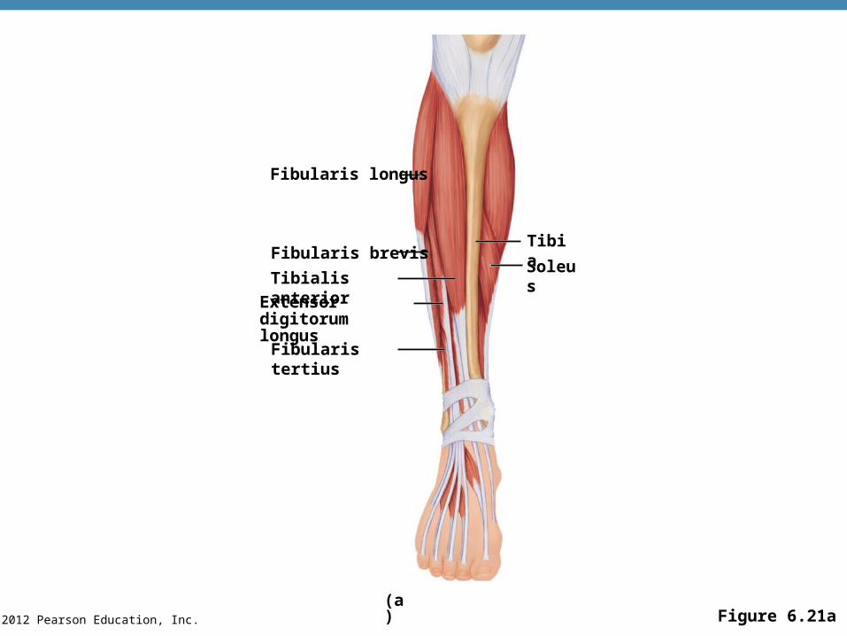
© 2012 Pearson Education, Inc. Figure 6.21a
Fibularis longus
Fibularis tertius
Tibialis anterior
Extensor digitorumlongus
Tibia
Soleus
(a)
Fibularis brevis
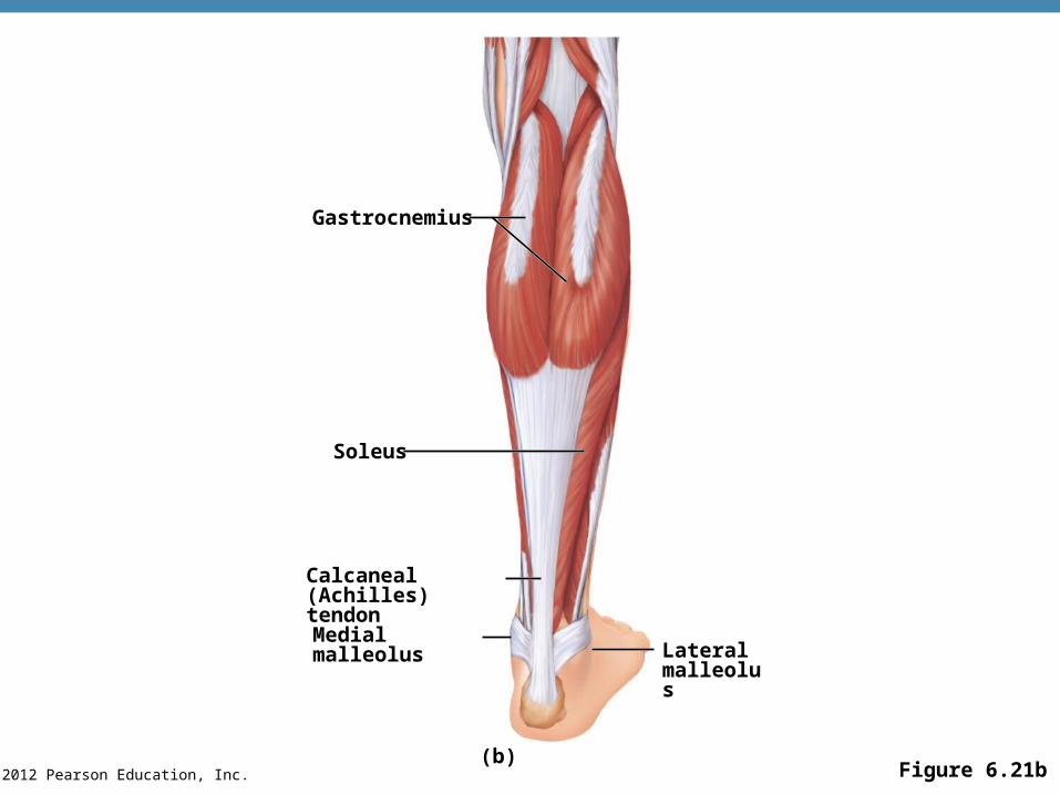
© 2012 Pearson Education, Inc. Figure 6.21b
Gastrocnemius
Soleus
Calcaneal (Achilles)tendon
Medial malleolusLateralmalleolus
(b)
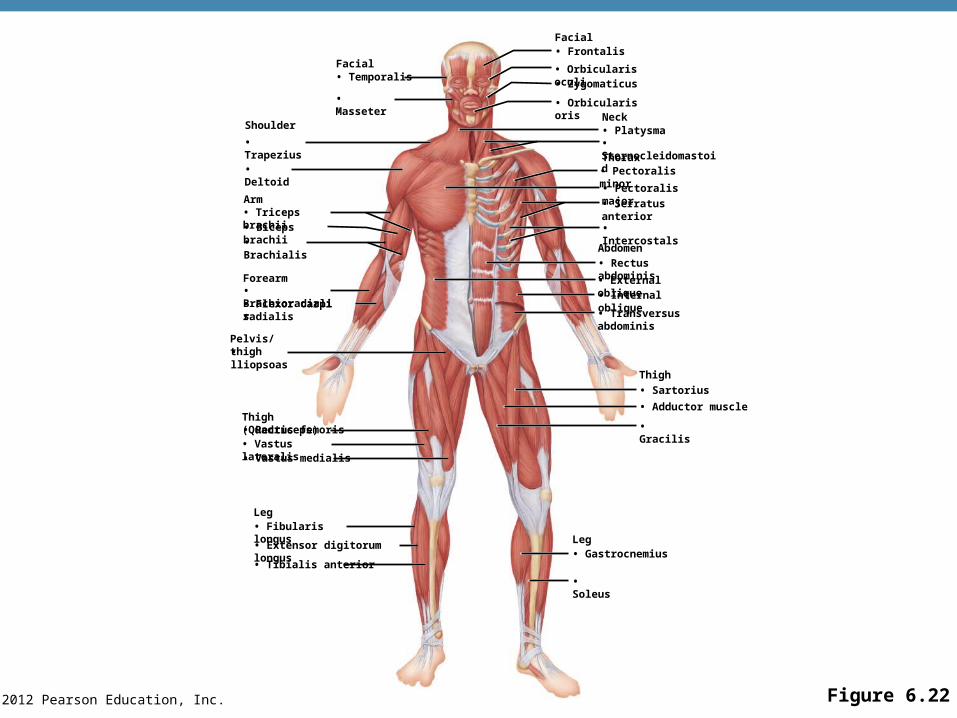
© 2012 Pearson Education, Inc. Figure 6.22
Facial• Temporalis
• Masseter
Shoulder
• Trapezius
• Deltoid
Arm• Triceps brachii• Biceps brachii
• Brachialis
Forearm• Brachioradialis• Flexor carpi radialis
Pelvis/thigh• lliopsoas
Thigh (Quadriceps)• Rectus femoris• Vastus lateralis• Vastus medialis
Leg• Fibularis longus
• Extensor digitorum longus
• Tibialis anterior
Facial• Frontalis
• Orbicularis oculi• Zygomaticus
• Orbicularis orisNeck• Platysma• SternocleidomastoidThorax• Pectoralis minor
• Pectoralis major
• Serratus anterior
• Intercostals
Abdomen
• Rectus abdominis
• External oblique• Internal oblique
• Transversus abdominis
Thigh
• Sartorius
• Adductor muscle
• Gracilis
Leg• Gastrocnemius
• Soleus
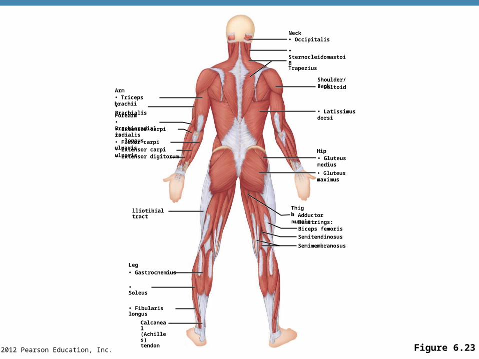
© 2012 Pearson Education, Inc. Figure 6.23
Arm• Triceps brachii
• Brachialis
Forearm• Brachioradialis• Extensor carpi radialis longus• Flexor carpi ulnaris
• Extensor carpi ulnaris• Extensor digitorum
lliotibial tract
Leg
• Gastrocnemius
• Soleus
• Fibularis longus
Calcaneal(Achilles)tendon
Neck• Occipitalis
• Sternocleidomastoid
• Trapezius
Shoulder/Back
• Deltoid
• Latissimus dorsi
Hip• Gluteus medius
• Gluteus maximus
Thigh• Adductor muscle• Hamstrings:
Biceps femoris
Semitendinosus
Semimembranosus
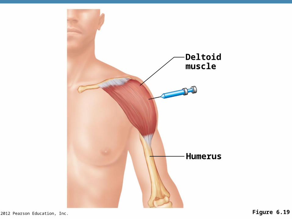
© 2012 Pearson Education, Inc. Figure 6.19
Deltoidmuscle
Humerus

© 2012 Pearson Education, Inc. Figure 6.20b
Sciatic nerve
Gluteus maximus
Safe area ingluteus medius
IIiac crest
Posterior superioriliac spine
(b)
© 2012 Pearson Education, Inc. Figure 6.20d
Vastuslateralis
(d)
Sartorius
Adductormuscles
Inguinalligament