Embed Size (px)
Citation preview

© 2009 Ryan Patrick Sullivan

ENGINEERING A FUNGAL L-ARABINOSE PATHWAY TOWARDS THE CO-
UTILIZATION OF HEMICELLULOSIC SUGARS FOR PRODUCTION OF XYLITOL
BY
RYAN PATRICK SULLIVAN
DISSERTATION
Submitted in partial fulfillment of the requirements
for the degree of Doctor of Philosophy in Chemical Engineering
in the Graduate College of the
University of Illinois at Urbana-Champaign, 2009
Urbana, Illinois
Doctoral Committee:
Professor Huimin Zhao, Chair
Professor Daniel Pack
Assistant Professor Nathan Price
Professor Hans Blaschek

ii
ABSTRACT
The biosynthesis of value-added products has shown great promise in recent years due to the
advances in molecular biology and protein engineering. Many advantages over chemical
synthesis include high selectivity and specificity, increased yield, and function under milder
conditions without the need for toxic metals, organic solvents, strong acids or bases, or high
pressures and temperatures. However, naturally occurring enzymatic pathways are often times
not particularly well suited towards obtaining high product yield due to the organism‘s evolution
not dependent on the production of such high levels of desired product. Xylitol is an attractive
pentose sugar alcohol that has many applications in the food, pharmaceutical, and high-value
based biochemical product industries. It is still, however, relatively expensive to produce, which
prevents its economical integration into the marketplace. To expand the possible routes of
xylitol synthesis, and provide a process which can utilize both hemicellulosic pentose sugars D-
xylose and L-arabinose from renewable plant biomass as feed substrates, my thesis research
focused on utilizing a fungal-derived biosynthetic pathway for production of xylitol. The
pathway consists of two NADPH-dependent reductases, xylose reductase (XR) and L-xylulose
reductase (LXR), and an NAD+-dependent L-arabinitol 4-dehydrogenase (LAD). The XR
enzyme converts D-xylose directly to xylitol, while the three enzyme in tandem convert L-
arabinose to xylitol. However, the cofactor imbalance presented with the pathway potentially
makes nicotinamide regeneration difficult for production in vivo. After cloning and
characterization of a highly active and stable NAD+-dependent L-arabinitol 4-dehydrogenase
(LAD) from Neurospora crassa, subsequent engineering via rational design and directed
evolution resulted in the isolation of the first known NADP+-dependent LAD enzyme. This
novel engineered LAD was then introduced into the fungal xylitol pathway and expressed in a

iii
model organism, E. coli, and the effect of the cofactor specificity alteration was evaluated in the
conversion of L-arabinose to xylitol. Further investigation of the cofactor balancing benefits
were applied to xylitol dehydrogenase (XDH) for full redox balancing of the initial steps of the
L-arabinose pathway, and investigations of the limiting steps in L-arabinose utilization conducted
in S. cerevisiae and E. coli led to further proposed engineering of LAD.

iv
To my loving family

v
ACKNOWLEDGEMENTS
First and foremost, many thanks to my advisor, Huimin Zhao, for his constant guidance, his
uncanny availability, and his great patience throughout the completion of my graduate career.
Thanks to my committee members, Daniel Pack, Nathan Price, and Hans Blaschek, for their
valued support. Thanks to every fellow lab member who was a part of the Zhao laboratory
during my graduate career. In particular, I would like to thank Michael Simurdiak and Tyler
Johannes for providing helpful instruction and training when I first joined the lab. Additional
thanks to Nikhil Nair, Fei Wen, Sheryl Rubin-Pitel, Matthew Desieno, Michael McLauchlan,
Zengyi Shao, and Hua Zhao for their intellectual insight and valued contributions. Special
thanks goes to the members of the IGB/EBI team of Jing Du, Byoungjin Kim, Amit Ghosh, Sijin
Li, Tae-Hee Lee, and Dawn Eriksen, who helped me tremendously during my final year of study.
Thanks to Brian Bae, Stacy Keller, and Satish Nair for their crystallization collaborative efforts.
Thanks to my undergraduate assistants Anu Biswas, Mark Laurenz, and Saroj Saha. Thanks to
the many friends I have gained and whose friendships I hope to sustain past my stay in Illinois,
particularly (and in no particular order) Matthew Cole and Jennifer Younker, Lon and Jessica
Chubiz, Matthew and Kristen Willis, John and Jennifer Schmidt, Mohan Karulkar, Eric Mock,
Matthew Langer, Fikhil Brushett and Karen Guralnick, Nicolas Londono, Esther Jeng, Tasha
Desai, Rachel Graff, Alice Hollister, Grant Pitel, Nate Gabrielson, Jonathan Silvestre and Lily
Wong, Doug Viehman, and Everett Scheer. To any names I may have missed throughout, please
receive my sincerest apology and know that you‘re in my heart always if not in my typing hands
at this particular moment. Finally, a special thanks to my family – my father Kevin, my mother
Caroline, and my sister Corrine – for always providing me with the strength and determination to
follow through with my aspirations, and for being my true inspiration.

vi
TABLE OF CONTENTS
CHAPTER 1 – Biosynthesis of Value-Added Products and Biofuels .............. 1
1.1. Introduction .......................................................................................................................... 1
1.2. Chemical Synthesis of Xylitol ............................................................................................. 2
1.3. Biotechnological Synthesis of Xylitol from Xylose ............................................................ 3
1.3.1. Bacterial Production of Xylitol ..................................................................................... 3
1.3.2. Fungal Production of Xylitol ........................................................................................ 3
1.3.3. Yeast Production of Xylitol .......................................................................................... 4
1.4. L-Arabinose as a Substrate for Xylitol Production .............................................................. 5
1.5. Protein Engineering ............................................................................................................. 8
1.6. Project Overview ................................................................................................................. 9
References ................................................................................................................................. 12
CHAPTER 2 – Cloning, Expression, Purification, and Characterization
of L-Arabinitol 4-Dehydrogenase from Neurospora crassa ............................ 18
2.1. Introduction ........................................................................................................................ 18
2.2. Results ................................................................................................................................ 19
2.2.1. N. crassa LAD Gene Identification ............................................................................ 19
2.2.2. Cloning, Expression, and Purification of N. crassa LAD .......................................... 21
2.2.3. Steady-state Kinetics ................................................................................................... 23
2.2.4. Temperature and pH Dependence ............................................................................... 26
2.2.5. Determination of Protein Mass and Quaternary Structure .......................................... 28
2.2.6. Cofactor Specificity .................................................................................................... 29
2.2.7. Homology Modeling ................................................................................................... 30
2.2.8. F59 Mutational Analysis ............................................................................................. 32
2.3. Discussion .......................................................................................................................... 32
2.4. Conclusions and Future Work ........................................................................................... 34
2.5. Materials and Methods ....................................................................................................... 35
2.5.1. Materials ..................................................................................................................... 35
2.5.2. RT-PCR and Cloning (Notebook #1, p. 42; Notebook #2, p. 51) .............................. 36

vii
2.5.3. Lysate Assay ............................................................................................................... 37
2.5.4. GST-tag Removal (Notebook #2, p. 67) ..................................................................... 38
2.5.5. Enzyme Kinetics (Notebook #2, pp. 79-102) ............................................................. 38
2.5.6. Thermal Dependence (Notebook #2, pp. 79-102) ...................................................... 39
2.5.7. pH Rate Profile (Notebook #2, pp. 79-102) ................................................................ 39
2.5.8. Determination of Protein Mass and Quaternary Structure(Notebook #2, p. 202) ...... 40
2.5.9. HPLC Analysis (Notebook #1, p. 186) ....................................................................... 40
2.5.10. Homology Modeling ................................................................................................. 40
References ..................................................................................................................................... 42
CHAPTER 3 – Engineering an NADP+-Dependent L-Arabinitol 4-
Dehydrogenase for Xylitol Production ..................................................................... 45
3.1. Introduction ........................................................................................................................ 45
3.2. Results ................................................................................................................................ 50
3.2.1. Reversal of Cofactor Specificity of N. crassa LAD by Rational Design ................... 50
3.2.2. Screening Procedure Validation and EP-PCR Results ............................................... 53
3.2.3. Kinetic Data of the Engineered ncLAD Mutants ........................................................ 54
3.3. Discussion .......................................................................................................................... 57
3.3.1. Protein Engineering Strategy ...................................................................................... 57
3.3.2. Properties of the ncLAD-3x Mutant ........................................................................... 60
3.3.3. Structural Analysis of the Mutations .......................................................................... 60
3.4. Conclusions ........................................................................................................................ 65
3.5. Materials and Methods ....................................................................................................... 66
3.5.1. Materials ..................................................................................................................... 66
3.5.2. Site-Directed and Saturation Mutagenesis .................................................................. 66
3.5.3. Error-Prone PCR Mutagenesis and Library Creation (Notebook #3, p. 130) ............. 68
3.5.4. Activity Assay Screening Procedure (Notebook #3, p. 136) ...................................... 70
3.5.5. DNA Sequencing and Analysis .................................................................................. 71
3.5.6. LAD Over-Expression and Purification (Notebook #4, p. 230-231) .......................... 72
3.5.7. HPLC Analysis of Products ........................................................................................ 73
3.5.8 Enzyme Kinetics (Notebook #4, p.232) ....................................................................... 73
References ................................................................................................................................. 75

viii
CHAPTER 4 – Application of an Engineered Fungal Pathway for Xylitol
Production from Hemicelluosic Sugars L-Arabinose and D-Xylose ............ 79
4.1. Introduction ........................................................................................................................ 79
4.2. Results ................................................................................................................................ 80
4.2.1. Strain Development .................................................................................................... 80
4.2.2. Operon Design ............................................................................................................ 82
4.2.3. Shake Flask Studies .................................................................................................... 85
4.2.3.1. Effect of Using Glucose as Co-Substrate and pH on Xylitol Production .......................... 85
4.2.3.2. Wild-type vs. Engineered Xylitol Pathway ........................................................................ 86
4.2.4. Bioreactor Studies ....................................................................................................... 88
4.2.4.1. Wild-Type vs. Engineered Pathway ................................................................................... 88
4.2.4.2. Engineered Xylitol Pathway – pH control vs. non-pH control .......................................... 92
4.2.4.3. WT vs. ENGR Pathway – Co-utilization ........................................................................... 94
4.3. Discussion .......................................................................................................................... 95
4.4. Conclusions and Outlook ................................................................................................... 98
4.5. Materials and Methods ....................................................................................................... 99
4.5.1. Materials ..................................................................................................................... 99
4.5.2. Gene Deletion (Notebook #3, p. 127) ......................................................................... 99
4.5.3. Mutant crp* Integration ............................................................................................ 100
4.5.4. Operon Design and Construction (Notebook #3, p. 231-234) .................................. 101
4.5.5. HPLC Analysis ......................................................................................................... 103
4.5.6. Shake Flask Studies (Notebook #4, p. 279) .............................................................. 103
4.5.7. Bioreactor Studies (Notebook #4, p. 158) ................................................................ 104
References ............................................................................................................................... 105
CHAPTER 5 – Fungal Arabinose Pathway Complementation Towards
Determining Limitations in Arabinose Utilization ............................................ 107
5.1. Introduction ...................................................................................................................... 107
5.2. Results .............................................................................................................................. 110
5.2.1. Cloning, Characterization, and Engineering of an NADP+-Dependent N. crassa
Xylitol Dehydrogenase ....................................................................................................... 110
5.2.2. Introduction of L-Arabinose Pathway into S. cerevisiae .......................................... 114

ix
5.2.3. E. coli Complementation Experiments ..................................................................... 116
5.3. Discussion ........................................................................................................................ 119
5.4. Conclusions ...................................................................................................................... 121
5.5. Materials and Methods ..................................................................................................... 121
5.5.1. Materials ................................................................................................................... 121
5.5.2. RT-PCR and Cloning ................................................................................................ 122
5.5.3. Cell Lysate Assay ..................................................................................................... 123
5.5.4. Enzyme Kinetics ....................................................................................................... 124
5.5.5. pH Profile .................................................................................................................. 124
5.5.6. Pathway Construction (S. cerevisiae) (Notebook #5, p. 101) ................................... 124
5.5.7. Yeast Transformation (S. cerevisiae) ........................................................................ 125
5.5.8. Pathway Construction (E. coli) (Notebook #4, p. 151) ............................................. 125
References ............................................................................................................................... 127

1
CHAPTER 1 – Biosynthesis of Value-Added Products and Biofuels
1.1. Introduction
Xylitol is a five-carbon sugar alcohol with a growing market as a sweetener that can be produced
by either chemical or biotechnological methods. The early research and development work of
xylitol began in Finland during the 1970‘s, and has been thoroughly reviewed by Mäkinen and
Peldyak1. In 1974, the Finnish Sugar Company Ltd. began the first large-scale production of
xylitol in Southern Finland. In 1975, the first sugar-free dental product was launched through
the introduction of a xylitol chewing gum (first in Finland, then the United States). Over the
next 25 years, global awareness of the significant advantages that xylitol offers to great-tasting
confectionery and gum products has steadily increased, especially the unique health and dental
benefits it provides. When continuously supplied in the diet, xylitol limits the tendency to
obesity, and its incorporation into food formulations results in improvement of the color and
taste of preparations without causing undesired changes in properties during storage. Along with
fructose, xylitol is the sugar recommended for diabetic patients, due to its human metabolism not
being insulin-mediated. In addition, the anticariogenic property of xylitol inhibits the growth of
oral bacteria so as to reduce plaque formation2, and it has also been reported that xylitol helps to
prevent acute otitis media (ear infection) in small children3. Recently the Department of Energy
(DOE) reported that xylitol was considered one of the top twelve sugar-derived building blocks
that can be subsequently converted to a number of high-value bio-based chemicals or materials4.
However, there is currently limited commercial production of xylitol, due mainly in part to its
comparatively high production cost (about 10 times that of alternatives sucrose or sorbitol) by
chemical reduction of D-xylose5.

2
1.2. Chemical Synthesis of Xylitol
Industrially, xylitol is produced by the chemical reduction of pure D-xylose, obtained from
hardwood hydrolysates, in the presence of a Raney nickel catalyst under high hydrogen
pressure6. The pure D-xylose is obtained using an industrial scale chromatographic method for
separating the different wood hemicellulosic sugars developed in Finland, where at the same
time the beneficial effects of xylitol in preventing dental caries were found. Pure and defined D-
xylose is the preferred starting material, which is difficult to obtain from the plant biomass.
Pretreatment and hydrolysis of lignocellulose from biomass yield a multi-component product
mixture that, in addition to the xylan (or D-xylose) fraction, contains other reducing sugars (such
as D-glucose from starch, L-arabinose from arabinan, etc.), acids, and various products of
decomposition of lignin and sugars7. The recovery of xylitol from the xylan fraction is about
50–60%, or roughly 8–15% of the raw material employed8. The value depends on the xylan
content of the raw material. There have been a number of patents filed on methods for obtaining
pure xylose and production of xylitol from xylan-containing materials6,9-13
. Drawbacks of the
chemical route of synthesis are the requirements of high pressure (up to 50 atm) and temperature
(80–140 C), use of an expensive catalyst, and use of extensive multi-step separation and
purification techniques, including mechanical filtration and chromatographic separation, to both
obtain pure initial D-xylose and to remove the other polyols and by-products after chemical
reduction.

3
1.3. Biotechnological Synthesis of Xylitol from Xylose
1.3.1. Bacterial Production of Xylitol
Xylitol has been reported to be produced by bacteria such as Enterobacter liquaefaciens14
,
Corynebacterium sp.15
, and Myobacterium smegmatis16
. In experiments leading to xylitol
production, an E. liquaefaciens strain was reported to reach 33.3 g xylitol L-1
in a fermentation
medium containing 100 g initial xylose L-1
, with a volumetric productivity of 0.35 g L-1
h-1
14
.
Fermentations with M. smegmatis (either as washed cells or as an immobilized form)
transformed D-xylose to xylitol at the same rate, giving 70% yield, and xylitol production from
D-xylose using commercial xylose isomerase from Bacillus coagulans and immobilized M.
smegmatis achieved a yield of 4 g xylitol from 10 g of D-xylose16
. Recombinant Lactococcus
lactis carrying the xylose reductase from Pichia stipitis and the xylose transporter from
Lactobacillus brevis gave xylitol productivity of 2.72 g L-1
h-1
during 20 h fermentations17
.
Some advantages of using bacterial hosts are a nearly quantitative yield of xylitol from xylose,
competitive volumetric productivity, and no requirement for aeration. However, conversions
typically do no proceed to completion at high initial xylose concentrations, requiring the
implementation of separation and recycling of D-xylose remaining in the product medium.
1.3.2. Fungal Production of Xylitol
Conversion of D-xylose to xylitol in a fermentation system using a fungal culture Petromyces
albertensis has been reported. In culture mediums containing 100 g L-1
initial D-xylose, cultures
reached 39.8 g L-1
xylitol after 10 days18
. After 160 h of anaerobic fermentation, Fusarium
oxysporum utilized almost 35 g L-1
of xylose and the maximum xylitol concentration (11.5 g L−1
)
was observed using ammonium as nitrogen source19
. Small concentrations of xylitol have also
been reported from cultured filamentous fungi including Penicillium, Aspergillus, Rhizopus,
4
Gliocladium, Byssochlamys, Myrothecium, and Neurospora when grown on D-xylose-based
media20
.
1.3.3. Yeast Production of Xylitol
Xylitol can be produced by natural xylose-assimilating yeasts, and varies a great deal with
species and aeration conditions. A wide range of yeast species belonging to the genus Candida
are well-known for their potential industrial applications, and include Candida tropicalis21-25
,
Candida guilliermondii26
, Candida boidinii27,28
, Candida parapsilosis29
, Candida maltosa30
, and
Candida peltata31,32
.. Candida tropicalis produced 8.5 g L−1
h−1
xylitol from D-xylose (yield of
0.85 mol mol-1
D-xylose, final concentration 180 g L-1
xylitol) in a cell recycling process in a
submerged membrane bioreactor with suction pressure and air sparging23
. When attempting to
ferment the processed hydrolysate without the added step of purifying substrate D-xylose, C.
tropicalis xylitol production was found to be 0.43 g/g and 0.45 g/g of D-xylose utilized with corn
fiber and sugarcane bagasse hydrolysate respectively, and after strain improvement by sub-
culturing, the production jumped to levels of 0.58 g/g and 0.65 g/g of D-xylose with corn fiber
hydrolysate and sugarcane bagasse hydrolysate respectively33
. The productivity for an
engineered C. tropicalis with an integrated NADH-preferring XR from Candida parapsilosis
was 5.1 g L-1
h-1 34
. Debaryomyces hansenii (Candida famata) produced 4.6 g L−1
h−1
xylitol
with a xylitol yield of 0.78 mol mol−1
from D-xylose and a final xylitol concentration of 221 g L-1
35. The ability of Candida peltata to ferment D-xylose to xylitol was evaluated and a maximum
xylitol yield of 0.56 g g-1
D-xylose was obtained when the yeast was cultivated in optimized
conditions. The yeast also produced ethanol (0.41 g g-1
in 40 h) from glucose (50 g L-1
) and
arabinitol (0.55 g g-1
in 87 h) from L-arabinose (50 g L-1
). Interestingly, moderate levels of
glucose (10 g L-1
), ethanol (7.5 g L-1
) and acetate (5 g L-1
) inhibited xylitol production by 61, 84

5
and 68%, respectively, whereas L-arabinose (10 g L-1
) had no inhibitory effect on xylitol
production31
. Another reoccurring phenomenon in most fermentation experiments was that
yeasts preferentially utilize glucose > D-xylose > L-arabinose from mixed substrates.
Xylitol production from D-glucose was also demonstrated using S. cerevisiae36
. A transketolase-
deficient strain (W303-1B tlk1 tlk2) growing on glucose accumulated D-xylulose 5-phosphate
intracellularly and released ribitol and pentose sugars D-ribose, D-ribulose, and D-xylulose into
the growth medium. Over-expressing the xylitol dehydrogenase from P. stipitis, which catalyzes
the reduction of D-xylulose to xylitol, and deletion of the xylulokinase-encoding gene (XKS1)
resulted in the production of xylitol and ribitol from D-xylulose and D-ribulose, respectively.
The yield of xylitol and ribitol formation was considerably lower than other methods, although
the authors did not attempt to optimize production conditions or metabolically engineer their
strain for improved PPP capacity.
1.4. L-Arabinose as a Substrate for Xylitol Production
Hemicellulose is the second most common polysaccharide in nature, representing 20-40% of
lignocellulosic (plant material) biomass. Hemicelluloses are heterogeneous polymers comprised
mostly of pentoses (D-xylose, L-arabinose), with minor contributions from hexoses (mannose,
glucose, galactose), and sugar acids37
. The utilization of the hemicellulose fraction for xylitol
production has been the subject of numerous research papers and patents9,33,37-41
, but only the D-
xylose component is converted to xylitol, with the other major component of hemicellulose, L-
arabinose, being converted to undesired byproduct L-arabinitol. However, D-xylose and L-
arabinose metabolism in yeasts and filamentous fungi conveniently share xylitol as an
intermediate (Figure 1.1). If the L-arabinose component of hemicellulose could also be utilized
for xylitol production, the economic feasibility by fermentative means could increase

6
dramatically. However, L-arabinose utilization by yeasts and filamentous fungi is poorly
characterized to date42
, and strains that do utilize L-arabinose typically produce large amounts of
L-arabinitol, instead of xylitol, along with biomass, suggesting a limitation earlier in the
utilization pathway43
.
One major issue that has yet to be resolved in utilization of L-arabinose as a substrate for xylitol
or ethanol is the subject of redox cofactor imbalance. The fungal pathway consists of two
oxidations and two reductions, which is redox neutral. However, the oxidations are NAD(H)-
linked and the reductions are NADP(H)-linked, so that there is an imbalance of the cofactors
involved in the process. In the view of xylitol production, the imbalance cannot be properly
regenerated by the cell‘s metabolism, leading to accumulation of L-arabinitol, the product of the
first enzyme in the pathway. In the view of fermentation, it remains an open question how
fungal micro-organisms cope with this cofactor imbalance. It has been suggested that NADPH is
mainly regenerated through the oxidative part of the pentose phosphate pathway44
. The
filamentous fungus Aspergillus niger exhibited higher activities of glucose 6-phosphate
dehydrogenase and 6-phosphogluconate dehydrogenase when growing on pentoses44
. In the
oxidative part of the pentose phosphate pathway, however, the reduction of NADP+ is coupled to
CO2 production. In this case the anaerobic conversion of L-arabinose to CO2 and ethanol is no
longer redox neutral, i.e. the fermentation of L-arabinose is inhibited by an accumulation of
reduced redox cofactors. Some attempts at engineering cofactor regeneration into these
processes have met with limited success, and those pertaining to xylose utilization are mentioned
below.
One proposed strategy of dealing with the cofactor imbalance was to integrate transhydrogenase
activity to facilitate the equilibrium between the coenzymes involved. Transhydrogenases

7
catalyze the reduction of NADP+ to NADPH with concomitant oxidation of NADH to NAD
+.
Yeasts are not believed to have such endogenous activity45
, so the expression of a cytosolic
transhydrogenase gene from Azotobacter yinlandii in S. cerevisiae was attempted and the
intracellular concentrations of the NAD(P) and NAD(P)H cofactors were measured46
. It was
hypothesized that since NADH could be consumed and NADPH produced by the enzyme,
expression of the gene encoding transhydrogenase could result in a decrease in glycerol
formation and the carbon flux through the pentose phosphate pathway, where there is a loss of
carbon in the form of carbon dioxide. This reduction in the carbon flux towards waste
components could be redirected towards formation of ethanol, leading to higher yield. However,
this was not found to be the case, as there was a resulting decrease in ethanol production and
increase in production of 2-oxogluarate and glycerol. Unfortunately, no mention was made of
the effects on pentose consumption or xylitol production.
Another strategy was to induce changes in the pentose phosphate pathway itself in order to try
and uncouple NADPH regeneration and CO2 production. In one study, the disruption of the
GND1 gene, one of the isogenes of 6-phosphogluconate dehydrogenase, or disruption of the
ZWF1 gene, which encodes glucose 6-phosphate dehydrogenase, blocked the pentose phosphate
pathway. These modifications resulted in a lower xylitol yield and higher ethanol yield than in
control strains. Also, xylitol production was shown to be strongly connected to the flux through
the oxidative part of the pentose phosphate pathway using metabolic flux analysis47
. A follow-
up to this approach expressed GDP1, which encodes for a fungal NADP+-dependent D-
glyceraldehyde-3-phosphate dehydrogenase (NADP-GAPDH), in a ZWF1 S. cerevisiae strain,
resulting in further stimulated xylose fermentation with respect to rate and yield, with increased
ethanol production under anaerobic conditions48
.

8
There was a recent publication during the course of the present thesis preparation by Bettiga et
al.49
that attempted at resolving the redox balance for L-arabinose utilization, which sought to
introduce a pathway that depends solely on NAD(H) using an engineered NADH-preferring
xylose reductase and a native NADH-preferring L-xylulose reductase along with NAD+-
dependent dehydrogenases for a cofactor balanced pathway. While the authors were focused on
improving pentose fermentation to ethanol, the attempt at improving the flux past L-arabinitol
using a cofactor-balancing strategy was shown to be effective, and will be discussed in Chapter
5. The goal of my work was to engineer NADP+-dependent L-arabinose pathway
dehydrogenases by means of rational design and directed evolution for application in xylitol or
biofuel production from hemicellulosic pentose sugars.
1.5. Protein Engineering
The tailoring of enzymes can be accomplished through two experimental routes. The first is
rational design, which targets specific residues of a protein for mutagenesis to predetermined
amino acid mutations, and is only applicable when there is detailed knowledge of the
relationships between the enzyme‘s structure and mechanism/function. And while an increasing
number of enzymes are being characterized, the majority do not have this depth of information
readily available, as it requires considerable effort to obtain. In the absence of this information,
the tailoring of an enzyme can still be accomplished through the second route - directed
evolution.
Directed evolution is the general term applied to the combined techniques of generation of a
library of protein mutants (or variants) and selection of a protein with desirable function from
within that library50
. It is an iterative Darwinian optimization process, whereby the fittest
variants are selected from an ensemble of mutants51
. Directed evolution can be used to target a
9
number of enzymatic characteristics, including activity, substrate specificity, thermal and
oxidative stability, enantioselectivity or enantiospecificity, pH optima or range, and tolerance to
solvent52
. While a typical directed evolution experiment focuses on a single enzymatic trait,
there are some examples of improving several traits simultaneously.
Choosing the appropriate methods of library generation and screening or selection is paramount
to the success of any directed evolution experiment. Library diversity can be created through
either mutagenesis (random or semi-rational) or gene recombination, and which of these methods
is chosen depends on many factors, such as the availabilities of homologous genes, structural
knowledge, and characteristic data of the enzyme of interest. The library size created is typically
very large (>104-6
), and close evaluation of each variant is not feasible. The need for a method to
find the improved ―needle in a haystack‖ enzyme becomes evident, with several strategies
including selection, enrichment, and high-throughput screening offering ways to sift through the
library clutter and find a variant with the desired enzymatic trait. However, once an evolved
enzyme has been found that exhibits improved characteristics, the artificial conditions in which
the selection method was carried out may result in an enzyme whose properties may not carry
over to the real biocatalytic process. Therefore, the more similar a screening system is to the
actual application process, the more likely it is to find an improved enzyme that will be
complementary to the application.
1.6. Project Overview
My research focused on two main projects: the engineering of an NADP+-dependent L-arabinitol
4-dehydrogenase and its application in xylitol biosynthesis from pentose sugars L-arabinose and
D-xylose, and engineering a cofactor balanced fungal L-arabinose pathway and analyzing its
effects on biofuel production. The first project focuses on the utilization of protein engineering

10
tools of rational and semi-rational design combined with directed evolution to reverse the
cofactor specificity of a single enzyme of a three-enzyme pathway in an attempt to improve flux
from L-arabinose substrate to product xylitol. The effect of this cofactor balancing is
investigated in a model organism, E. coli, and shown to enhance xylitol production from L-
arabinose and D-xylose. The engineering of an NADP+-dependent xylitol dehydrogenase and
introduction of the full L-arabinose assimilation pathway is established, with investigation into
the limiting factors of growth complementation in S. cerevisiae and E. coli when using the
cofactor dependent fungal pathway, with insights leading to further projects past the scope of this
thesis.
Chapter 2 describes the cloning and characterization of a novel L-arabinitol 4-dehydrogenase
(LAD) from Neurospora crassa, which is one of the most active and stable LADs reported.
Similar to all other characterized LADs, the enzyme is strictly NAD+-dependent. The high
stability is attributed to the structural zinc ion, and the substrate specificity of the LAD is
analyzed further by mutational analysis by comparison of active site residues to homologous
xylitol and sorbitol dehydrogenases. The enzyme is highly active and stable, acts on several five
carbon sugar alcohol substrates, and operates over a wide pH range, although the activity in
acidic pHs drops considerably, which is an issue that is addressed in its usage in the xylitol
production strategy in Chapter 4.
Chapter 3 describes the application of rational and semi-rational design followed by directed
evolution to reverse the cofactor specificity of L-arabinitol 4-dehydrogenase from NAD+ to
NADP+. Using the engineered enzyme along with NADPH-dependent xylose reductase and L-
xylulose reductase was hypothesized to alleviate the cofactor imbalance of the pathway such that
conversion of L-arabinose and/or D-xylose to xylitol along with a cosubstrate would produce

11
more desired product xylitol than the wild-type pathway. By using bioinformatics tools for
BLAST search and sequence alignment, along with homology modeling of the enzyme of
interest, key residues involved in the cofactor specificity were identified and subjected to site-
directed mutagenesis and saturation mutagenesis and screened for improved NADP+ activity.
When rational design sites were exhausted, the enzyme kinetics were further improved by
implementation of a directed evolution strategy following the validation of a proper screening
procedure. This resulted in the first known NADP+-dependent LAD enzyme.
Chapter 4 describes the application of expressing xylose reductase, wild-type or engineered L-
arabinitol 4-dehydrogenase, and L-xylulose reductase for xylitol biosynthesis from L-arabinose
and D-xylose. The effects of pH-control as well as substrate sugar ratios are also addressed.
Chapter 5 describes the cloning, characterization of xylitol dehydrogenase (XDH) from N.
crassa, and the engineering of an NADP+-dependent XDH by rational design. With the
engineered LAD created in Chapter 4, the cofactor imbalance associated with the reductases and
dehydrogenases of the initial L-arabinose pathway is relieved, and the subsequent introduction of
the pathway into S. cerevisiae and E. coli paves the way for investigation into the limiting factors
of L-arabinose utilization with the fungal-based pathway.

12
References
(1) Peldyak, J.; Makinen, K. K. Xylitol for caries prevention J Dent Hyg 2002, 76, 276-85.
(2) Makinen, K. K. Xylitol: the sugar that does not cause tooth decay Refuat Hapeh
Vehashinayim 1978, 27, 36-7.
(3) Uhari, M.; Tapiainen, T.; Kontiokari, T. Xylitol in preventing acute otitis media Vaccine
2000, 19 Suppl 1, S144-7.
(4) Werpy, T.; Petersen, G. Top Value Added Chemicals From Biomass: Volume 1 - Results
of Screening for Potential Candidates from Sugars and Synthesis Gas, U.S. Department
of Energy, National Renewable Energy Laboratory, 2004.
(5) Parajo, J. C.; Dominguez, H.; Dominguez, J. Biotechnological production of xylitol. Part
1: Interest of xylitol and fundamentals of its biosynthesis Bioresour Technol 1998, 65,
191-201.
(6) Melaja, A.; L, H. US, 1977.
(7) Mayer, G.; Kulbe, K. D.; Nidetzky, B. Utilization of xylitol dehydrogenase in a combined
microbial/enzymatic process for production of xylitol from D-glucose Appl Biochem
Biotechnol 2002, 98-100, 577-89.
(8) Winkelhausen, E.; Kuzmanova, S. Microbial conversion of D-xylose to xylitol J Ferment
Bioeng 1998, 86, 1-14.
(9) Sinner, M.; Dietrichs, H.; Puls, J.; Schweers, W.; Brachthauser, K. US, 1988.
(10) Melaja, A.; Virtanen, J.; Heikkila, H. US, 1978.
(11) Jaffe, G.; Szkrybalo, W.; Weinert, P. US, 1974.
(12) Melaja, A.; Hamalainen, L. US, 1978.
(13) Steiner, K.; Lindlar, R. US, 1971.
13
(14) Yoshitake, J.; Ishizaki, H.; Shimamura, M.; Imai, T. Xylitol production by an
Enterobacter species Agri Biol Chem 1973, 37, 2261-2267.
(15) Rangaswamy, S.; Agblevor, F. A. Screening of facultative anaerobic bacteria utilizing D-
xylose for xylitol production Appl Microbiol Biotechnol 2002, 60, 88-93.
(16) Izumori, K.; Tuzaki, K. Production of xylitol from D-xylulose by Mycobacterium
smegmatis J Ferment Technol 1988, 66, 33-36.
(17) Nyyssola, A.; Pihlajaniemi, A.; Palva, A.; von Weymarn, N.; Leisola, M. Production of
xylitol from D-xylose by recombinant Lactococcus lactis J Biotechnol 2005, 118, 55-66.
(18) Dahiya, J. Xylitol production by Petromyces albertensis grown on medium containing D-
xylose Can J Microbiol 1991, 37, 14-18.
(19) Panagiotou, G.; Christakopoulos, P.; Olsson, L. The influence of different cultivation
conditions on the metabolome of Fusarium oxysporum J Biotechnol 2005, 118, 304-15.
(20) Chiang, C.; Knight, S. G. Metabolism of D-xylose by moulds Nature 1960, 188, 79-81.
(21) Ko, B. S.; Rhee, C. H.; Kim, J. H. Enhancement of xylitol productivity and yield using a
xylitol dehydrogenase gene-disrupted mutant of Candida tropicalis under fully aerobic
conditions Biotechnol Lett 2006.
(22) Rao, R. S.; Jyothi, C. P.; Prakasham, R. S.; Rao, C. S.; Sarma, P. N.; Rao, L. V. Strain
improvement of Candida tropicalis for the production of xylitol: biochemical and
physiological characterization of wild-type and mutant strain CT-OMV5 J Microbiol
2006, 44, 113-20.
(23) Kwon, S. G.; Park, S. W.; Oh, D. K. Increase of xylitol productivity by cell-recycle
fermentation of Candida tropicalis using submerged membrane bioreactor J Biosci
Bioeng 2006, 101, 13-8.

14
(24) Kim, T. B.; Lee, Y. J.; Kim, P.; Kim, C. S.; Oh, D. K. Increased xylitol production rate
during long-term cell recycle fermentation of Candida tropicalis Biotechnol Lett 2004,
26, 623-7.
(25) Lopez, F.; Delgado, O. D.; Martinez, M. A.; Spencer, J. F.; Figueroa, L. I.
Characterization of a new xylitol-producer Candida tropicalis strain Antonie Van
Leeuwenhoek 2004, 85, 281-6.
(26) Roberto, I. C.; Felipe, M. G. A.; de Mancilha, I. M.; Vitolo, M.; Sato, S.; da Silva, S. S.
Xylitol production by Candida guillermondii as an approach for the utilization of
agroindustrial residues Bioresour Technol 1995, 51, 255-257.
(27) Suryadi, H.; Katsuragi, T.; Yoshida, N.; Suzuki, S.; Tani, Y. Polyol production by culture
of methanol-utilizing yeast J Biosci Bioeng 2000, 89, 236-40.
(28) Ko, C. H.; Chiu, P. C.; Yang, C. L.; Chang, K. H. Xylitol conversion by fermentation
using five yeast strains and polyelectrolyte-assisted ultrafiltration Biotechnol Lett 2008,
30, 81-6.
(29) Oh, D. K.; Kim, S. Y.; Kim, J. H. Increase of xylitol production rate by controlling redox
potential in Candida parapsilosis Biotechnol Bioeng 1998, 58, 440-4.
(30) Guo, C.; Zhao, C.; He, P.; Lu, D.; Shen, A.; Jiang, N. Screening and characterization of
yeasts for xylitol production J Appl Microbiol 2006, 101, 1096-104.
(31) Saha, B.; Bothast, R. Production of xylitol by Candida peltata J Ind Microbiol
Biotechnol 1999, 22, 633-636.
(32) Park, S. M.; Sang, B. I.; Park, D. W.; Park, D. H. Electrochemical reduction of xylose to
xylitol by whole cells or crude enzyme of Candida peltata J Microbiol 2005, 43, 451-5.

15
(33) Rao, R. S.; Jyothi Ch, P.; Prakasham, R. S.; Sarma, P. N.; Rao, L. V. Xylitol production
from corn fiber and sugarcane bagasse hydrolysates by Candida tropicalis Bioresour
Technol 2006, 97, 1974-8.
(34) Lee, J. K.; Koo, B. S.; Kim, S. Y. Cloning and characterization of the xyl1 gene,
encoding an NADH-preferring xylose reductase from Candida parapsilosis, and its
functional expression in Candida tropicalis Appl Environ Microbiol 2003, 69, 6179-88.
(35) Dominguez, J. M.; Cao, N.; Gong, C. S.; Tsao, G. T. Dilute acid hemicellulose
hydrolysates from corn cobs for xylitol production by yeast Bioresour Technol 1997, 61,
85-90.
(36) Toivari, M. H.; Ruohonen, L.; Miasnikov, A. N.; Richard, P.; Penttila, M. Metabolic
engineering of Saccharomyces cerevisiae for conversion of D-glucose to xylitol and other
five-carbon sugars and sugar alcohols Appl Environ Microbiol 2007, 73, 5471-6.
(37) Saha, B. C. Hemicellulose bioconversion J Ind Microbiol Biotechnol 2003, 30, 279-91.
(38) Parajo, J. C.; Dominguez, H.; Dominguez, J. Biotechnological production of xylitol. Part
3: Operation in culture media made from lignocellulose hydrolysates Bioresour Technol
1998, 66, 25-40.
(39) Hallborn, J.; Walfridsson, M.; Airaksinen, U.; Ojamo, H.; Hahn-Hagerdal, B.; Penttila,
M.; Kerasnen, S. Xylitol production by recombinant Saccharomyces cerevisiae
Biotechnology (N Y) 1991, 9, 1090-5.
(40) Jin, Y. S.; Cruz, J.; Jeffries, T. W. Xylitol production by a Pichia stipitis D-xylulokinase
mutant Applied Microbiology and Biotechnology 2005, 68, 42-45.

16
(41) Rodrigues, R. C.; Sene, L.; Matos, G. S.; Roberto, I. C.; Pessoa, A., Jr.; Felipe, M. G.
Enhanced xylitol production by precultivation of Candida guilliermondii cells in
sugarcane bagasse hemicellulosic hydrolysate Curr Microbiol 2006, 53, 53-9.
(42) Verho, R.; Putkonen, M.; Londesborough, J.; Penttila, M.; Richard, P. A novel NADH-
linked L-xylulose reductase in the L-arabinose catabolic pathway of yeast J Biol Chem
2004, 279, 14746-51.
(43) McMillan, J. D.; Boynton, B. L. Arabinose utilization by xylose-fermenting yeasts and
fungi Appl Biochem Biotechnol 1994, 45-46, 569-84.
(44) Witteveen, C. F. B.; Busink, R.; van de Vondervoort, P.; Dijkema, C.; Swart, K.; Visser,
J. L-arabinose and D-xylose catabolism in Aspergillus niger J Gen Microbiol 1989, 135,
2163-2171.
(45) Bruinenberg, P. M. The NADP(H) redox couple in yeast metabolism Antonie Van
Leeuwenhoek 1986, 52, 411-29.
(46) Nissen, T. L.; Anderlund, M.; Nielsen, J.; Villadsen, J.; Kielland-Brandt, M. C.
Expression of a cytoplasmic transhydrogenase in Saccharomyces cerevisiae results in
formation of 2-oxoglutarate due to depletion of the NADPH pool Yeast 2001, 18, 19-32.
(47) Jeppsson, M.; Johansson, B.; Hahn-Hagerdal, B.; Gorwa-Grauslund, M. F. Reduced
oxidative pentose phosphate pathway flux in recombinant xylose-utilizing
Saccharomyces cerevisiae strains improves the ethanol yield from xylose Appl Environ
Microbiol 2002, 68, 1604-9.
(48) Verho, R.; Londesborough, J.; Penttila, M.; Richard, P. Engineering redox cofactor
regeneration for improved pentose fermentation in Saccharomyces cerevisiae Appl
Environ Microbiol 2003, 69, 5892-7.

17
(49) Bettiga, M.; Bengtsson, O.; Hahn-Hagerdal, B.; Gorwa-Grauslund, M. F. Arabinose and
xylose fermentation by recombinant Saccharomyces cerevisiae expressing a fungal
pentose utilization pathway Microb Cell Fact 2009, 8, 40.
(50) Yuan, L.; Kurek, I.; English, J.; Keenan, R. Laboratory-directed protein evolution
Microbiol Mol Biol Rev 2005, 69, 373-92.
(51) Roodveldt, C.; Aharoni, A.; Tawfik, D. S. Directed evolution of proteins for heterologous
expression and stability Curr Opin Struct Biol 2005, 15, 50-6.
(52) Hibbert, E. G.; Baganz, F.; Hailes, H. C.; Ward, J. M.; Lye, G. J.; Woodley, J. M.; Dalby,
P. A. Directed evolution of biocatalytic processes Biomol Eng 2005, 22, 11-9.
18
CHAPTER 2 – Cloning, Expression, Purification, and Characterization of
L-Arabinitol 4-Dehydrogenase from Neurospora crassa
2.1. Introduction
As detailed in Chapter 1, the route through which L-arabinose is converted to xylitol in the
fungal pathway passes through three key enzymes: xylose reductase (XR), L-arabinitol 4-
dehydrogenase (LAD), and L-xylulose reductase (LXR). There are many examples of XRs that
have been characterized from various source organisms, including a highly active XR
characterized by Ryan Woodyer and Michael Simurdiak in Professor Zhao‘s laboratory from
Neurospora crassa, a filamentous fungus known to ferment xylose1. With the first enzyme of
the process already available, the focus turned to the remaining two enzymes that would
complete the process. However, there are relatively few cases of the other two enzymes being
isolated and identified. The cloning and characterization of the LXR from N. crassa was done
by Nikhil Nair in the Zhao laboratory. However, this enzyme did not possess high affinity
towards substrate L-xylulose, with an unusually high Km,L-xylulose value of > 275 mM. It was also
recently challenged that the homologous LXR1 from Hypocrea jecorina was instead a D-
mannitol 2-dehydrogenase (MDH) based on phylogenetic analysis and deletion experiments
showing deletion of lxr1 did not affect growth on L-arabinose, whereas MDH activity levels
dropped2. Due to the inconclusive identity of N. crassa LXR and less than desirable kinetic
parameters, the reported LXR from Aspergillus niger was selected for the xylitol production
process due to its higher affinity towards L-xylulose (Km,L-xylulose of 17 mM)3.
The final enzyme remaining, L-arabinitol 4-dehydrogenase (LAD, EC 1.1.1.12), is commonly
found in yeasts and filamentous fungi and catalyzes the second step of the fungal L-arabinose

19
metabolic pathway by oxidizing L-arabinitol to L-xylulose with concomitant NAD+ reduction.
LAD is purportedly a fungal orthologue of the eukaryotic sorbitol dehydrogenase (SDH)4 and
belongs to the family of zinc-containing alcohol dehydrogenases. Several LADs have
successfully been cloned and expressed3,5,6
. However, they are not optimal for in vivo enzymatic
production of xylitol due to their poor stability and/or activity. This chapter will focus on the
cloning, expression, purification, and characterization of a novel LAD from N. crassa with high
activity and stability for use in xylitol production from L-arabinose and D-xylose. This enzyme
serves as the template for engineering altered coenzyme specificity that will be detailed further in
Chapter 3.
2.2. Results
2.2.1. N. crassa LAD Gene Identification
LADs from Hypocrea jecorina (GenBank accession number AF355628.1) and Aspergillus
oryzae (AB116938.2) were used as templates for a protein BLAST search
(www.ncbi.nlm.nih.gov). Utilizing the whole-genome sequence of N. crassa7, a postulated
LAD-encoding gene, hypothetical protein NCU00643.1 (EAA36547.1), was discovered which
had the greatest sequence identity (~80%). This protein (referred to as N. crassa LAD hereafter)
had significant homology (72 to 80% identity) with other LADs (Fig. 2.1). Among the
conserved residues were those that formed the active site and the structural Zn2+
-binding site8
and the glycine fingerprint9 found in polyol dehydrogenases, as well as the majority of those
shown to bind substrate in the SDH homologues4.

20
Neurospora crassa MASSAS-------------KTNIGVFTNPQHDLWISEASPSLESVQKGEE
Hypocrea jecorina MSPSAVDDAPKATGAAISVKPNIGVFTNPKHDLWISEAEPSADAVKSGAD
Aspergillus niger MATATVLE-----------KANIGVFTNTKHDLWVADAKPTLEEVKNGQG
Aspergillus oryzae MATATVLE-----------KANIGVYTNTNHDLWVAESKPTLEEVKSGES
Aspergillus fumigatus MATATTTVLE---------KPNIGVYTNPKHDLWIAESTPTLEDVKSGNG
:.:: *.****:**.:****:::: *: : *:.*
Neurospora crassa LKEGEVTVAVRSTGICGSDVHFWKHGCIGPMIVECDHVLGHESAGEVIAV
Hypocrea jecorina LKPGEVTIAVRSTGICGSDVHFWHAGCIGPMIVEGDHILGHESAGEVIAV
Aspergillus niger LQPGEVTIEVRSTGICGSDVHFWHAGCIGPMIVTGDHILGHESAGQVVAV
Aspergillus oryzae LKPGEVTVQVRSTGICGSDVHFWHAGCIGPMIVTGDHILGHESAGEVIAV
Aspergillus fumigatus LKPGEVTIEVRSTGICGSDVHFWHAGCIGPMIVEGDHILGHESAGQVIAV
*: ****: **************: ******** **:*******:*:**
Neurospora crassa HPSVKSIKVGDRVAIEPQVICNACEPCLTGRYNGCERVDFLSTPPVPGLL
Hypocrea jecorina HPTVSSLQIGDRVAIEPNIICNACEPCLTGRYNGCEKVEFLSTPPVPGLL
Aspergillus niger APDVTSLKPGDRVAVEPNIICNACEPCLTGRYNGCENVQFLSTPPVDGLL
Aspergillus oryzae ASDVTHLKPGDRVAVEPNIPCHACEPCLTGRYNGCEKVLFLSTPPVDGLL
Aspergillus fumigatus APDVTSLKPGDRVAIEPNIPCHACEPCLTGRYNGCLNVAFLSTPPVDGLL
. *. :: *****:**:: *:************* .* ******* ***
Neurospora crassa RRYVNHPAVWCHKIGNMSYENGAMLEPLSVALAGLQRAGVRLGDPVLICG
Hypocrea jecorina RRYVNHPAVWCHKIGNMSWENGALLEPLSVALAGMQRAKVQLGDPVLVCG
Aspergillus niger RRYVNHPAIWCHKIGDMSYEDGALLEPLSVSLAGIERSGLRLGDPCLVTG
Aspergillus oryzae RRYVNHPAVWCHKIGDMSYEDGALLEPLSVSLAAIERSGLRLGDPVLVTG
Aspergillus fumigatus RRYVNHPAVWCHKIGDMSFEDGALLEPLSVSLAAIERSGLRLGDPCLITG
********:******:**:*:**:******:**.::*: ::**** *: *
Neurospora crassa AGPIGLITMLCAKAAGACPLVITDIDEGRLKFAKEICPEVVTHKVER-LS
Hypocrea jecorina AGPIGLVSMLCAAAAGACPLVITDISESRLAFAKEICPRVTTHRIEIGKS
Aspergillus niger AGPIGLITLLSARAAGASPIVITDIDEGRLEFAKSLVPDVRTYKVQIGLS
Aspergillus oryzae AGPIGLITLLSARAAGATPIVITDIDEGRLAFAKSLVPDVITYKVQTNLS
Aspergillus fumigatus AGPIGLITLLSAKAAGATPLVITDIDEGRLQFAKSLVPEVRTYKVQFGLS
******:::*.* **** *:*****.*.** ***.: * * *:::: *
Neurospora crassa AEESAKKIVESFG--------GIEPAVALECTGVESSIAAAIWAVKFGGK
Hypocrea jecorina AEETAKSIVSSFG--------GVEPAVTLECTGVESSIAAAIWASKFGGK
Aspergillus niger AEQNAEGIINVFNDGQGSGPGALRPRIAMECTGVESSVASAIWSVKFGGK
Aspergillus oryzae AEDNAAGIIDAFNDGQGSAPDALKPKLALECTGVESSVASAIWSVKFGGK
Aspergillus fumigatus AEEQANAIINVFNDGQGSGPDALRPRLALECTGVESSVASAIWSVKFGGK
**: * *:. *. .:.* :::********:*:***: *****
Neurospora crassa VFVIGVGKNEIQIPFMRASVREVDLQFQYRYCNTWPRAIRLVENGLVDLT
Hypocrea jecorina VFVIGVGKNEISIPFMRASVREVDIQLQYRYSNTWPRAIRLIESGVIDLS
Aspergillus niger VFVIGVGKNEMTVPFMRLSTWEIDLQYQYRYCNTWPRAIRLVRNGVIDLK
Aspergillus oryzae VFVIGVGKNEMKIPFMRLSTQEIDLQYQYRYCNTWPRAIRLVRNGVISLK
Aspergillus fumigatus VFVIGVGKNEMTIPFMRLSTQEIDLQYQYRYCNTWPRAIRLVQNGVINLK
**********: :**** *. *:*:* ****.*********:..*::.*.
Neurospora crassa RLVTHRFPLEDALKAFETASDPKTGAIKVQIQSLE------------
Hypocrea jecorina KFVTHRFPLEDAVKAFETSADPKSGAIKVMIQSLD------------
Aspergillus niger KLVTHRFLLEDAIKAFETAANPKTGAIKVQIMSSEDDVKAASAGQKI
Aspergillus oryzae KLVTHRFLLEDALKAFETAADPKTGAIKVQIMSNEEDVKGASA----
Aspergillus fumigatus RLVTHRFALEDALKAFETAANPKTGAIKVQIMSSEEDVKAASATQ--
::***** ****:*****:::**:***** * * :
Figure 2.1. Protein sequence alignment of N. crassa LAD with four other closely related LAD
sequences from filamentous fungi and yeast. Residues highlighted in gray represent the four
conserved residues that make up the catalytic zinc binding tetrad. Residues highlighted in red
represent the four conserved cysteine residues that make up the proposed structural zinc binding
tetrad.

21
2.2.2. Cloning, Expression, and Purification of N. crassa LAD
RT-PCR performed on total RNA isolated from L-arabinose-induced N. crassa 10333 showed
the expected size of gene product (~1.1 kb). The RT-PCR product was cloned into the pGEX-
4T-3 vector using EcoRI and NotI restriction sites and was transformed into E. coli BL21 (DE3).
This construct (pGEX-lad1) expressed N. crassa LAD as an N-terminal GST-tagged fusion with
a thrombin cleavage site. Cell lysates of IPTG-induced cultures of these cells were prepared,
analyzed by SDS-PAGE, and assayed for LAD activities. The construct produced soluble GST-
tagged N. crassa LAD at ~16% of the total soluble cellular proteins (Fig. 2.2), which was then
purified in a single step with a GST-Bind kit according to manufacturer‘s protocol. The purified
protein was desalted by ultrafiltration with several washes of 50 mM morpholinepropanesulfonic
(MOPS) buffer (pH 7.25). After digesting with biotinylated thrombin, the enzyme was incubated
with streptavidin agarose to remove the thrombin and then passed through GST-Bind resin again
to remove the cleaved GST-tag. GST-tagged LAD cleaved with thrombin was used for
characterization purposes, as it had about 65% greater specific activity than the tagged LAD
enzyme. LAD stocks were stored frozen with 10% (v/v) glycerol at −80 °C. Protein
concentrations were determined by the Bradford method10
by using an estimated extinction
coefficient (San Diego Supercomputer Center Biology Workbench [http://workbench.sdsc.edu])
of 35.3 mM−1
cm−1
at 280 nm with similar results. The purity of the protein was analyzed by an
SDS-PAGE gel stained with Coomassie brilliant blue (Fig. 2.2). The final yield of protein was
30 mg/L of culture (~9 mg/g of E. coli) of greater than 95% pure LAD with a molecular mass of
~39 kDa, consistent with the predicted value of 39.6 kDa.

22
Figure 2.2. Overexpression and purification of recombinant N. crassa LAD. Lane 1, the
molecular weight marker proteins (size in kDa is shown); lane 2, cell-free crude extract; lane 3,
purified LAD enzyme with N-terminal GST-tag; lane 4, purified LAD enzyme with GST-tag
removed by thrombin cleavage.

23
2.2.3. Steady-state Kinetics
Purified N. crassa LAD displayed activity with NAD+ as the preferred cofactor (Table 2.1),
although there was small yet detectable activity with NADP+, which was verified later by high
performance liquid chromatography (HPLC) as shown in Figure 2.5. This is the first reported
detection of NADP+ utilization by an LAD, although it is still considered a strongly NAD
+-
dependent enzyme.
Table 2.2 displays the kinetic constants of several other sugar substrates accepted by N. crassa
LAD. D-Arabinitol, adonitol, xylitol, D-sorbitol, and D-mannitol were all examined as alternative
substrates for N. crassa LAD with NAD+ as the cofactor. Of those, only the five carbon sugar
alcohols acted as substrates, with Km values of 80 mM and 280 mM for adonitol and xylitol,
respectively. This pattern of substrate promiscuity is similar to those of LADs isolated from
other sources5,6
. Kinetic parameters of characterized LADs from Hypocrea jecorina, Aspergillus
niger, and Aspergillus oryzae are displayed in Table 2.33-6
.

24
TABLE 2.1. Kinetic parameters for N. crassa LADa.
N. crassa LAD with
indicated coenzyme
Km for NAD(P)
(mean SD) (M)
kcat (mean SD)
(min-1
)
kcat/Km for NAD(P)
(M-1
min-1
)
Km for L-arabinitol
(mean SD) (mM)
NAD 174 24 1,206 54 6.9 16 3
NADP - - 4.3 x 10⁻⁵
-
a All assays were performed at 25 C in 50 mM Tris, pH 8.0
TABLE 2.2. Kinetic parameters for N. crassa LAD with other substratesa.
N. crassa LAD with
indicated substrate
kcat (mean SD)
(min-1
)
Km (mean SD)
(mM)
kcat/Km
(mM-1
min-1
) % Activity
L-Arabinitol 1,210 30 18 2 67 100
Xylitol 970 40 290 27 3.3 4.9
Adonitol 1,080 30 35 3 31 46
D-Arabinitol - - NDb 0
D-Sorbitol -c -
c -
c 0
c
D-Mannitol - - ND 0
a All assays were performed at 25C in 50 mM Tris, pH 8.0, at saturated NAD
+
concentration b ND, not detected
c trace activity at 2 M D-sorbitol concentration, possibly due to substrate contamination

25
TABLE 2.3. Kinetic parameters of LAD from various source organisms.
Organism (reference) Specific activity
(U/mg)
kcat
(min-1
)
Km, L-arabinitol
(mM)
kcat/Km, L-arabinitol
(mM-1
min-1
)
Km, NAD+
(M)
N. crassa (this work) 31 1,206 16 75 174
H. jecorina 5 1.6 N/A
a 40 N/A 180
H. jecorina 4 0.013 51 4.5 11 N/A
A. niger 3 96 N/A 89 N/A 50
A. oryzae 6 0.04 N/A N/A N/A N/A
a N/A, not determined
TABLE 2.4. Kinetic parameters of F59 mutantsa.
Substrate Enzyme kcat (mean SD)
(min-1
)
Km (mean SD)
(mM)
kcat/Km
(mM-1
min-1
)
L-Arabinitol WT 1,210 30 18 2 67
F59Y 840 30 42 5 20
F59S 60 3 62 9 0.97
F59A -b > 400 0.12
Xylitol WT 970 40 290 27 3.3
F59Y - b > 880 1.2
F59S - b > 1,400 0.04
F59A - b > 1,850 0.01
Adonitol WT 1,080 30 35 3 31
F59Y 1,420 50 193 11 7.4
F59S 120 5 430 48 0.28
F59A - b > 1,110 0.03
a All assays were performed at 25 C in 50 mM Tris, pH 8.0.
b Saturation of substrate was not reached.

26
2.2.4. Temperature and pH Dependence
The data show the optimum temperature to be between 45 and 55 °C (Fig. 2.3A). At higher
temperatures, the enzyme inactivates rapidly, and at lowered temperatures, the rate increases
with temperature according to the Arrhenius equation. Utilizing the Arrhenius equation to fit the
data from 12 to 30 °C, the energy of activation for L-arabinitol oxidation by N. crassa LAD was
determined to be 47 kJ/mol. The stability for N. crassa LAD was relatively high, as it retained
activity at room temperature for longer than one month and at 4 °C for several months. Figure
2.3B shows the results of thermal inactivation of N. crassa LAD at 50 °C, which followed a first-
order exponential decay with a half-life of 36 min. Interestingly, when tested at a slightly lower
temperature of 45 °C, the enzyme was able to retain ~70% of its activity after 3 hr. The high
stability can potentially be attributed to the conserved structural zinc binding residues. A similar
thermostabilizing mechanism was also found in alcohol dehydrogenase11
and engineered xylitol
dehydrogenase12
.
The pH range for N. crassa LAD activity was large, with >25% of the activity occurring with pH
values of 7.0 to 10.5 (Fig. 2.3C). The pH optimum was around pH 9.5, and >60% of the activity
remained in the 2 pH-unit span from 8.0 to 10.0.

27
Figure 2.3. A) kcat dependence on temperature. N. crassa LAD was assayed at different
temperatures from 12 to 65 °C at saturating concentrations of 200 mM L-arabinitol and 2 mM
NAD+. B) Thermal inactivation of LAD at 50 °C. The heat inactivation at 50 °C was irreversible
and followed first-order kinetics with a half-life of 45 min. C) pH rate profile. Saturating
concentrations of 200 mM L-arabinitol and 2 mM NAD+ were used to measure the activity in a
universal buffer at various pH values from 7.0 to 11.0.
10 20 30 40 50 60 700
10
20
30
40
50
A
kca
t (s
-1)
Temperature (oC)
0 25 50 75 100 125 150 175 20020
30
40
50
60
70
80
90
100 B
Resid
ua
l A
ctivity (
%)
Time (min)
7 8 9 10 11
20
40
60
80
100C
No
rma
lize
d S
pe
cific
Activity (
%)
pH

28
2.2.5. Determination of Protein Mass and Quaternary Structure
Based on the standardized retention times of a Bio-Rad molecular mass standard, the molecular
mass of the native N. crassa LAD was calculated from its retention time to be ~129 kDa (Fig.
2.4). Monomerization was induced in the presence of 15% SDS, causing N. crassa LAD to elute
as a single peak with a retention time corresponding to a molecular mass of ~39 kDa. The data
suggest that the native LAD is a noncovalently linked tetramer, which is typical for fungal
derived zinc-containing alcohol dehydrogenases13
.
Figure 2.4. HPLC size exclusion chromatography. A size exclusion standard was used to
calibrate a Bio-Sil SEC-250, 300 x 7.8 mm column with a mobile phase of 0.1 M Na2HPO4, 0.15
M NaCl, 0.01 M NaN3, pH 6.8 at a flow rate of 1 ml/min. The standard proteins are represented
by closed squares. LAD samples (represented by open squares) with and without 15% SDS were
injected separately and fitted to the standard curve.

29
2.2.6. Cofactor Specificity
The cofactor specificity of N. crassa LAD was examined by HPLC. The separation of NAD+,
NADP+, NADH, and NADPH (Figs. 2.5A and 2.5B) was carried out as previously described
14.
No discernible cross-contamination of oxidized cofactors was observed (data not shown). 20 L
reaction mixtures consisting of equal parts of 1 mM NAD(P)+ and 25 mM L-arabinitol in 50 mM
Tris-HCl (pH 8.0) were set up, and following addition of approximately 1 g of enzyme, the
reaction was allowed to proceed for 20 min at 37 °C. When NAD+ was used as the cofactor, the
products were analyzed by HPLC, and a single peak (UV 340 nm) was observed that had the
same retention time as authentic NADH (Fig. 2.5C). The same process was carried out for
NADP+ as the cofactor, and a small yet detectable peak was observed with a retention time
corresponding to an authentic sample of NADPH (Fig. 2.5D). This indicated the strong
preference for NAD+ as the cofactor of N. crassa LAD.

30
Figure 2.5. HPLC analysis of the reaction products of N. crassa LAD. A) Cofactor stocks, 260
nm; B) Cofactor stocks, 340 nm; C) NAD+ reaction mix products, 340 nm; D) NADP
+ reaction
mix before (gray) and after (black) LAD enzyme addition, 340 nm (zoomed in).
2.2.7. Homology Modeling
The structural model of N. crassa LAD was verified for consistency with known protein folds
and allowed ϕ and ψ angles. The Profiles3-D check resulted in a self-compatibility score of
139.94, which compares well to the scores of 150.53 and 145.49 for the coordinates from 1PL8
and 1E3J, respectively. The ProStat check of ϕ and ψ angles were determined to be 81.2%
within their core expected values, comparing well to the 83.3 and 82.4% for the same analysis of
PDB structures 1PL8 and 1E3J, respectively. This model was very similar to the human SDH
crystal structure in overall fold and binding of coenzyme. The only major deviation between the
backbone of these two structures is the N-terminal region of amino acids 1 through 8. However,

31
this may be due to the different conformations of the N-terminus between being in solution and
forming dimerization contacts found to be present in SDH4. The conserved catalytic zinc
binding residues C53, H78, E79, and E163 have similar orientations and locations in the N.
crassa LAD model (Fig. 2.6). When comparing proposed substrate binding residues from SDH
to N. crassa LAD, the majority—S55, F127, T130, E163, R308, Y309—are strictly conserved
and configured in similar orientations. However, one substrate-binding residue, F59, was
different from the homologous tyrosine residue in SDH, making the N. crassa LAD-binding
pocket slightly more hydrophobic (Fig 2.6B).
Figure 2.6. A) Homology model of N. crassa LAD with bound NAD+ and catalytic zinc ion,
built using the Insight II and MOE programs. The catalytic zinc ion (Zn2+
), four catalytic zinc
binding residues, and NAD+ cofactor are colored by atom type. B) Active site of N. crassa LAD
with docked catalytic zinc (shown in space-fill), cofactor NAD+, and substrate L-arabinitol
(colored green) coordinated with the active zinc. Key residues located within the substrate
binding pocket are labeled.
B
F127 Y309
R308
T130
NAD+
F59
E163
Zn S55 C53
H78
A
E163
Zn E79
H78
NAD+
C53

32
2.2.8. F59 Mutational Analysis
Enzyme activity assays were carried out for three mutants of N. crassa LAD (F59A, F59S, and
F59Y) to study the effect of mutation of this active site, putative substrate-binding residue
homologous to tyrosine in other SDHs. All assays were carried out similar to substrate
specificity profile for the wild-type enzyme (see ―Steady-state kinetics‖ in ―Materials and
Methods‖), with the cofactor NAD+ held at saturating concentration of 2 mM for all assays. The
mutants were still found to have activity with L-arabinitol, xylitol, and adonitol, and their kinetic
parameters are displayed in Table 2.4. D-Sorbitol was also tested but showed no significant
activity over the wild-type N. crassa LAD.
2.3. Discussion
Hypothetical protein NCU00643.1 (EAA36547.1) from N. crassa was found to encode an LAD
of 363 amino acid residues with a calculated Mr of 39,245. Sequence alignment with other
reported LADs shows high homology (~70–80%), with conserved regions for Zn2+-binding,
cofactor binding, and active site residues. Comparison with several mammalian SDHs (mouse,
rat, bovine, sheep, and human) showed ~40% sequence identity, whereas comparison with
xylitol dehydrogenases (Hypocrea jecorina, Aspergillus oryzae, Candida tropicalis, Pichia
stipitis, and Aspergillus fumigatus Af293) was ~30% sequence identity.
Kinetic parameters of characterized LADs from H. jecorina, Aspergillus niger, and A. oryzae are
displayed in Table 2.3. It should be noted that only the LAD enzyme from H. jecorina by Pail et
al.4 was purified to homogeneity, whereas the others were characterized either as partially
purified enzymes3,5
or as cell-free extracts.6 The Km value of N. crassa LAD was 16 mM f or L-
arabinitol, which when compared to LADs from H. jecorina and A. niger, is one of the lower

33
values reported of characterized LADs. The values of 45 mM5 and 4.5 mM
4 were reported for
the same H. jecorina LAD, although the enzymes were purified from 15
different heterologous
hosts (S. cerevisiae and E. coli, respectively), with the former LAD being obtained by cleavage
of a GST-fusion protein. In addition, it is difficult to determine the effect of the larger Km,NAD+ of
N. crassa LAD compared to other LADs, as no other characterizations reported both kcat and Km
values for the cofactor. With a specific activity of the purified N. crassa LAD equal to about 31
U/mg, it is almost 20-fold greater than H. jecorina LAD purified from S. cerevisiae heterologous
expression and orders of magnitude higher than other LADs except for A. niger LAD, which
shows about three-fold greater specific activity than that of N. crassa LAD. However, it was also
reported that the purified A. niger LAD was highly unstable, with rapidly diminishing activity at
4 °C and complete loss of activity after freeze-thawing of the enzyme.3 In contrast, N. crassa
LAD is quite stable and does not markedly lose activity when frozen repeatedly.
There has been no in-depth study of the substrate binding residues for LAD, but the enzyme has
been postulated to be a fungal orthologue of the eukaryotic D-SDHs4, which have been
investigated more thoroughly. Based on these reports, the active site substrate-binding residues
are all strictly conserved in all LADs characterized to date (see Figure 2.6). When comparing
these residues in N. crassa LAD to SDHs, only F59 was not conserved, which instead was a
tyrosine residue in all of the SDHs examined. Mutational studies of this position were examined
for F59A, F59S, and F59Y, to determine what effects this residue has on substrate specificity
alteration, and are shown in Table 2.4. Replacement of the native F59 residue with the
homologous tyrosine found in SDH decreased the catalytic efficiency toward all active
substrates. The ability of LAD to bind each substrate markedly decreased as the size of the
amino acid at position 59 was decreased. Although these results suggest this residue is important

34
for binding and catalysis for the active substrates of the wild-type LAD, it did not confer the
enzyme the ability to accept D-sorbitol as a substrate. This confirms previously reported
hypotheses that the amino acids flanking the active site cleft may be responsible for the activity
and affinity patterns between LAD and SDH4. The results were also recently confirmed in
mutational analysis studies of A. niger LAD15
. An important finding of this study was the
discovery of mutation F59Y, which abolished xylitol activity but did not eliminate L-arabinitol
activity, although the kcat was lowered by ~33%. It is possible this mutation could be necessary
for the production of xylitol from L-arabinose from the combination of XR, LAD, and LXR, as
LAD could continue to convert xylitol to D-xylulose. However, as will be discussed in Chapter
4, the low LAD activity with xylitol does not prevent xylitol from being detected from the three
enzyme pathway.
2.4. Conclusions and Future Work
In conclusion, a gene from N. crassa encoding an LAD was cloned and purified in high yields.
The enzyme is highly active and stable, acts on several five carbon sugar alcohol substrates, and
operates over a wide pH range. This enzyme should prove to be a useful starting point for the
ultimate goal of the production of xylitol and ethanol from L-arabinose derived from renewable
resources. However, the newly characterized N. crassa LAD still has a strict preference for
NAD+ over NADP
+ as the native coenzyme. In order to alleviate some of the cofactor
regeneration issues that were mentioned in Chapter 1, engineering of the cofactor specificity of
N. crassa LAD will need to be implemented to generate a successful strategy towards in vitro
xylitol production and possibly in vivo ethanol fermentation. The next chapter will focus on
efforts to engineer the crucial cofactor-binding residues to introduce NADP+ activity into N.
crassa LAD.

35
2.5. Materials and Methods
2.5.1. Materials
The Neurospora crassa genomic sequence and LAD protein sequences were accessed from the
National Center for Biotechnology Information (NCBI, www.ncbi.nlm.nih.gov). Neurospora
crassa 10333 was obtained from the American Type Culture Collection (ATCC). Escherichia
coli BL21(DE3), GST-Bind resin, biotinylated thrombin, and streptavidin agarose were
purchased from Novagen (Madison, WI). E. coli WM1788 was kindly provided by Professor
William Metcalf at the University of Illinois (Urbana, IL). GST-fusion expression vector pGEX-
4T-3 was purchased from Amersham Biosciences. SuperScript One-Step RT-PCR with
Platinum
Taq kit was obtained from Invitrogen (Carlsbad, CA). Shrimp alkaline phosphatase,
and PCR grade dNTPs were obtained from Roche Applied Sciences (Indianapolis, IN). Phusion
High-Fidelity DNA Polymerase and DNA-modifying enzymes DNase I, NdeI, SacI, and T4
DNA ligase and their appropriate buffers were purchased from New England Biolabs (NEB)
(Beverly, MA). L-arabinitol, D-arabinitol, adonitol, xylitol, D-sorbitol, D-mannitol, ampicillin,
kanamycin, isopropyl -D-thiogalactopyranoside (IPTG), NADH, NADP+, and NADPH were
purchased from Sigma (St. Louis, MO). NAD+ was a gift from Jülich Fine Chemicals (Jülich,
Germany). Other required salts and reagents were purchased from Fisher (Pittsburgh, PA) or
Sigma-Aldrich. The QIAprep spin plasmid mini-prep kit, QIAquick gel purification kit, RNeasy
midiprep kit, and QIAquick PCR purification kit were purchased from Qiagen (Valencia, CA).
Various oligonucleotide primers were obtained from Integrated DNA Technologies (Coralville,
IA). SDS-PAGE gel materials, electrophoresis equipment, protein size markers, size exclusion
standards (catalog number 151-1901) and Bio-Sil SEC-250, 300 x7.8 mm column were
purchased from Bio-Rad (Hercules, CA).

36
2.5.2. RT-PCR and Cloning (Notebook #1, p. 42; Notebook #2, p. 51)
N. crassa 10333 was grown on rich potato media at 30 °C for 24 h and induced with 150 mM L-
arabinose for 2 hr. Because the predicted gene contained one intron, reverse transcription PCR
(RT-PCR) was utilized to isolate the processed gene. Total ribonucleic acid (RNA) was purified
from collected cells (RNeasy purification kit, Qiagen) and treated with DNase I to remove
residual genomic DNA. RT-PCR was performed using SuperScript™ One-Step RT-PCR with
Platinum® Taq (Invitrogen) following the manufacturer‘s guidelines. A control reaction
consisted of the same protocol except for the SuperScript enzyme mix was heated to 95 ºC for
ten minutes to thermally inactivate the reverse transcriptase enzyme and the reverse transcription
thermocycler step was omitted. The primers used for the RT-PCR were: forward 5′-GTA GCT
ACG TCA GAA TTC CAT GGC TTC TAG CGC TTC C-3′ and reverse 5′-GCT GAT TCT
GCG GCC GCT TAC TCC AGA CTC TGG ATC-3′. The forward primer contained an EcoRI
restriction site (shown in bold), while the reverse primer contained a NotI restriction site (shown
in bold) and stop codon (italicized). The resulting RT-PCR product was isolated by a QIAquick
agarose gel purification kit and amplified by an additional 20 cycles of PCR. The product was
digested with EcoRI and NotI restriction enzymes and purified again by agarose gel
electrophoresis. It was then ligated into pGEX-4T-3 which had been previously prepared by
EcoRI and NotI digestion, dephosphorylation by shrimp alkaline phosphatase, and gel
purification. The ligation mixture was precipitated with n-butanol and resuspended in water. The
new construct was used to transform E. coli WM1788 by electroporation. Positive clones were
selected on Luria–Bertani (LB) solid media with ampicillin at 37 °C overnight. All colonies were
then removed from the plates and grown to saturation in 5 mL liquid LB from which the
plasmids were purified using a QIAprep spin plasmid miniprep kit, which were used to transform

37
E. coli BL21(DE3) by heat shock. Positive clones were selected on LB solid media with
ampicillin, picked individually, and assayed for LAD activity by the cell lysate assay described
below. Plasmids were sequenced using the BigDye® Terminator sequencing method and an ABI
PRISM 3700 sequencer (Applied Biosystems, Foster City, CA). Sequencing results determined
at the Biotechnology Center of the University of Illinois showed that the product had four silent
mutations compared with the predicted sequence from the NCBI database.
2.5.3. Lysate Assay
E. coli BL21 (DE3) harboring the pGEX-4T-3-derived vector was grown to a maximum optical
density at 600 nm (OD600) at 37 °C with shaking at 250 rpm. Fifty microliters was used to
inoculate a new culture, which was grown at 37 °C with shaking at 250 rpm until an OD600 of
~0.6 was reached. The cultures were then induced with 0.3 mM IPTG and shaken at 250 rpm at
25°C for 4 h. One milliliter of cells was harvested by centrifugation and lysed by resuspension
in 1 mL of 1 mg/mL lysozyme/50 mM Tris-HCl (pH 8.0). The cells were frozen at −80 °C and
thawed at room temperature. The resulting lysate was vortexed thoroughly and centrifuged to
remove cell debris. Ten microliters of the lysate was used in a kinetic assay described below in
the Enzyme Kinetics Section with 200 mM L-arabinitol and 2 mM NAD+ as the substrates in 50
mM Tris (pH 8.0). To determine soluble and insoluble expression, lysozyme was utilized as the
lysis reagent for the induced and normalized cells following the manufacturer‘s
recommendations, and samples were subsequently analyzed by SDS-PAGE.

38
2.5.4. GST-tag Removal (Notebook #2, p. 67)
The N-terminal GST-tag was removed by incubation with biotinylated thrombin overnight at 4
°C, incubation with streptavidin agarose for 30 min at 4 °C to remove thrombin, and passing
mixture through GST-Bind resin to remove GST-tag, leaving five residues (GlySerProAsnSer)
attached to the N terminus of the N. crassa LAD sequence. To determine the effect of removal
of the GST-tag, the purified LAD was incubated with and without thrombin at 4 °C overnight.
Complete cleavage of the 25.7 kDa tag was verified by SDS-PAGE. The specific activities of the
cleaved and noncleaved samples were compared. It was determined that removal of the GST-tag
enhanced activity by about 65%. Because of this significant difference in activity, the cleaved
enzyme was used in all subsequent assays.
2.5.5. Enzyme Kinetics (Notebook #2, pp. 79-102)
Initial rates were determined by measuring the initial rate of reaction as observed by the increase
of absorbance at 340 nm using a Varian Cary 100 Bio UV-visible spectrophotometer (Varian,
Palo Alto, CA) at 25 °C in 50 mM Tris-HCl (pH 8.0). N. crassa LAD kinetic measurements
with substrate L-arabinitol and cofactor NAD+ were taken in a five-by-five matrix format, with
substrate and cofactor concentrations varied from below their Km to tenfold higher than their Km.
The kinetic data were analyzed with a modified version of Cleland‘s program16
. Vmax and Km for
both L-arabinitol and NAD+ were obtained by fitting the data to a sequential ordered mechanism
with NAD+ binding first, based on the proposed mechanism for sorbitol dehydrogenase
17:
v = VmaxAB/(KiaKB+KAB+KBA+AB) (Eq. 1)

39
where v is the initial velocity, Vmax is the maximum velocity, KA and KB are the Michaelis-
Menten constants for NAD+ and L-arabinitol, respectively, A and B are the concentrations of
NAD+ and L-arabinitol, respectively, and Kia is the dissociation constant for NAD
+.
N. crassa LAD kinetic measurements with alternate substrates were taken with varied
concentrations of substrate sugar alcohol while keep NAD+ concentration saturating at 2 mM.
The data were used to calculate the kinetic constants by fitting the Michaelis-Menten equation
using Origin 5.0. N. crassa LAD displayed typical Michaelis-Menten type kinetics with respect
to all substrates tested except D-sorbitol. The data represent averages of assays performed in
duplicate or triplicate on two separate occasions.
2.5.6. Thermal Dependence (Notebook #2, pp. 79-102)
The optimal temperature of activity was determined by assaying N. crassa LAD activities at
temperatures ranging from 12 to 65 °C in saturating substrate conditions. Thermal inactivation
of N. crassa LAD was studied by incubating at varied elevated temperatures in 50 mM Tris-HCl
(pH 8.0), with samples removed at various times and assayed for activity in saturating substrate
conditions.
2.5.7. pH Rate Profile (Notebook #2, pp. 79-102)
Activity was measured at pH values between 7.0 and 11.0 under saturating concentrations of
NAD+ (2 mM) and L-arabinitol (200 mM) in a universal buffer (50 mM
morpholineethanesulfonic acid (MES) / 50 mM Tris / 50 mM glycine). Ionic strength of the
universal buffer was taken into consideration according to previously established buffer
compositions.18

40
2.5.8. Determination of Protein Mass and Quaternary Structure(Notebook #2, p. 202)
The quaternary structure of native N. crassa LAD was determined using an Agilent 1100 series
HPLC system with a Bio-Sil SEC-250 column (300 by 7.8 mm) and a mobile phase of 0.1 M
Na2HPO4, 0.15 M NaCl, and 0.01 M NaN3, pH 6.8. Monomerization was induced in the
presence of 15% SDS and loaded onto the HPLC in similar fashion to the native enzyme to
determine the N. crassa LAD monomer size.
2.5.9. HPLC Analysis (Notebook #1, p. 186)
The enzyme (1 g) was incubated in a mixture of buffer and 25 mM L-arabinitol with 1 mM
NAD(P)+ at 37 C for 20 min. The sample was eluted on a Zorbax 150 mm x 3.0 mm C-18 (3.5
m) column with a UV detector (Agilent 1100 series). The eluent consisted of two elements: 0.1
M KH2PO4 containing 5 mM tetrabutylammonium hydrogen sulfate (pH 5.5) (buffer A) and
100% methanol (buffer B). The most suitable gradient was an initial isocratic step for 6 min at
93% buffer A, a gradient for 5 min from 7 to 30% buffer B, and a final isocratic step for 5 min at
30% buffer B.
2.5.10. Homology Modeling
Insight II was used to prepare the model (Insight II, version 2000; Accelrys Inc., San Diego, CA)
and MOE (Chemical Computing Group Inc., Montreal, Canada) was used for optimization. To
verify the model, the overall fold was checked using Profiles3-D (Insight II), and the allowed
states for and angles and bond distances were checked using ProStat (Insight II), both with
default settings. The Profiles3-D (Insight II, default parameters) check resulted in a self-
compatibility score of 139.94, which compares well to the scores of 150.53 and 145.49 for the
coordinates from 1PL8 and 1E3J, respectively. The ProStat check of and angles were

41
determined to be 81.2% within their core expected values, comparing well to the 83.3% and
82.4% for the same analysis of PDB structures 1PL8 and 1E3J, respectively.
42
References
(1) Woodyer, R.; Simurdiak, M.; van der Donk, W. A.; Zhao, H. Heterologous expression,
purification, and characterization of a highly active xylose reductase from Neurospora
crassa Appl Environ Microbiol 2005, 71, 1642-7.
(2) Metz, B.; de Vries, R. P.; Polak, S.; Seidl, V.; Seiboth, B. The Hypocrea jecorina (syn.
Trichoderma reesei) lxr1 gene encodes a D-mannitol dehydrogenase and is not involved
in L-arabinose catabolism FEBS Lett 2009, 583, 1309-13.
(3) de Groot, M. J.; Prathumpai, W.; Visser, J.; Ruijter, G. J. Metabolic control analysis of
Aspergillus niger L-arabinose catabolism Biotechnol Prog 2005, 21, 1610-6.
(4) Pail, M.; Peterbauer, T.; Seiboth, B.; Hametner, C.; Druzhinina, I.; Kubicek, C. P. The
metabolic role and evolution of L-arabinitol 4-dehydrogenase of Hypocrea jecorina Eur J
Biochem 2004, 271, 1864-72.
(5) Richard, P.; Londesborough, J.; Putkonen, M.; Kalkkinen, N.; Penttila, M. Cloning and
expression of a fungal L-arabinitol 4-dehydrogenase gene J Biol Chem 2001, 276, 40631-
7.
(6) Suzuki, T.; Tran, L. H.; Yogo, M.; Idota, O.; Kitamoto, N.; Kawai, K.; Takamizawa, K.
Cloning and expression of NAD+-dependent L-arabinitol 4-dehydrogenase gene (ladA) of
Aspergillus oryzae J Biosci Bioeng 2005, 100, 472-4.
(7) Galagan, J. E.; Calvo, S. E.; Borkovich, K. A.; Selker, E. U.; Read, N. D.; Jaffe, D.;
FitzHugh, W.; Ma, L. J.; Smirnov, S.; Purcell, S.; Rehman, B.; Elkins, T.; Engels, R.;
Wang, S.; Nielsen, C. B.; Butler, J.; Endrizzi, M.; Qui, D.; Ianakiev, P.; Bell-Pedersen,
43
D.; Nelson, M. A.; Werner-Washburne, M.; Selitrennikoff, C. P.; Kinsey, J. A.; Braun, E.
L.; Zelter, A.; Schulte, U.; Kothe, G. O.; Jedd, G.; Mewes, W.; Staben, C.; Marcotte, E.;
Greenberg, D.; Roy, A.; Foley, K.; Naylor, J.; Stange-Thomann, N.; Barrett, R.; Gnerre,
S.; Kamal, M.; Kamvysselis, M.; Mauceli, E.; Bielke, C.; Rudd, S.; Frishman, D.;
Krystofova, S.; Rasmussen, C.; Metzenberg, R. L.; Perkins, D. D.; Kroken, S.; Cogoni,
C.; Macino, G.; Catcheside, D.; Li, W.; Pratt, R. J.; Osmani, S. A.; DeSouza, C. P.; Glass,
L.; Orbach, M. J.; Berglund, J. A.; Voelker, R.; Yarden, O.; Plamann, M.; Seiler, S.;
Dunlap, J.; Radford, A.; Aramayo, R.; Natvig, D. O.; Alex, L. A.; Mannhaupt, G.;
Ebbole, D. J.; Freitag, M.; Paulsen, I.; Sachs, M. S.; Lander, E. S.; Nusbaum, C.; Birren,
B. The genome sequence of the filamentous fungus Neurospora crassa Nature 2003, 422,
859-68.
(8) Karlsson, C.; Jornvall, H.; Hoog, J. O. Zinc binding of alcohol and sorbitol
dehydrogenases Adv Exp Med Biol 1995, 372, 397-406.
(9) Lesk, A. M. NAD-binding domains of dehydrogenases Curr Opin Struct Biol 1995, 5,
775-83.
(10) Bradford, M. M. A rapid and sensitive method for the quantitation of microgram
quantities of protein utilizing the principle of protein-dye binding Anal Biochem 1976,
72, 248-54.
(11) Eklund, H.; Nordstrom, B.; Zeppezauer, E.; Soderlund, G.; Ohlsson, I.; Boiwe, T.;
Soderberg, B. O.; Tapia, O.; Branden, C. I.; Akeson, A. Three-dimensional structure of
horse liver alcohol dehydrogenase at 2.4 Å resolution J Mol Biol 1976, 102, 27-59.
44
(12) Watanabe, S.; Kodaki, T.; Makino, K. Complete reversal of coenzyme specificity of
xylitol dehydrogenase and increase of thermostability by the introduction of structural
zinc J Biol Chem 2005, 280, 10340-9.
(13) Nordling, E.; Jornvall, H.; Persson, B. Medium-chain dehydrogenases/reductases (MDR).
Family characterizations including genome comparisons and active site modeling Eur J
Biochem 2002, 269, 4267-76.
(14) Woodyer, R.; van der Donk, W. A.; Zhao, H. Relaxing the nicotinamide cofactor
specificity of phosphite dehydrogenase by rational design Biochemistry 2003, 42, 11604-
14.
(15) Rutten, L.; Ribot, C.; Trejo-Aguilar, B.; Wosten, H. A.; de Vries, R. P. A single amino
acid change (Y318F) in the L-arabitol dehydrogenase (LadA) from Aspergillus niger
results in a significant increase in affinity for D-sorbitol BMC Microbiol 2009, 9, 166.
(16) Cleland, W. W. Statistical analysis of enzyme kinetic data Methods Enzymol 1979, 63,
103-38.
(17) Lindstad, R. I.; Hermansen, L. F.; McKinley-McKee, J. S. The kinetic mechanism of
sheep liver sorbitol dehydrogenase Eur J Biochem 1992, 210, 641-7.
(18) Ellis, K. J.; Morrison, J. F. Buffers of constant ionic strength for studying pH-dependent
processes Methods Enzymol 1982, 87, 405-26.

45
CHAPTER 3 – Engineering an NADP+-Dependent L-Arabinitol 4-
Dehydrogenase for Xylitol Production
3.1. Introduction
The goal of my research project is to introduce the first three enzymes of L-arabinose metabolism
into a model organism, Escherichia coli, and address the bottlenecks in the conversion of L-
arabinose and D-xylose to xylitol, with one of the main issues being the nicotinamide cofactor
imbalance involved. Xylose reductase (XR) and L-xylulose reductase (LXR) are preferably
NADPH-dependent, whereas L-arabinitol 4-dehydrogenase (LAD) is NAD+-dependent. This
results in the formation of byproduct nicotinamide cofactors NADP+ and NADH, which must be
regenerated by an organism‘s metabolism (Figure 3.1A). In an attempt to reduce the burden
placed on the cell to regenerate these accumulating cofactors, the use of an NADP+-dependent
LAD would alleviate the imbalance partially (Figure 3.1B). The resultant pathway would then
require one equivalent of NADP+ to be regenerated, which has been shown to be possible in
xylitol production from D-xylose alone in E. coli.1,2
However, there has been no report to date of
a naturally occurring NADP+-dependent LAD. It was then proposed to attempt to engineer the
NAD+-dependent ncLAD (described in Chapter 2) into an NADP
+-dependent ncLAD. With the
newly engineered enzyme, it was hypothesized that the partial alleviation of the cofactor
imbalance could potentially lead to improved xylitol production from D-xylose and L-arabinose
in a microbial host.

46
Figure 3.1. Xylitol production scheme using D-xylose and/or L-arabinose as starting
substrate. (A) Wild-type pathway shows inherent difficulties associated with redox
cofactor imbalance. (B) Engineered pathway is designed with an NADP+-dependent
LAD (in red) to regenerate NADPH and eliminate the need for NAD+ regeneration.
XYLITOL
Xylose reductase
(XR)
XYLITOL L-arabinitol
Engineered NADP+-
dependent LAD
L-xylulose
NADPH
NADPH
NADP+
NADP+
NADPH
NADP+
NADPH
NADP+
Engineered LAD
or central
metabolism w/
cosubstrate
XR or LXR
L-xylulose reductase
(LXR)
D-xylose L-arabinose (B)
XYLITOL
Xylose reductase
(XR)
XYLITOL L-arabinitol
L-arabinitol
4-dehydrogenase
(LAD) L-xylulose
NADPH
NADH
NAD+
NADP+
NADPH
NADP+
NADPH
NADP+
L-xylulose reductase
(LXR)
D-xylose L-arabinose (A)
Engineered LAD
or central
metabolism w/
cosubstrate

47
There exists a large body of literature describing the alteration of nicotinamide cofactor
specificity3-10
, including two review articles outlining the typical determinants and evolution of
nicotinamide binding sites11,12
. Yet despite the prevalence of these attempts, altering cofactor
specificity remains a challenge, as very few examples exist where catalytic efficiency for the
initially disfavored cofactor has been improved such that it reaches the same level of efficiency
as with the physiological cofactor. Comparison of the strategies required to achieve efficient use
of the non-physiological cofactor in these enzymes indicates that there is no clear formula for
success, as mutations found in some enzymes do not necessarily translate to the same activity
effect in another template enzyme. As shown in Figure 3.2, NAD(H) and NADP(H) differ only
in the phosphate group esterified at the 2‘-position of adenosine ribose, and therefore there are a
limited number of amino acid residues interacting with this characteristic moiety that are suitable
as the first candidates for protein engineering using site-directed mutagenesis. The ability of
polyol dehydrogenases to discriminate between NAD+ and NADP
+ has been established to lie in
the amino acid sequence of the -- motif of the coenzyme binding domain. The primary
determinant of NAD+ specificity is the presence of an aspartate residue, which forms double
hydrogen bonds to both the 2‘- and 3‘-hydroxyl groups in the ribosyl moiety of NAD+ and
induces negative electrostatic potential to the binding site. Commonly, this residue in NADP+-
dependent dehydrogenases is replaced by a smaller and uncharged residue such as Gly, Ala, and
Ser, accompanied by the concurrent presence of an arginine residue that forms a positive binding
pocket for the 2‘-phosphate group of NADP+. Despite the prevalence of this information, the
full reversal of coenzyme specificity, in terms of having a mutant enzyme catalytically efficient
as the wild-type, has rarely been achieved.

48
Figure 3.2. Structural comparison of nicotinamide cofactors. NAD+ (top) and NADP
+
(bottom) are shown colored by atom. Although these two cofactors tend to adapt slightly
different bound conformations, the only structural difference is the 2‘-phosphate group
found on the adenine ribose of NADP+.
Sorbitol dehydrogenases (SDHs) are typically NAD+-dependent, but there is one reported case
13
from silverleaf whitefly Bemisia argentifolii that is the only known enzyme showing a strict
preference to NADP+, and a crystal structure of the enzyme is available (Protein Data Bank
[PDB] ID:1E3J)14
. Again, the homologous residues of the motif binding the signature phosphate
moiety of NADP+ has the characteristics of a smaller residue (serine) and positively charged
Nicotinamide end Adenine end
NAD+
NADP+

49
residue (arginine) in the cofactor binding pocket. N. crassa LAD (ncLAD) shares homology
with NADP+-dependent B. argentifolii SDH (43% identity, 61% positive), so it was expected
that similar mutations would result in reversal of cofactor specificity.
Xylitol dehydrogenases are also strongly NAD+-dependent as well, but there have been several
reports of successful reversal of cofactor specificity towards NADP+. Watanabe et al. described
the engineering of NAD+-dependent XDH from yeast Pichia stipitis (psXDH), which resulted in
completely reversing the coenzyme specificity to generate a novel NADP+-dependent XDH
mutant by multiple site-directed mutagenesis4. However, the mutations produced decreased
thermostability compared with the wild-type enzyme, so the authors focused on a structural zinc
atom that is not contained in native PsXDH. Introduction of specific cysteine ligands into the
enzyme gave an additional zinc-binding site and improved thermostability. Furthermore, the
catalytic efficiency of NADP+-dependent PsXDH was also improved by the introduction of an
additional zinc atom. Their best NADP+-dependent mutant XDH had three mutations
(D207A/I208R/F209S) in the cofactor binding pocket near the phosphate moiety of NADP+,
with three additional mutations (S96C/S99C/Y102C) used to introduce a second structural zinc
later to stabilize their mutant enzyme. The ncLAD enzyme already possesses the structural zinc
motif and has been shown to contain two zinc ions per monomer (catalytic zinc and structural
zinc), so it was promising that reversal of cofactor specificity in ncLAD could be attained
without loss of stability.
A double mutant (D38S/M39R) of the NAD+-dependent short-chain xylitol dehydrogenase from
Gluconobacter oxydans (goXDH) was constructed that was able to use NADP+ exclusively. In
fact, the affinity for NADP+ was higher (Km = 206 M) than the wild-type enzyme for NAD
+
(Km = 348 M), with only a slight drop in kcat. While the goXDH is a short-chain xylitol

50
dehydrogenase with a different fold and mechanism for catalysis than medium-chain sugar
alcohol dehydrogenases (including ncLAD), the mutations conferring cofactor specificity are of
the same type, with amino acids aspartate and methionine mutated to a smaller residue serine and
positively-charged arginine, respectively.
To the best of our knowledge, there has been no report of a naturally-occurring or engineered
NADP+-dependent LAD enzyme in literature. Therefore, initial attempts at reversing the
cofactor specificity of N. crassa LAD were focused on determining the important residues
involved in differentiating between NAD+ and NADP
+ based on results from homologous
enzymes sorbitol dehydrogenase (XDH) and xylitol dehydrogenase (SDH). This work describes
the successful engineering of a novel NADP+-dependent LAD enzyme using both rational design
and directed evolution.
3.2. Results
3.2.1. Reversal of Cofactor Specificity of N. crassa LAD by Rational Design
N. crassa LAD (ncLAD) shares homology with Pichia stipitis XDH (35% identity, 52%
positive), and it was hypothesized that mutating the homologous residues to those that were
reported to be important for cofactor specificity alteration from NAD+ to NADP
+ would result in
an efficient way to generate an NADP+-dependent ncLAD enzyme for later use in xylitol
production or other value-added products from L-arabinose. Therefore, a triple mutant ncLAD
(D211A/I212R/D213S) was constructed and tested for cofactor activity. However, there was no
detectable activity with either NAD+ or NADP
+ in the cell lysate activity assay, and later it was
determined that the mutant was not found to be expressed in the soluble fraction following SDS-
PAGE analysis (data not shown). A more systematic approach was deemed necessary to analyze

51
each residue in order to gain insight into which mutations would be important in reversing the
cofactor specificity. Single mutants of LAD were generated as described in the Materials and
Methods section, and consisted of mutating D211 to smaller amino acid residues (Ala, Gly, Ser,
Thr, or Val), I212 to a basic residue (Lys or Arg), and D213 to serine. All of these were
expressed in E. coli and SDS-PAGE analysis was performed to determine the solubility of these
mutant LADs. However, the solubility of these mutants was variable, with D211S, I212R,
I212K and D213S being the most soluble, followed by D211A and D211G at about half of the
wild-type enzyme level. The solubilities of D211T and D211V were dramatically reduced
compared to that of the wild type, although purification of these LADs from cell lysate did result
in a small amount of recovered enzyme (~0.1-0.2 mg /g cells). All mutant enzymes were
purified to homogeneity and then analyzed by HPLC to determine the effects of the mutations,
and three single mutants D211A, D211G, and D211S all showed improvements in NADP+
activity.
Based on the single mutant studies, engineering of ncLAD for reversal of cofactor specificity
from NAD+ to NADP
+ was continued, starting from the single mutants D211A, D211G, and
D211S. Position 212 in engineered NADP+-dependent xylitol dehydrogenase is an arginine
residue, and has been shown to be arginine in both sorbitol dehydrogenase14
, engineered
phosphite dehydrogenase9, and engineered formate dehydrogenase
15 to confer NADP
+
specificity/activity. The double mutants ncLAD-AR, -GR, and -SR were created, expressed, and
purified. HPLC analysis showed further improved activity towards NADP+, with the largest
gains in activity being with ncLAD-AR and ncLAD-SR.
The last rational design attempt at incorporating homologous residues from other NADP+-
dependent enzymes was D213S. Introduction of this mutation in the background of ncLAD-AR
52
was attempted to begin with, which resulted in an enzyme that was not expressed in the soluble
fraction, so the mutation was placed into the remaining two double mutants, ncLAD-SR and
ncLAD-GR, and also resulted in significantly decreased expression. Although no increased
activity was found, the decrease in activity suggested this position was important for binding
NADP+.
Saturation mutagenesis libraries at residue D213X on both the ncLAD-AR and ncLAD-SR
templates were created and screened for activity as detailed in the Materials and Methods
Section. Positive mutants with >25% activity than the parent from the ncLAD-SR screen were
found, with none from the ncLAD-AR parent library, and were selected and verified by tube
assay to have improved activity. All clones verified were sequenced and found to have an
asparagine residue in position 213. This triple mutant was named ncLAD-SRN. As shown in
Table 3.1, ncLAD-SRN displayed almost 1.4-fold higher kcat than parent ncLAD-SR, but
suffered a 3-fold loss in affinity as the Km,NADP rose from 0.48 mM for ncLAD-SR to 1.45 mM
for ncLAD-SRN. The overall catalytic efficiency decreased 2-fold. Unfortunately, the ncLAD-
SRN mutant was also significantly less stable than the ncLAD-SR parent, with high amount of
precipitated enzyme occurring during the buffer exchange and concentration steps of the
purification protocol (Materials and Methods Section). This could explain why the measured
catalytic efficiency of ncLAD-SRN was lower than ncLAD-SR, because the screen should have
resulted in improvement of this property. Despite having generated a successful LAD mutant
(ncLAD-SR) with specificity now towards NADP+ and minimal activity towards NAD
+, the low
affinity for NADP+ was still of major concern. The intracellular concentrations of NADP
+ in E.
coli range from 0.1 mM – 0.4 mM16-18
, based on converting reported values with estimated
intracellular E. coli cell volumes19,20
. It would be desired to have a mutant LAD with Km values

53
at or below these levels for optimal performance. Saturation mutagenesis libraries of residues
surrounding the rational design targets (210X, 214X, 215X) were also generated and screened,
without any positive hits found. Therefore attention was shifted to attempt directed evolution on
ncLAD-SR template.
3.2.2. Screening Procedure Validation and EP-PCR Results
Before attempting to screen a mutant library, particularly a larger-size library generated from
error-prone PCR (EP-PCR), it is particularly important to verify the reproducibility of the
screening procedure21,22
. Variability inherent in every screen arises from various factors such as
varying cell growth rates, expression of enzyme from multiple-copy vectors, cell lysis efficiency,
pipetting errors, etc. These variations must be minimized in order to eliminate false positives
and simplify data interpretation.
The production of NAD(P)H at 340 nm is commonly used to measure the activity of
dehydrogenases. Therefore, a 96-well plate screening assay was developed for high-throughput
analysis of a library of error-prone PCR mutants of ncLAD-SR. E. coli BL21(DE3) cells
harboring plasmid pET-28a ncLAD-SR were used to inoculate 96-well plates and following
growth, induction, and lysing steps, the reproducibility of NADP+ activity measurement was
tested by calculating the coefficient of variance (CV).21
The assay was confirmed to be
sufficient with a coefficient of variance (CV) of approximately 15%.
After the screening procedure was validated, the optimization of the diversity of the error-prone
library was addressed. The diversity of a mutant library is dependent on the level of
mutagenesis, which can be modified by varying the concentration of Mn2+
in the PCR reaction21
.
Three different libraries of ncLAD-SR, created using 0.10, 0.15, and 0.20 mM Mn2+
were tested

54
with the validated screening procedure. Based on these results, an optimal concentration of 0.15
mM Mn2+
resulted in 40-50% of the clones being inactive, which correlates to approximately 1-2
amino acid substitutions per gene. ncLAD has 364 amino acids, so a library of this size can have
7,280 single amino acid substitutions. However, due to codon degeneracy and that in most cases
only a single nucleotide within a single codon is altered, only about 6 amino acids can be
accessed by EP-PCR. This results in a library of about 2,200 potentially different clones. A
library size of 3-5 times this size must be screened in order to have a 95% confidence level of
screening all possible variants, which can easily be screen with the 96-well plate assay.
Therefore, after screening a little more than 6,000 clones, a final mutant LAD, ncLAD-3x, was
discovered and characterized.
3.2.3. Kinetic Data of the Engineered ncLAD Mutants
The kinetic parameters of the parent wild-type and engineered mutant ncLAD enzymes towards
substrates NAD+, NADP
+, and L-arabinitol are listed in Table 3.1. The best first round rational
design generated mutant, D211S, showed a dramatic decrease in activity towards NAD+, with
minimal yet detectable activity increase towards NADP+. The best second round rational design
mutant, D211S/I212R, displayed a significant reversal in cofactor specificity, although still had
~5-fold lower kcat/Km than the wild-type had with NAD+. With rational design targets exhausted,
a round of EP-PCR and screening via 96-well plate format resulted in the discovery of ncLAD-
3x, which contained a third mutation S348T. In addition, activity with NAD+ was also kept low,
only slightly above background activity. This result was promising, as the goal of the
engineering effort was to not only improve activity towards NADP+, but to also minimize
activity with NAD+, such that the cofactor imbalance in the wild-type xylitol pathway could be
handled by inclusion of a suitable co-substrate to regenerate the excess NADP+. However, as

55
shown in Table 3.2, an unfortunate artifact of the screening procedure was the subsequent loss of
affinity for substrate L-arabinitol of the mutant ncLAD-3x (Km,L-arabinitol of ~290 mM) compared
to the wild-type (Km,L-arabinitol of ~12 mM). As a result, the catalytic efficiency is also
substantially reduced from 64 mM-1
min-1
for the wild-type to 4 mM-1
min-1
ncLAD-3x. This loss
of affinity and catalytic efficiency came about despite keeping the L-arabinitol concentration of
the screening assay at 20 mM, which was near the wild-type Km,L-arabinitol value. Intermediate
mutants ncLAD-S and ncLAD-SR could not be saturated for sufficient kinetic parameter
analysis.

56
Table 3.1. Kinetic parameters of ncLAD mutants for nicotinamide cofactors.
NAD+ NADP
+
Enzyme kcat Km kcat/Km kcat Km kcat/Km
min-1
mM mM-1
min-1
min-1
mM mM-1
min-1
ncLAD-wt 650
± 25
0.14
± 0.03
4740 -a >8 <30
ncLAD-S 515
± 21
2.9
± 0.3
175 -a >5 <40
ncLAD-SR 191
± 17
3.6
± 0.7
53 494
± 26
0.48
± 0.08
1030
ncLAD-
SRN
-a >5 <100 714
± 8
1.45
± 0.05
496
ncLAD-3x -a >5 <100 1210
± 44
0.55
± 0.09
2190
aNot determined, saturation of cofactor not reached
All assays run at 25 C, 50 mM Tris-HCl pH 8.0
All enzymes were purified and characterized with N-His6Tag
Table 3.2. Kinetic parameters of ncLAD mutants for substrate L-arabinitol.
Enzyme kcat Km kcat/Km
min-1
mM mM-1
min-1
ncLAD-wt 736 ± 110 11.6 ± 0.9 64
ncLAD-S -a > 1000 -
a
ncLAD-SR -a > 500 -
a
ncLAD-3x 1190 ± 390 292 ± 110 4
aNot determined, saturation of cofactor not reached
All assays run at 25 C, 50 mM Tris-HCl pH 8.0
All enzymes were purified and characterized with N-His6Tag

57
3.3. Discussion
3.3.1. Protein Engineering Strategy
Initial attempts to alter the cofactor specificity of N. crassa LAD were conducted in similar
fashion to the successful cofactor reversal of xylitol dehydrogenase4. However, when
introducing similar mutations and generating a triple mutant (D211A/I212R/D213S), a cell lysate
assay resulted in no significant activity increase from the background in converting NADP+.
One possible reason behind this result was that the mutations introduced were not suitable for
cofactor specificity change in the context of N. crassa LAD. Another possibility was that the
cell lysate assay was not sensitive enough to detect the subtle increase in activity that could result
from mutagenesis, as the wild type enzyme did not show any detectable cell lysate activity on its
own towards NADP+. SDS-PAGE analysis also showed that the triple mutant was not expressed
in the soluble fraction (data not shown), which could have also been a reason for no increased
activity being detected. A more systematic approach was then taken (Figure 3.3) to engineer an
NADP+-dependent LAD enzyme.

58
Figure 3.3. Lineage and protein level mutations of the engineered LAD variants. Mutant
enzymes outlined were purified and kinetics characterized in Table 3.1.
59
Another attempt was to develop a screening method to determine beneficial mutations
incorporated by site-directed saturation mutagenesis of the proposed important residues that
conferred cofactor specificity. However, the lack of sensitivity in the high-throughput 96-well
plate assay resulted in no positive mutants being identified with increased affinity for NADP+. A
third attempt at improving NADP+ affinity was based on HPLC analysis in which the generation
of NADPH could be detected with much higher sensitivity when compared to the 96-well plate
or test tube based activity assays. As mentioned in Chapter 2, HPLC analysis was able to show
that the wild type N. crassa LAD did indeed possess activity with NADP+, although with an
exceedingly low kcat/Km value of ~6.5 x 10⁻⁷
M-1
min-1
. Single mutants of LAD were generated
as described in the Materials and Methods section, and consisted of mutating D211 to smaller
amino acid residues (Ala, Gly, Ser, Thr, or Val), I212 to a basic residue (Lys or Arg), and D213
to serine. The best mutants (D211A, D211G, and D211S) were then subjected to site-directed
mutagenesis for introduction of arginine at position 212, with improvements found in ncLAD-
AR and ncLAD-SR. Targeting position 213 with replacement with serine did not improve
activity and resulted in insoluble protein. However, the double mutants provided a reproducible
positive control in a 96-well plate formatted screening procedure, and saturation mutagenesis at
position 213 was performed. The best mutant found had a D213N mutation, but this enzyme was
found to be less stable than previous rounds, with noticeable amounts of white precipitate
forming during purification and concentration, leading to kinetic parameter analysis that did not
match the screening results. Therefore, the best double mutant, ncLAD-SR, was used in an
error-prone PCR library screen in an attempt to enhance NADP+-activity and improve solubility
and stability. The final mutant, ncLAD-3x, contained the two rational design mutations
(D211A/I212R) and a third mutation S348T. It should be noted that a second round of EP-PCR

60
was attempted with ncLAD-3x as the parent, but no improvement in NADP+-dependent activity
was able to be found.
3.3.2. Properties of the ncLAD-3x Mutant
This work resulted in the engineering of an NADP+-dependent LAD, ncLAD-3x. From the
cofactor kinetics, the kcat value of 1210 min-1
was close to 2-fold higher than the wild type value
of 650 min-1
. And while the Km,NADP+ value of 0.55 mM was slightly higher than the template
ncLAD-SR Km,NADP+ value of 0.48 mM, the catalytic efficiency for NADP+ experienced an
almost 2-fold increase from 1030 to 2190 mM-1
min-1
. The decrease in affinity for substrate L-
arabinitol, going from Km,arabinitol of 20 mM for the wild-type to Km,arabinitol of 200 mM for
ncLAD-3x, was an unfortunate artifact of the screen. Although the concentration of L-arabinitol
was kept at a low level (25 mM) in the screening procedure, the improvement in the NADP+-
activity apparently masked this effect.
3.3.3. Structural Analysis of the Mutations
There is currently not a crystal structure of L-arabinitol 4-dehydrogenase available from any
source organism. Therefore, a homology model of wild-type ncLAD was built using Insight II
software (Accelrys, San Diego, CA) during the engineering efforts. The NAD+-dependent
human sorbitol dehydrogenase (SDH, PDB ID:1PL823
) and NADP+-dependent SDH from
silverleaf whitefly, Bemisia argentifolii (PDB ID:1E3J14
) were used as templates, sharing 44%
and 43% homology with ncLAD, respectively. The resulting model was docked with NAD+ and
the catalytic zinc ion and subjected to energy minimization by using the Molecular Operating
Environment (MOE, Chemical Computing Group, Montreal, Canada) program. Based on this
structural model, the molecular basis for reversal of cofactor specificity was further probed.

61
Figure 3.4. Homology model illustration of cofactor binding interactions of ncLAD-wt.
Only the adenine portion of NAD(P) is shown for clarity. A) LAD bound with NAD+ in
the active site, showing favorable interaction through hydrogen bonding between D211
and the 2‘-hydroxyl of the adenine ribose. B) With NADP+ bound, however, D211 and
the adenine ribose 2‘-phosphate of NADP+ are electrostatically repulsed, resulting in poor
binding and significantly reduced activity due to the strong steric hindrance.
Firstly, the docked NAD+ cofactor in the wild-type molecule was changed to NADP
+ using the
builder tool in MOE and then subjected to energy minimization. When the phosphate moiety of
NADP+ is present in the wild-type model, there are both unfavorable electrostatic repulsions and
steric interactions that prevent proper binding of the NADP+ cofactor (Figure 3.4). As in most
dehydrogenase domains that bind NAD+, the space that would be taken up by the extra
phosphate of NADP+ is occupied by the conserved hydrogen bond between the aspartate in the
binding site (D211 in N. crassa LAD) and the adenosine ribose of the NAD+ 24
.
During the course of the engineering efforts, the crystal structure of ncLAD-wt was obtained in
collaboration with Brian Bae in Dr. Satish Nair's group (manuscript in preparation). Figure 3.5A
shows the comparison of the monomeric structure of ncLAD in the homology model versus the

62
crystal structure. The catalytic domain (left-hand side of the figure) shows slightly more
deviation, particularly among the loop structures, between the model and the crystal structure
than with the cofactor binding domain (right hand side of the figure). This is to be expected as
the template structures used for the modeling were either with NAD+ bound (1PL8) or with no
cofactor ligand bound (1E3J), and the enzyme is known to have a hinge region between the two
domains that closes when ligand is bound. However, as shown in Figure 3.5B, the residues in
the cofactor binding site as well as the docked NAD+ ligand in the homology model were very
closely arranged as in the crystal structure, with only a slight turn in the I212 residue being the
most noticable difference. Regardless, the model was deemed suitable enough for the earlier
cofactor binding analysis, but the S348 residue was pointed in the opposite direction entirely, so
the crystal structure was used to analyze the mutations found in ncLAD-3x.

63
Figure 3.5. (A) Comparison of the monomeric ncLAD subunit in the homology model (shown in
red) and the crystal structure (shown in blue). The structural zinc ion is only shown for the
crystal structure as this was not docked into the homology due to the engineering efforts focusing
on the cofactor binding site. (B) Cofactor binding domain of homology model (shown as lines)
and the crystal structure (shown as sticks).

64
The three mutations in ncLAD-3x were introduced into the crystal structure using MOE and
energy minimized, and the engineered cofactor binding site is shown in Figure 3.6. The D211A
substitution served to open up the cofactor binding pocket for the phosphate moiety to enter, as
well as remove the negatively charged electrostatic repulsion of aspartate with phosphate. The
second rational design target residue, I212R, introduced a positively charged residue to balance
the negative charge of the phosphate moiety of NADP+. The final mutation found from directed
evolution screening, S348T, is located between two alpha helices approximately 6.9 angstroms
from the phosphate moiety of the docked NADP+ molecule, and does not appear to form any
direct contact with the cofactor. The side chain atoms are actually positioned away from the
phosphate moiety. However, it is possible this mutation alters the positioning of surrounding
helix structures to enable more space for the extra phosphate moiety to position itself in the
cofactor binding pocket, along with making the pocket too large for the wild-type‘s preferential
cofactor, NAD+, to be held securely. It should be noted that the improvement of ncLAD-3x over
the double mutant was mostly attributed to an increase in kcat. The mutation S348T also is
located on the opposite end of the alpha helix in close proximity to the catalytic zinc. Therefore,
it is entirely possible that the S348T mutation served to alter the secondary structure of the
enzyme such that the catalytic site was modified to improve the turnover number and/or reduce
the affinity for substrate L-arabinitol. Saturation mutagenesis and subsequent screening of the
library did not result in finding a mutant with improved NADP+ activity, so no efforts were made
to generate and characterize other mutants at this site to determine the true nature of the
mutation‘s effect.

65
Figure 3.6. Illustration of cofactor binding interactions of ncLAD-3x with NADP+, based off of
the ncLAD-wt crystal structure.
3.4. Conclusions
The cofactor specificity of L-arabinitol 4-dehydrogenase from Neurospora crassa was
successfully reversed from NAD+ to NADP
+ preference. Two mutations, D211S and I212R, are
similar to those found in other NADP+-dependent dehydrogenase enzymes, and a third mutation,
S348T, is the first of its position to be described to influence LAD activity. Modeling the third
mutation showed the difficulty in choosing this position to mutate rationally, showing that
directed evolution can aid in cases where rational design targets have been exhausted. The

66
combined rational design and directed evolution approaches resulted in the isolation of ncLAD-
3x with 2-fold improvement in kcat, and a catalytic efficiency with NADP+ close to the wild-type
with NAD+. With a novel NADP
+-dependent LAD developed, attention can be shifted towards
designing and implementing an engineered fungal pathway for production of xylitol from
hemicellulosic sugars, which will be addressed in Chapter 4.
3.5. Materials and Methods
3.5.1. Materials
Escherichia coli BL21 (DE3) and pET-28a were purchased from Novagen (Madison, WI).
Ampicillin, kanamycin, isopropyl--D-thiogalactopyranoside (IPTG), NAD, NADP, NADH, and
NADPH were obtained from Sigma-Aldrich (St. Louis, MO). Other salts and reagents were
purchased from either Fisher (Pittsburg, PA) or Sigma-Aldrich. Co2+
Talon immobilized metal
affinity resin was purchased from Clontech BD biosciences (San Jose, CA). Amicon® Ultra-15
filter devices were purchased from Millipore. Restriction enzymes and T4 DNA ligase were
purchased from New England Biolabs (Beverly, MA). Kits for plasmid purification, gel and
column purification of DNA fragments were obtained from Qiagen (Valencia, CA). DNA
oligonucleotide primers were obtained from Integrated DNA Technologies (Coralville, IA).
3.5.2. Site-Directed and Saturation Mutagenesis
A megaprimer PCR method for site-directed mutagenesis25
was utilized to introduce site-specific
mutations using purified plasmid pET-28a ncLAD-wt as the template. Two oligonucleotide
primers flanking the LAD gene were used in combination with the mutagenic primers listed in
Table 3.3. For the construction of each mutant, the first step was the creation of the megaprimer
piece, using the forward mutagenic primer and reverse flanking primer. After PCR purification,

67
a second PCR reaction was run, this time with the forward flanking primer and the megaprimer
piece from the previous reaction utilized as primers for amplification. The correctly sized gene
products (~1.1 kb) were purified from the agarose gel after DNA electrophoresis and another
round of amplification was run with the flanking primers to increase product yield when
necessary. Products of the correct size were isolated by gel electrophoresis, digested with NdeI
and SacI, and ligated into the NdeI-SacI digested, dephosphorylated pET-28a(+) vector as N-
terminal His6-Tag fusions. Chemically competent E. coli BL21(DE3) were then transformed
with the ligation mixtures by heat shock treatment and grown on agar plates containing 50
g/mL kanamycin. Several colonies were picked and clones were first analyzed by colony PCR
to ensure the gene insert was incorporated into the pET-28a vector. Cultures of the clones with
correct sized inserts were grown again and the subsequently isolated plasmids were sequenced in
both directions at the Biotechnology Center of the University of Illinois using the Big DyeTM
Terminator sequencing method and an ABI PRISM® 3700 sequencer (Applied Biosystems,
Foster City, CA). Sequencing of the mutant genes was conducted to confirm correct mutations
were introduced and to eliminate the chance of PCR-introduced random mutations being
incorporated into the final DNA construct. Colonies with plasmids containing the correct mutant
genes were used for protein expression and purification.

68
Table 3.3. Primers sequences used in site-directed mutagenesis and saturation mutagenesis of ncLAD.
Restriction sites are shown in bold. Codons underlined encode the mutations incorporated. Primer Name DNA Sequence (5‘→3‘)
Fwd ncLAD NdeI 5‘-GTA GCT ACG TCA CAT ATG GCT TCT AGC GCT TCC AAG-3‘
Fwd ncLAD D211A 5‘-CTT GTC ATT ACC GCC ATT GAC GAA GGC CGC TTG-3‘
Fwd ncLAD D211G 5‘-CTT GTC ATT ACC GGN ATT GAC GAA GGC CGC TTG-3‘
Fwd ncLAD D211S 5‘-CTT GTC ATT ACC TCN ATT GAC GAA GGC CGC TTG-3‘
Fwd ncLAD D211T 5‘-CTT GTC ATT ACC ACN ATT GAC GAA GGC CGC TTG-3‘
Fwd ncLAD D211V 5‘-CTT GTC ATT ACC GTN ATT GAC GAA GGC CGC TTG-3‘
Fwd ncLAD I212R 5‘-CTT GTC ATT ACC GAC CGT GAC GAA GGC CGC TTG-3‘
Fwd ncLAD D213S 5‘-CTT GTC ATT ACC GAC ATT TCC GAA GGC CGC TTG-3‘
Fwd ncLAD D211A/I212R 5‘-CTT GTC ATT ACC GCC CGT GAC GAA GGC CGC TTG-3‘
Fwd ncLAD D211G/I212R 5‘-CTT GTC ATT ACC GGN CGT GAC GAA GGC CGC TTG-3‘
Fwd ncLAD D211S/I212R 5‘-CTT GTC ATT ACC TCN CGT GAC GAA GGC CGC TTG-3‘
Fwd ncLAD D211A/I212R/D213S 5‘-CTT GTC ATT ACC GCC CGT TCN GAA GGC CGC TTG-3‘
Fwd ncLAD D211G/I212R/D213S 5‘-CTT GTC ATT ACC GGN CGT TCN GAA GGC CGC TTG-3‘
Fwd ncLAD D211S/I212R/D213S 5‘-CTT GTC ATT ACC TCN CGT TCN GAA GGC CGC TTG-3‘
Fwd ncLAD D211S/I212R/D213X 5‘-CTT GTC ATT ACC TCN CGT NNS GAA GGC CGC TTG-3‘
Rev ncLAD SacI 5‘-AGC TGA TAG CGA GCT CTT ACT CCA GAC TCT GGA TC-3‘
3.5.3. Error-Prone PCR Mutagenesis and Library Creation (Notebook #3, p. 130)
Random mutagenesis was carried out using error-prone PCR26
. The following stock solutions
were prepared for the PCR protocol. A stock solution of 10X Mutagenic Buffer was prepared
containing 70 mM MgCl2, 500 mM KCl, 100 mM Tris-HCl (pH 8.3 @ 25 °C), and 0.1%

69
(wt/vol) gelatin. A stock solution of 5 mM MnCl2 was prepared by dilution of a stock of 250
mM MnCl2. For a mutagenesis rate of ~1-2 amino acid substitutions per gene product, it was
established by an initial experiment with varied MnCl2 concentrations and subsequent
sequencing of a number of the resultant clones that a final concentration of 0.15 mM MnCl2 was
to be included in the PCR mixture. A 10X stock solution of unbalanced dNTPs was prepared
containing 2 mM dATP, 2 mM dGTP, 10 mM dCTP, and 10 mM dTTP.
In a 0.6 mL thin-wall PCR tube, 10 L of 10X Mutagenic Buffer, 3 L of 5 mM MnCl2, 50 ng
parent template plasmid DNA, 50 pmole of T7-fwd primer (5‘-
TAATACGACTCACTATAGGG-3‘), 50 pmole of T7-rev primer (5‘-
GCTAGTTATTGCTCAGCGG-3‘), 10 L of 10X unbalanced dNTPs, and an amount of water
to bring the mixture volume to 99 L. 1 L of Taq DNA Polymerase (5 U/mL, Roche Applied
Science, Indianapolis, IN) was added and mixtures were gently mixed by pipetting. The error-
prone PCR reaction was carried out in an MJ Research PTC-200 thermal cycler (Watertown,
MA) with the following program:
Step 1 = 94 °C for 4:00
Step 2 = 94 °C for 0:45
Step 3 = 55 °C for 0:45
Step 4 = 72 °C for 1:45
Step 5 = Goto 2, 18 times
Step 6 = 72 °C for 5:00
Step 7 = 4 °C for ever
Step 8 = End

70
PCR products were run on a 1% Tris Acetate EDTA gel and purified by using a QiaQuick® Gel
Extraction Kit (Qiagen, Valencia, CA). PCR products were then digested with NcoI and SacI
and ligated into pET-28a vector digested with the same two restriction enzymes. Digested pET-
28a was also dephosphorylated with alkaline phosphatase (New England Biolabs, Ipswich, MI)
prior to ligation to lower background of subsequent transformation. Ligation reactions of 10 L
final volume contained 100 ng vector, 60 ng insert (3:1 insert to vector molar ratio), 1X T4 DNA
Ligase Buffer, and 0.5 U T4 DNA Ligase, and were incubated at either room temperature for 1 h
or overnight at 16 °C. The ligated library was transformed into freshly prepared
electrocompetent E. coli BL21(DE3) cells, which were then plated onto Luria-Bertani (LB) agar
plates containing 50 g/mL kanamycin. Viable colonies each contained one mutant gene, and
were used to inoculate media-containing 96-well plates for activity screening, as described below
in Section 3.5.4.
3.5.4. Activity Assay Screening Procedure (Notebook #3, p. 136)
To identify LAD mutants that showed improved NADP+ activity while keeping NAD
+ activity
limited, a 96-well plate based screening procedure was developed. Library colonies were
selected with sterile toothpicks and placed into single wells to inoculate 96-well plates
containing 100 L LB media supplemented with 50 g/mL kanamycin. The plates were secured
in place by placement in plastic pipette tip boxes and incubated at 37 °C for five hours with
shaking (250 rpm). The cell cultures were then induced with IPTG (final concentration of 0.3
mM IPTG) followed by incubation at 30 °C for 12-16 hours overnight. Plates were wrapped
with wetted towels to prevent too much evaporation from occurring to the outside wells of the
96-well plates. After protein expression, the plates were centrifuged at 4,000 rpm for 15 minutes
and the media was decanted. Cell pellets were lysed by resuspension in 100 uL lysis buffer

71
consisting of 100 mM phosphate buffer (pH 7.0) with 1 mg/mL lysozyme. Adhesive labels were
placed on the 96-well plates and vortexed until cell pellets were uniformly resuspended, and then
followed by a freeze-thawing step. 96-well plates containing cell lysates were then centrifuged
at 4,000 rpm at 4 °C for 15 minutes, and 20 mL of cell lysates was transferred to a fresh 96-well
plate. Addition of 100 L of activity assay mix comprised of 100 mM KH2PO4 buffer (pH 7.0),
25 mM L-arabinitol, and 0.125 mM NADP+ was immediately followed by monitoring the change
in absorbance at 340 nm for 5 minutes in a SpectraMax 340PC microplate reader (Molecular
Devices, Sunnyvale, CA) to determine the initial activities of each library mutant. Mutants with
increased activity towards NADP+ were found by comparison to the parent enzyme, which was
inoculated in triplet for each 96-well plate screened. One well was also reserved for the wild-
type enzyme to serve as a negative control for the assay. Positive mutants with the improved
activity towards NADP+ were verified by scale up of the 96-well screening procedure to a 14 mL
tube assay variation.
3.5.5. DNA Sequencing and Analysis
Plasmid DNA from E. coli BL21(DE3) was isolated using QIAprep® spin Miniprep kit (Qiagen,
Valencia, CA). Sequencing reactions contained 50 ng of template plasmid DNA, 10 pmole of
primer, 1X BigDye sequencing buffer, and the BigDye reagent (Applied Biosystems, Foster
City, CA). Reactions were carried out in an MJ Research PTC-200 thermal cycler (Watertown,
MA). Samples were submitted to the Biotechnology Center at the University of Illinois for
sequencing on an ABI PRISM® 3700 sequencer (Applied Biosystems, Foster City, CA). The
sequencing reaction program was as follows:
Step 1 = 96 °C for 2:00
Step 2 = 96 °C for 0:20

72
Step 3 = 50 °C for 0:10
Step 4 = 60 °C for 3:50
Step 5 = Goto 2, 30 times
Step 6 = 4 °C for ever
Step 7 = End
3.5.6. LAD Over-Expression and Purification (Notebook #4, p. 230-231)
Protein purification of wild-type or mutant LAD enzymes was carried out in a protocol described
elsewhere with some modifications.27
E. coli BL21(DE3) pET-28a ncLAD cells were grown in
2 L baffled flasks containing 600 mL Luria-Bertani (LB) medium supplemented with kanamycin
(50 g/mL). Cells were grown at 37 °C to OD600 ~0.6 and then induced with IPTG to a final
concentration of 0.3 mM. The culture was incubated at 30 °C for an additional 6 hours, after
which the cells were harvested by centrifugation (15 min at 10,000 rpm) and resuspended in 20
mL Buffer A, composed of 20 mM Tris-HCl (pH 7.2 at 4 C), 15% glycerol, and 0.5 M NaCl.
Lysozyme (1 mg/mL) was added to the resuspended cells and the sample was incubated at room
temperature for 15 minutes and then freeze-thawed. The cells were further lysed by French
press, cell debris was removed by centrifugation at 13,000 rpm for 15 minutes (twice), and cell
lysate was filtered with 0.45 m syringe filters prior to affinity column loading. The N-His6-
tagged enzymes were purified using immobilized metal affinity chromatography (IMAC).
TALONTM
Superflow Co2+
resin (Clontech, Mountain View, CA) was charged and equilibrated
with buffer A according to manufacturer‘s instructions. The clarified supernatant was loaded
onto the resin and washed with Buffer A. Non-specifically binding proteins were removed by
washing the column with Buffer A with 10 mM imidazole (Buffer B). His6-tagged proteins were
eluted from the column with Buffer A with 250 mM imidazole (elution buffer). Proteins were
concentrated using a Millipore Amicon Ultra-15 centrifugal filter device (MWCO = 10 kDa) at

73
4,000 rpm at 4 °C, washed three times with 50 mM 4-(2-Hydroxyethyl)piperazine-1-
ethanesulfonic acid (HEPES) buffer (pH 7.2) containing 15% glycerol, and concentrated.
Concentrated proteins were stored at -80 °C. Protein concentration was determined using Bio-
Rad Protein Assay with BSA as standard.
3.5.7. HPLC Analysis of Products
The mutant enzyme (10 g) was incubated in a mixture of buffer and 250 mM L-arabinitol with
2.5 mM NAD(P)+ at 37 C for 30 min. The sample was eluted on a Zorbax 150 mm x 3.0 mm
C-18 (3.5 m) column with a UV detector (Agilent 1100 series). The eluent consisted of two
elements: 0.1 M KH2PO4 containing 5 mM tetrabutylammonium hydrogen sulfate (pH 5.5)
(buffer A) and 100% methanol (buffer B). The most suitable gradient was an initial isocratic
step for 6 min at 93% buffer A, a gradient for 5 min from 7 to 30% buffer B, and a final isocratic
step for 5 min at 30% buffer B.
3.5.8 Enzyme Kinetics (Notebook #4, p.232)
The kinetic rate constants for the purified N-His6tagged wild-type and mutant LAD enzymes
were determined as described elsewhere28
. Briefly, the initial rates of reaction were determined
by monitoring the increase in absorbance of NAD(P)H ( = 6.22 mM-1
cm-1
) at 340 nm. Initial
rates were measured at 25 °C using a Varian Cary UV 100 Bio UV-visspectrophotometer.
Reactions were initiated by addition of ~1-20 g purified LAD enzyme with N-terminal His6tag.
Enzyme concentrations were determined with the Bio-Rad Protein Assay (Bio-Rad, Hercules,
CA) with bovine serum albumin (Sigma, St. Louis, MO) as standard. For determining kinetics,
L-arabinitol or NAD(P)+
concentrations were varied from below the Km value to at least 5-times
higher than the Km value, with saturating concentrations of the other substrate/cofactor present.

74
The Michaelis-Menton equation was fitted to the data using the non-linear curve fitting with least
squares regression analysis in OriginPro 8 (OriginLab Corporation, Northampton, MA) to
determine kcat and Km values.

75
References
(1) Nair, N. U.; Zhao, H. Evolution in reverse: engineering a D-xylose-specific xylose
reductase Chembiochem 2008, 9, 1213-5.
(2) Cirino, P. C.; Chin, J. W.; Ingram, L. O. Engineering Escherichia coli for xylitol
production from glucose-xylose mixtures Biotechnol Bioeng 2006, 95, 1167-76.
(3) Metzger, M. H.; Hollenberg, C. P. Amino acid substitutions in the yeast Pichia stipitis
xylitol dehydrogenase coenzyme-binding domain affect the coenzyme specificity Eur J
Biochem 1995, 228, 50-4.
(4) Watanabe, S.; Kodaki, T.; Makino, K. Complete reversal of coenzyme specificity of
xylitol dehydrogenase and increase of thermostability by the introduction of structural
zinc J Biol Chem 2005, 280, 10340-9.
(5) Ehsani, M.; Fernandez, M. R.; Biosca, J. A.; Dequin, S. Reversal of coenzyme specificity
of 2,3-butanediol dehydrogenase from Saccharomyces cerevisae and in vivo functional
analysis Biotechnol Bioeng 2009, 104, 381-9.
(6) Zeng, Q. K.; Du, H. L.; Wang, J. F.; Wei, D. Q.; Wang, X. N.; Li, Y. X.; Lin, Y. Reversal
of coenzyme specificity and improvement of catalytic efficiency of Pichia stipitis xylose
reductase by rational site-directed mutagenesis Biotechnol Lett 2009, 31, 1025-9.
(7) Andreadeli, A.; Platis, D.; Tishkov, V.; Popov, V.; Labrou, N. E. Structure-guided
alteration of coenzyme specificity of formate dehydrogenase by saturation mutagenesis to
enable efficient utilization of NADP+ Febs J 2008, 275, 3859-69.
(8) Flores, H.; Ellington, A. D. A modified consensus approach to mutagenesis inverts the
cofactor specificity of Bacillus stearothermophilus lactate dehydrogenase Protein Eng
Des Sel 2005, 18, 369-77.

76
(9) Woodyer, R.; van der Donk, W. A.; Zhao, H. Relaxing the nicotinamide cofactor
specificity of phosphite dehydrogenase by rational design Biochemistry 2003, 42, 11604-
14.
(10) Wiegert, T.; Sahm, H.; Sprenger, G. A. The substitution of a single amino acid residue
(Ser-116 --> Asp) alters NADP-containing glucose-fructose oxidoreductase of
Zymomonas mobilis into a glucose dehydrogenase with dual coenzyme specificity J Biol
Chem 1997, 272, 13126-33.
(11) Carugo, O.; Argos, P. NADP-dependent enzymes. I: Conserved stereochemistry of
cofactor binding Proteins 1997, 28, 10-28.
(12) Carugo, O.; Argos, P. NADP-dependent enzymes. II: Evolution of the mono- and
dinucleotide binding domains Proteins 1997, 28, 29-40.
(13) Wolfe, G. R.; Smith, C. A.; Hendrix, D. L.; Salvucci, M. E. Molecular basis for
thermoprotection in Bemisia: structural differences between whitefly ketose reductase
and other medium-chain dehydrogenases/reductases Insect Biochem Mol Biol 1999, 29,
113-20.
(14) Banfield, M. J.; Salvucci, M. E.; Baker, E. N.; Smith, C. A. Crystal structure of the
NADP(H)-dependent ketose reductase from Bemisia argentifolii at 2.3 A resolution J
Mol Biol 2001, 306, 239-50.
(15) Serov, A. E.; Popova, A. S.; Fedorchuk, V. V.; Tishkov, V. I. Engineering of coenzyme
specificity of formate dehydrogenase from Saccharomyces cerevisiae Biochem J 2002,
367, 841-7.

77
(16) Lilius, E.-M.; Multanen, V.-M.; Toivonen, V. Quantitative extraction and estimation of
intracellular nicotinamide nucleotides of Escherichia coli Analytical Biochemistry 1979,
99, 22-27.
(17) Andersen, K. B.; von Meyenburg, K. Charges of nicotinamide adenine nucleotides and
adenylate energy charge as regulatory parameters of the metabolism in Escherichia coli J
Biol Chem 1977, 252, 4151-6.
(18) Walton, A. Z.; Stewart, J. D. Understanding and improving NADPH-dependent reactions
by nongrowing Escherichia coli cells Biotechnol Prog 2004, 20, 403-11.
(19) Bakker, E. P. Accumulation of thallous ions (Tl+) as a measure of the electrical potential
difference across the cytoplasmic membrane of bacteria Biochemistry 1978, 17, 2899-
904.
(20) Kashket, E. R. Effects of aerobiosis and nitrogen source on the proton motive force in
growing Escherichia coli and Klebsiella pneumoniae cells J Bacteriol 1981, 146, 377-84.
(21) Salazar, O.; Sun, L. Directed Enzyme Evolution Screening and Selection Methods;
Humana Press Inc.: Totowa, NJ, 2009.
(22) Wang, T. W.; Zhu, H.; Ma, X. Y.; Zhang, T.; Ma, Y. S.; Wei, D. Z. Mutant library
construction in directed molecular evolution: casting a wider net Mol Biotechnol 2006,
34, 55-68.
(23) Pauly, T. A.; Ekstrom, J. L.; Beebe, D. A.; Chrunyk, B.; Cunningham, D.; Griffor, M.;
Kamath, A.; Lee, S. E.; Madura, R.; McGuire, D.; Subashi, T.; Wasilko, D.; Watts, P.;
Mylari, B. L.; Oates, P. J.; Adams, P. D.; Rath, V. L. X-ray crystallographic and kinetic
studies of human sorbitol dehydrogenase Structure 2003, 11, 1071-85.
78
(24) Lesk, A. M. NAD-binding domains of dehydrogenases Curr Opin Struct Biol 1995, 5,
775-83.
(25) Sarkar, G.; Sommer, S. S. The "megaprimer" method of site-directed mutagenesis
Biotechniques 1990, 8, 404-7.
(26) Zhao, H.; Arnold, F. H. Directed evolution converts subtilisin E into a functional
equivalent of thermitase Protein Eng 1999, 12, 47-53.
(27) Woodyer, R.; Simurdiak, M.; van der Donk, W. A.; Zhao, H. Heterologous expression,
purification, and characterization of a highly active xylose reductase from Neurospora
crassa Appl Environ Microbiol 2005, 71, 1642-7.
(28) Sullivan, R.; Zhao, H. Cloning, characterization, and mutational analysis of a highly
active and stable L-arabinitol 4-dehydrogenase from Neurospora crassa Appl Microbiol
Biotechnol 2007, 77, 845-52.

79
CHAPTER 4 – Application of an Engineered Fungal Pathway for Xylitol
Production from Hemicelluosic Sugars L-Arabinose and D-Xylose
4.1. Introduction
While there are numerous strategies that have been developed to convert D-xylose to xylitol (see
Chapter 1), there have been very limited number of reports on converting L-arabinose to xylitol
in literature, as the major focus tends to be towards conversion of both D-xylose and L-arabinose
to biofuels and other alternative value-added chemicals. To the best of our knowledge, only one
report by Sakakibara et al. described the development of a novel pathway for bioconversion of L-
arabinose to xylitol1. The three-enzyme pathway consisted of three reactions: isomerization of L-
arabinose to L-ribulose by L-arabinose isomerase from E. coli (AraA), epimerization of L-
ribulose to L-xylulose by D-psicose 3-epimerase from Rhizobium radiobacter (DPE), and
reduction of L-xylulose to xylitol by an NADH-dependent L-xylulose reductase from
Ambrosiozyma monospora (LXR). When this pathway was expressed under the control of PBAD
promoter in an E. coli strain deficient in L-arabinose degradation, the xylitol yields were 0.72 g/g
initial arabinose, and 0.95 g/g consumed L-arabinose. Low amounts of ribitol (1.0 g/L) and L-
arabinitol (0.1 g/L) were also produced from initial concentration of 20.1 g/L L-arabinose.
However, in order for the pathway to efficiently produce xylitol, a co-substrate glycerol was
necessary to regenerate the NADH used by the LXR enzyme. In addition, the pathway could not
incorporate D-xylose conversion to xylitol, although the authors hinted that they were attempting
to incorporate a separate pathway involving xylose isomerase (XylA), to convert D-xylose to D-
xylulose, followed by xylitol dehydrogenase, to convert D-xylulose to xylitol, while deleting
endogenous xylB to prevent xylose assimilation in their pathway expressing strain.

80
In this chapter, we discuss how the application of an NADP+-dependent L-arabinitol 4-
dehydrogenase (LAD) in the bioconversion of xylose and arabinose to xylitol improves the flux
towards desired product over the wild-type pathway using an NAD+-dependent LAD. In
addition, the effects of altering pentose sugar and co-substrate concentrations and the detrimental
effect of acidic pH are discussed. This work represents the first example of an engineered,
entirely fungal-derived system for xylitol production from both xylose and arabinose sugars
present in hemicellulosic hydrolysates. Further optimization is necessary to improve
productivity, and these issues will be addressed.
4.2. Results
4.2.1. Strain Development
In order to effectively compare the wild-type vs. the engineered xylitol pathway, a couple of
necessary characteristics of the expression strain were: (1) the ability to uptake pentose sugars as
well as co-substrate simultaneously, and (2) the inability to assimilate xylose or arabinose to
prevent shuttling pentose substrates to biomass or undesired products.
The co-utilization of both pentose sugars along with co-substrate sugar, in this case D-glucose, to
regenerate NADPH cofactor was first addressed. As a result of cyclic AMP receptor protein
(CRP)-dependent control of xyl genes2, E. coli exhibits diauxic growth characteristics such that
glucose is preferentially assimilated before xylose2-5
. Catabolite repression refers to the
phenomenon in which the transcription of sensitive operons is reduced by certain carbon sources
in the medium, most prominently by glucose (‗glucose effect‘). E. coli exhibits diauxic growth
in sugar mixtures due to CRP-mediated catabolite repression and inducer exclusion related to
phosphotransferase system enzyme activity. Replacement of the native crp gene with a

81
catabolite repression mutant (referred to as crp*) enables co-utilization of glucose and other
sugars in E. coli6-8
. With these desired properties satisfied, the mutant crp* was incorporated
into the xylitol expression strain presented in this work.
In xylose metabolism by E. coli, the bacterial xylAB operon consists of two genes; xylA (D-
xylose isomerase) and xylB (D-xylulokinase). These genes encode for enzymes that convert D-
xylose to D-xylulose and D-xylulose to D-xylulose-5-phosphate, respectively. The deletion of the
xylA gene was performed using the red system9, such that the resultant xylitol pathway
expression strain could not use D-xylose as a carbon source for growth, leaving any internalized
D-xylose substrate available for xylitol production. This strain was constructed by Nikhil Nair
using the crp* mutant strain as the parent, and was labeled as HZ1450. The presence of the xylB
gene was later determined not be to a major issue, as the deletion of xylA resulted in a polar
effect that dramatically lowered XylB activity to levels such that expression of xylA on a multi-
copy plasmid could not complement the xylA deletion and restore cell growth on D-xylose (see
Chapter 5).
The bacterial araBAD operon consists of three genes; araB (L-ribulokinase), araA (L-arabinose
isomerase), and araD (L-ribulose-5-phosphate 4-epimerase). These genes encode for enzymes
that convert L-arabinose directly to D-xylulose 5-phosphate through L-ribulose and L-ribulose 5-
phosphate.10
The deletion of the araBAD operon was performed using the red system9, such
that the resultant xylitol pathway expression strain would not be able to use L-arabinose as a
carbon source for cell growth, leaving any internalized L-arabinose substrate available solely for
xylitol production. The final strain used in the expression of all xylitol constructs (C600 crp*
xylA araBAD) was named HZ1173.

82
4.2.2. Operon Design
The xylitol pathway that is required to perform this bioconversion is also composed of three
enzymes; xylose reductase (XR), L-arabinitol dehydrogenase (LAD), and L-xylulose reductase
(LXR). Therefore, it was strategized to design the xylitol operon after the araBAD operon
structure, with the same promoter region, intergenic regions between the 1st and 2
nd and between
the 2nd
and 3rd
genes, and neglect the terminator region which would already be present in the
expression vector chosen as the backbone. However, one difference from the araBAD structure
was implemented in the xylitol operon, and that was the order of the genes involved. Typically,
the first gene of an operon is translated and expressed the most of all the genes in the operon.
There are concerns that ncLAD-wt (or ncLAD-3x for the engineered pathway) is the rate limiting
enzymatic step due to the accumulation of L-arabinitol in most L-arabinose utilizing species,
which may be due to enzyme expression, kinetics compared to the other pathway enzymes, its
pH optimum falling in the basic pH range when intracellular pH is typically neutral to acidic,
etc.. Therefore, LAD was placed first in the operon, followed by LXR, and lastly XR, which had
the highest activity towards its substrates than the other enzymes.
The final design of the xylitol operon is illustrated in Figure 4.1a, and consisted of the following
placed in order; (1) the araBp promoter, (2) ncLAD-wt gene for the wild-type pathway operon or
ncLAD-3x gene for the engineered pathway operon, (3) intergenic region 1 (linkBA), (4) the
anLXR gene, (5) intergenic region 2 (linkAD), and finally, (6) the XR gene. The operons were
digested with NsiI and EcoRI sites that had been designed into the flanking primers, and ligated
into pTrc99a digested with the same restriction enzymes (Figure 4.1b). The constructs were
verified to be correct by restriction digest and colony PCR amplification of the genes of the
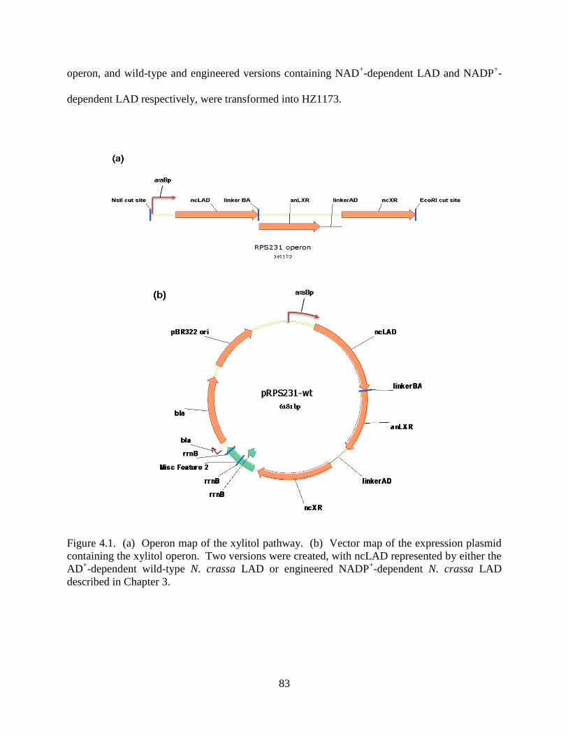
83
operon, and wild-type and engineered versions containing NAD+-dependent LAD and NADP
+-
dependent LAD respectively, were transformed into HZ1173.
Figure 4.1. (a) Operon map of the xylitol pathway. (b) Vector map of the expression plasmid
containing the xylitol operon. Two versions were created, with ncLAD represented by either the
AD+-dependent wild-type N. crassa LAD or engineered NADP
+-dependent N. crassa LAD
described in Chapter 3.

84
The resulting strains were designated as HZE2 (with wild-type xylitol synthesis operon 231-wt)
and HZE3 (with engineered xylitol synthesis operon 231-3x). When the strain were grown and
induced with 1% L-arabinose for four hours at 30 °C and analyzed by SDS-PAGE, the levels of
LAD were highest, followed by LXR, and little detection of XR was visible (Figure 4.2). While
the absence of XR expression from the gel staining was alarming at first, the activity check with
cell lysates verified that XR activity was present over a negative control lysate from a strain with
only empty vector.
Figure 4.2. SDS-PAGE analysis of xylitol synthesis pathway expressing strains. HZE1 =
HZ1173 + pTrc99a empty vector. HZE2 = wild-type pathway expression strain. HZE3 =
engineered pathway expression strain. Only soluble fractions of cell lysates are shown.

85
4.2.3. Shake Flask Studies
4.2.3.1. Effect of Using Glucose as Co-Substrate and pH on Xylitol Production
In using glucose as the co-substrate for xylitol production, the glycolysis rate at high sugar
concentrations often exceeds respiratory capacity, leading to build-up of intermediate
metabolites. In E. coli, this results in the accumulation of acetate, leading to an acidic
environment that results in decreased growth rates as well as lowered recombinant protein
production. Previous work in a similar crp* strain showed that at 18 g/L glucose concentration,
the acetate production is significant, accumulating up to 70 mM.11
When HZE3 was grown in 30
g/L glucose and 10 g/L arabinose, the pH dropped to 5.1 within 24 hours, and growth was
inhibited. Further initial studies attempting xylitol production resulted in minimal xylitol
production by the wild-type strain (HZE2) and engineered strain (HZE3) (data not shown).
As discussed in Chapter 2, the three enzymes involved in the pathway for conversion of L-
arabinose to xylitol have very different pH profiles. This raises the concern that the optimal
activity of LAD is around pH 9.0-9.5, with a significant decrease in activity found at pH 6.8
(~20% maximum activity), which is the reported intracellular pH of E. coli12
. While it was
reported that there exists an intracellular pH homeostasis in E. coli that serves to keep the
intracellular pH at a relatively constant level regardless of external pH13
, a shift in so much as
half of a pH unit around physiological pH could drastically affect the LAD activity.
Bioreactors are typically pH-controlled in order to allow for high cell densities to be reached.
However, pH-control by acid or base addition during shake flask experimentation is not possible
aseptically. Therefore, flask studies were then run which incorporated the addition of a buffering
component (MOPS) to the media in an attempt to offset the acetate buildup and allow for

86
improved cell growth and potentially xylitol production. As shown in Figure 4.3, the
extracellular pH dropped dramatically in cultures containing 2% or more glucose, while pHext
remained relatively constant at pH 7.2 for 1% glucose. The final cell densities also increased
with increasing buffering capacity. Therefore, 1% glucose was chosen as the amount used in
further shake flask studies.
Figure 4.3. Effect of glucose concentration on extracellular pH and OD600 of cultures of HZE3.
3% glucose, squares; 2% glucose, circles; 1% glucose, triangles. pHext values shown in filled
symbols, OD600 shown in empty symbols.
4.2.3.2. Wild-type vs. Engineered Xylitol Pathway
Cultures of HZE2 and HZE3 were compared in M9 + tryptone media with 100 mM MOPS
added as a buffering component. Glucose was added in excess of 3:1 (w/v). As shown in Figure
4.4, both pathways consumed all glucose and arabinose in 20 hours. However, the wild-type
pathway showed xylitol production up to ~10 mM after 33 hours (approximately 50% molar
yield from L-arabinose) whereas the engineered pathway showed minimal production with less

87
than 1 mM (<5% molar yield) xylitol produced after 56 hours. One possible explanation for the
slower xylitol production in the engineered pathway is the inherent 10-fold reduced affinity for
ncLAD-3x for substrate L-arabinitol (see Chapter 3). The maximum amount of L-arabinitol that
can be formed is equal to the starting L-arabinose concentration (~20 mM), which is close to the
Km,arabinitol value for ncLAD-wt (16 mM), but much less than ncLAD-3x (Km,arabinitol = 292 mM).
Unfortunately, the amount of starting arabinose is limited in flask studies due to the fixed 1% co-
substrate glucose level restraint in order to keep the media properly buffered and prevent acetate
accumulation and resultant pH drop. It was found that adding more arabinose while keeping a
constant 1% glucose concentration results in insufficient conversion of arabinose (data not
shown). In order to test whether L-arabinose levels were limiting, the pathways were compared
in pH-controlled bioreactors.
Figure 4.4. Xylitol production profiles from substrate L-arabinose with glucose co-substrate in
shake flasks. (A) Wild-type xylitol pathway and (B) Engineered xylitol pathway.

88
4.2.4. Bioreactor Studies
4.2.4.1. Wild-Type vs. Engineered Pathway
Figure 4.5 shows the xylitol production profiles from 0.4 L batch controlled bioreactor cultures
in M9 media + tryptone supplemented with 20 g/L glucose and 20 g/L L-arabinose using strain
HZE2 (Fig. 4.5A) and HZE3 (Fig. 4.5B). Media was maintained at pH 7.0 by automatic addition
of 5 N NaOH. After depletion of the initial glucose, additional glucose was added from a sterile
60% stock. Although residual arabinose was still left in the media, the wild-type pathway
showed generation of L-arabinitol (up to 25 mM) and xylitol (up to 13 mM) only until ~50 hours
and then the conversion is stalled. The engineered pathway containing the NADP+-dependent
LAD, however, shows slow yet steady conversion of L-arabinose to L-arabinitol (up to 50 mM)
and finally to xylitol (up to 30 mM) over the course of the time prior to glucose addition at 96
hours. After the glucose spiking at 96 hours, both strains continued to produce L-arabinitol and
xylitol, although the wild-type pathway did so at a modest rate compared to the engineered
pathway.

89
Figure 4.5. Comparison of 0.4 L (working volume) fermentation profiles for (A) HZE2 and (B)
HZE3. The initial culture contained 2% glucose and 2% arabinose in minimal media + tryptone.
Additional glucose was added at the 96 hour mark. pH was controlled at 7.0.

90
Figure 4.6 shows the results of comparing the wild-type xylitol pathway vs. engineered xylitol
pathway in M9 + tryptone media supplemented with 3% glucose:1% arabinose. The higher ratio
of cosubstrate:substrate resulted in full conversion of arabinose after two days, and also resulted
in different product profiles than when using an equal weight percent ratio. The wild-type xylitol
pathway (Figure 4.6A) showed xylitol production for two days, but then xylitol concentration
began to decrease afterwards while the arabinitol concentration continues to increase. The
engineered pathway (Figure 4.6B) showed conversion of arabinose to arabinitol within two days,
and slowly afterwards the xylitol accumulation began to increase with decreasing arabinitol.
Figure 4.6. Comparison of 0.4 L (working volume) fermentation profiles for (A) HZE2 and (B)
HZE3. The initial culture contained 3% glucose and 1% arabinose in minimal media + tryptone.
pH was controlled at 7.0.
Further analysis of the samples is necessary to determine whether the arabinitol peak detected,
particularly after xylitol production begins, is either the L-arabinitol enantiomer or possibly the
D-arabinitol enantiomer. LAD has low activity with xylitol (see Chapter 2) for conversion to D-
xylulose, and LXR or another endogenous reductase could conceivably convert D-xylulose to D-
arabinitol, although this activity has not been measured with LXR in vitro. In case the xylitol

91
was converted to D-xylulose and then to D-xylulose-5-phosphate by xylB, which was not deleted
in the expression strain, it would be taken up into the pentose phosphate pathway and used for
biomass production. However, the OD600 of the cultures did not deviate after glucose was
consumed (data not shown), and it was verified by testing growth of the strains on minimal
media plates supplemented with arabinose that neither HZE2 nor HZE3 could grow, which
indicated that the endogenous pathways were indeed removed and that the xylitol concentration
decrease seen in the wild-type pathway (Figure 4.6A) is not due to conversion to D-xylulose and
subsequent conversion to D-xylulose-5-phosphate by xylB. It is worth noting that negligible
xylulose was found in the cultures during the experimental run in both cases, indicating LAD is
still a limiting conversion step in the xylitol production process from L-arabinose. Also
unfortunate is that xylitol production in this case was faster and of higher yield in the wild-type
pathway than in the engineered pathway. It is difficult to ascertain why this result occurred.
One possible explaination is the decrease in expression levels of ncLAD-3x in HZE3 compared
to ncLAD-wt in HZE2 (Figure 4.2). Another possibility is the apparent decrease in affinity of
ncLAD-3x for substrate arabinitol compared to the wild-type enzyme (see Chapter 3). To test
whether a higher arabinose concentration would overcome the reduced affinity, an attempt at
xylitol production in a bioreactor setting with 4% arabinose was attempted, but results were
inconclusive due to cultures stalling production in both strains after 24 hours prior to sugar
addition, and another run could not be completed by the submission deadline for this disseration.
Further optimization of this process is apparent, utilizing both buffered flasks studies combined
with pH controlled bioreactor runs, to determine the proper conditions for optimal xylitol
production and the effects of the cofactor balancing.

92
4.2.4.2. Engineered Xylitol Pathway – pH control vs. non-pH control
Figure 4.7 shows the profile of HZE3 when run in a bioreactor without pH control (Fig. 4.7A)
and with pH control (Fig. 4.7B). Starting levels of glucose were 2% and arabinose was
decreased to 1% in an attempt to fully convert L-arabinose at least to L-arabinitol, and this
purpose was served in the pH controlled run. As seen from the uncontrolled profile, within 24
hours cells cease to grow and L-arabinose conversion as well as glucose consumption is halted.
L-arabinitol accumulates to ~16 mM, but no xylitol production is seen during the course of the
fermentation. When pH control is applied, cell density reaches a much higher level (OD600 ~13),
and L-arabinose if fully converted to L-arabinitol within 36 hours, where xylitol production is
starting to be noticed. L-arabinitol is slowly converted to xylitol (up to 24 mM) until the end of
the reactor run, but is never fully converted nor do levels drop below that of xylitol. While
optimization of the xylitol pathway is still an issue in terms of productivity, the effect of the pH
control is dramatic and the engineered pathway continued to show steady xylitol production from
L-arabinose.

93
Figure 4.7. Comparison of 0.4 L (working volume) fermentation profiles for (A) uncontrolled
and (B) pH-stat controlled HZE3 @ pH 7.0. The initial culture contained 2% glucose and 1%
arabinose in minimal media + tryptone.

94
4.2.4.3. WT vs. ENGR Pathway – Co-utilization
As shown in Figure 4.8, the extension of the fungal xylitol pathway for co-utilization of pentose
sugars xylose and arabinose resulted in a similar effect seen in the arabinose only fermentations.
Similar to the effect seen in the flask experiments and arabinose-only fermentation, the wild-type
pathway (Figure 4.8A) again showed a halting of xylitol production after 48 hours, while the
engineered pathway (Figure 4.8B) again showed slow yet steady production of xylitol from L-
arabinitol.
Figure 4.8. Comparison of 0.4 L (working volume) fermentation profiles for co-utilization of
xylose and arabinose by (A) HZE2 and (B) HZE3. The initial culture contained 3% glucose,
0.5% xylose, and 0.5% arabinose in minimal media + tryptone. Cultures were run at 30 °C, pH
7.0.

95
4.3. Discussion
In this work, we have studied the possibility of developing a more efficient pathway for xylitol
production from L-arabinose and D-xylose, the two major components of hemicellulosic biomass
hydrolysates. The pathway consists of three reactions, the first of which, xylose reductase,
catalyzes the reduction of D-xylose directly to the desired product xylitol, and also converts L-
arabinose to L-arabinitol. L-arabinitol is subsequently converted to L-xylulose by L-arabinitol 4-
dehydrogenase and finally to xylitol by L-xylulose reductase. However, the reductases catalyze
preferentially NADPH-dependent reactions, whereas the dehydrogenase is strictly NAD+-
dependent. Therefore, the xylitol pathway is redox imbalanced and the cellular metabolism is
relied on to regenerate the cofactors in order to enhance flux towards product xylitol. In an
attempt to alleviate the burden placed upon the cell to regenerate NADP+ and NADH, it was
proposed to engineer the LAD enzyme to be NADP+-dependent (as discussed in Chapter 3),
which consolidates the xylitol pathway to a single cofactor pair, NADP(H). This effectively
reduces the cell metabolism‘s workload to only be needed to regenerate NADPH, which can be
accomplished through either the pentose phosphate pathway or citric acid cycle with the addition
of an appropriate co-substrate, in this case D-glucose.
In order to test the effects of utilizing an NADP+-dependent LAD in an engineered xylitol
pathway, E. coli was chosen as a suitable host organism. E. coli is an ideal organism for
industrial production of chemicals due to its ability to assimilate both hexose and pentose sugars,
rapid growth rates, ease of manipulation, and inexpensive growth medium requirements14
.
Most E. coli strains (including K-12) can uptake D-xylose and L-arabinose, but do not naturally
synthesize or metabolize xylitol. However, E. coli will grow readily on pentose sugar substrates
D-xylose and L-arabinose, and it has been previously established that xylitol production by

96
introduction of heterologously expressed enzymes that convert D-xylose to xylitol is severely
limited when endogenous pathways for D-xylose utilization are present. In order to prevent the
assimilation of pentose sugars by the pathway-expressing strain, the endogenous pathway genes
for D-xylose (xylAB) and L-arabinose (araBAD) utilization were deleted.
For the engineered pathway, even though the NADP+-dependent LAD enzyme partially relieves
the cofactor imbalance, there are still an odd number of enzymes involved in the conversion of
either D-xylose or L-arabinose to xylitol, so regeneration of NADPH is still necessary by the
cell‘s metabolism. NADPH can be regenerated by the pentose phosphate pathway and the citric
acid cycle when using co-substrate D-glucose. However, due to catabolite repression effects of
using glucose with other sugars, the expression strain needed to be tailored to utilization these
sugars simultaneously. Different strategies of catabolite de-repression for the co-utilization of
the glucose, xylose, and arabinose were tested by Nikhil Nair in the Zhao laboratory. He found
that incorporation of a mutant cyclic AMP-independent receptor protein (CRP*) showed
efficient simultaneous assimilation of all three sugars, although xylose was shown to be the
slowest15
. For all experiments run for conversion of L-arabinose to xylitol in this chapter, the
simultaneously consumption of glucose, arabinose, and/or xylose was confirmed using a crp*
expression strain.
The effect of engineering and NADP-dependent LAD for use in the xylitol production pathway
varied depending on the reaction conditions. In bioreactor runs involving L-arabinose
conversion alone with equal weight percent co-substrate glucose (Fig. 4.4) as well as co-
utilization of D-xylose and L-arabinose (Fig. 4.6), it appeared the engineered pathway was better
suited for xylitol production. Whereas the wild-type pathway experienced stalling of xylitol
production, the engineered pathway with improved cofactor balancing showed slow yet continual

97
production of xylitol from the accumulated L-arabinitol. However, when monitoring the
conversion more closely with flask studies, there appears to be an initial generation of xylitol in
the wild-type pathway followed by conversion of xylitol to an unknown by-product. The xylitol
conversion was not noticeable in the engineered pathway experiments, but the reactions were not
carried out past five or six days and it is possible that the reaction is simply slower and the
xylitol conversion does not occur until later in the reaction. Further analysis of the samples is
currently being conducted to determine if the by-product is D-arabinitol, which could not be
separated from L-arabinitol by the HPLC method used to detect all sugars in the culture broth.
There were no detectable amounts of accumulating L-xylulose in the fermentation broths, which
suggests that the LXR reaction is not limiting in xylitol formation and that LAD is still likely the
bottleneck in the process. Fermentation with both D-xylose and L-arabinose as substrates
showed full conversion of xylose to xylitol and arabinose to arabinitol, with slow steady
conversion of arabinitol to xylitol in the engineered pathway compared to conversion of xylitol
to an unknown byproduct in the wild-type pathway, as seen with buffered flask studies and
arabinose-only fermentation.
The effect of pH-stat control is illustrated strongly in Figure 4.5. Without addition of base to
counter the acetate production, cell growth halted after 24 hours and stayed around OD600 levels
of ~5.5 to 6.0. Low levels of L-arabinitol (~16 mM) were produced in the first 24 hours, but all
xylitol pathway production shut down after this point and no xylitol was produced. In contrast,
utilizing a pH-stat to keep levels at pH 7.0 resulted in enhanced growth of the expression strain,
resulting in more than twice the amount of biomass produced. L-arabinose was fully converted
the first enzyme, XR, to L-arabinitol within 24 hours and xylitol production was noticed after 48

98
hours. L-arabinitol was then slowly converted to xylitol for the remainder of the bioreactor run,
although the process did not go to completion within 162 hours.
4.4. Conclusions and Outlook
This chapter illustrated the application of a fungal L-arabinose pathway for co-utilization of D-
xylose and L-arabinose for xylitol production. While this represents a novel route of xylitol
biosynthesis, the potential added benefit of introducing an engineered NADP+-dependent LAD
from Chapter 3 to attempt to alleviate the cofactor imbalance inherent in the pathway is not yet
established. There remain unsolved issues with low productivity that requires further
optimization of the pathway. Also, the depletion of xylitol in the wild-type pathway has yet to
be elucidated, and work is currently investigating whether byproduct D-arabinitol is
accumulating instead, which could lead to a novel biosynthetic route to a different sugar alcohol
than what was originally intended. Regardless, this groundwork represents an excellent strategy
for improving utilization of both pentose sugars present in hemicellulosic materials for synthesis
of value-added product xylitol. Chapter 5 will discuss work dedicated towards extending this
engineering strategy for further downstream products attainable by fermentation of pentose
sugars such as bioethanol.

99
4.5. Materials and Methods
4.5.1. Materials
Escherichia coli C600 was kindly provided by the Coli Genetics Stock Center (CGSC, Yale
University). Plasmid pTrc99a was obtained from Amersham Biosciences. Restriction enzymes,
NsiI and EcoRI, T4 DNA Ligase, and Phusion DNA polymerase were purchased from New
England Biolabs (NEB, Beverly, MA). All media components were purchased from Becton-
Dickinson (BD, Sparks, MD). Oligonucleotide primers were purchased from Integrated DNA
Technologies (IDT, Coralville, IA). All DNA purification kits were obtained from Qiagen
(Valencia, CA). Wizard Genomic DNA Purification Kit was purchased from Promega
(Madison, WI). Cells were maintained on Luria-Bertani (LB) plates containing 1.5% agar and
the appropriate antibiotic.
4.5.2. Gene Deletion (Notebook #3, p. 127)
For xylitol production, E. coli K-12 C600 derivative strains were used. Deletions were
performed using the red system.9 Briefly, PCR product containing cat flanked by FRT (Flp
recognition target) and 45-50 nucleotides of sequence identical to the target locus (using forward
primer ―H1-P1 fwd araBp‖ with sequence 5‘-AGC GGA TCC TAC CTG ACG CTT TTT ATC
GCA ACT CTC TAC TGT GTA GGC TGG AGC TGC TTC-3‘ and reverse primer ―H2-P2 rev
araD‖ with sequence 5‘-TTC GCC GTT GAT TTC TGC GTC GGT CAT TTT GCG GGT GCA
GCA GTG GAA TAT GGA TAT CCT CCT TAG-3‘ in the amplification reaction) was
transformed into cells expressing red recombinase proteins (encoded on pKD46). Gene
replacement was selected on chloramphenicol plates and verified by functional assay and PCR.

100
Table 4.1. Strains and plasmids used in this study. Strains/plasmids Relevant Characteristics Source/Reference
Strains
C600 F- tonA21 thi-1 thr-1 leuB6 lacY1 glnV44
rfbC1 fhuA1 -
CGSC, Yale University
ET23 Source of crp*::Tn10 Eppler and Boos, 1999
HZ1302 C600 crp*::Tn10 Nair and Zhao, 2009
HZ1450 C600 crp*:: Tn10 xylA Nair and Zhao, 2009
HZ1173 C600 crp*:: Tn10 xylA araBAD This work
HZE2 HZ1173 with pRPS231-wt This work
HZE3 HZ1173 with pRPS231-3x This work
Plasmids
pTrc99a Amp, pBR322-derived plasmid Amersham Pharmacia
pRPS231-wt pTrc99a with LAD-wt, XR, LXR operon under araBp
promoter
This work
pRPS231-3x pTrc99a with LAD-3x, XR, LXR operon under araBp
promoter
This work
The resistance marker was then removed by the expression of Flp recombinases from a thermo-
inducible promoter on a temperature sensitive plasmid (pCP20). Flp recombinase plasmid loss
and cat loss occurred simultaneously and was verified by the sensitivity to ampicillin and
chloramphenicol. Deletion of xylA was previously performed by Nikhil Nair, and deletion of
araBAD was verified by PCR using cell lysates as template and appropriate flanking primers
―Fwd minus 275 araC‖ (5‘-AAA CCA ATT GTC CAT ATT GCA TCA GA-3‘) and ―Rev araD
stop‖ (5‘-TTA CTG CCC GTA ATA TGC CTT CGC GC-3‘). It should be noted that araB and
araA were fully deleted, while araD was truncated to avoid any possible downstream conflicts of
the araBAD operon.
4.5.3. Mutant crp* Integration
The crp* mutant was generated by Nikhil Nair in the Zhao laboratory by P1 transduction from
crp*-containing plasmid ET23 (obtained from Patrick C. Cirino, Penn State University) and
selecting for TetR integrants
6. Verification of glucose de-repression was first done by blue/white
screening on LB plates containing 10 g/L glucose. Strong induction of lacZ in presence of

101
glucose indicated de-repressed phenotype. Finally, monitoring of sugar co-utilization in shake
flasks was used to verify de-repression.
4.5.4. Operon Design and Construction (Notebook #3, p. 231-234)
Overlap extension PCR (OE-PCR)16
was used to construct the xylitol synthesis operons. Operon
231-wt consisted of the following in order: an arabinose-inducible promoter from E. coli C600
genomic DNA (araBp), wild-type N. crassa L-arabinitol 4-dehydrogenase (ncLAD-wt), A. niger
L-xylulose reductase (anLXR), and N. crassa xylose reductase (ncXR). The linker region
between E. coli genes araB and araA (10 bp) was used as the linker region between ncLAD-wt
and anLXR, and was designed into the primers used for amplification. The linker region
between E. coli genes araA and araD (284 bp) was used as the linker region between anLXR and
ncXR, and was amplified from E. coli C600 genomic DNA, with sequences listed in Table 4.3.
All primers used for operon construction are listed in Table 4.2. Operon 231-3x was constructed
in a similar fashion to operon 231-wt, except that engineered N. crassa L-arabinitol 4-
dehydrogenase (ncLAD-3x) was used in place of ncLAD-wt. Operons 231-wt and 231-3x were
digested with NsiI and EcoRI and ligated into identical sites of vector pTrc99a with T4 DNA
Ligase. Ligation mixtures were transformed into DH5 and selected on Luria Broth (LB) agar
plates supplemented with 100 g/mL ampicillin. Colonies were inoculated into 5 mL LB
medium supplemented with 100 g/mL ampicillin, and plasmids were isolated from the
overnight culture using the Plasmid Miniprep Kit from Qiagen (Valencia, CA). Plasmids
isolated were then subjected to restriction digestion by NsiI and EcoRI, after which the reaction
mixtures were loaded onto 1% agarose gels and checked for correct restriction digestion pattern
by DNA electrophoresis.

102
Table 4.2. Primers sequences used in construction of the xylitol synthesis operons. Forward
primer names consist of directionality, restriction site or overhang, and the name of the region
being amplified. For example, ―Fwd NsiI araBp‖ stands for the forward primer used to
amplify araBp with an NsiI cut site in front of the promoter (5‘-end). Reverse primer names
consist of directionality, the region being amplified, and the restriction site or overhang. For
example, ―Rev araBp ncLAD‖ stands for the reverse primer used to amplify araBp, with a 3‘-
end overhang sequence homologous to ncLAD. Restriction sites shown with underlines. Primer Name DNA Sequence (5‘→3‘)
Fwd NsiI araBp TTGATCGGTATGCATCCGCCATTCAGAGAAGAAAC
Rev araBp ncLAD GAAGCGCTAGAAGCCATCGTTTCACTCCATCC
Fwd araBp ncLAD GGATGGAGTGAAACGATGGCTTCTAGCGCTTC
Rev ncLAD-linkBA-
anLXR
GAGGGAATGGAGATAGGCATTATCGTGTCCTTACTCCAGACTCTGGATC
Fwd ncLAD-linkBA-
anLXR
GATCCAGAGTCTGGAGTAAGGACACGATAATGCCTATCTCCATTCCCTC
Rev anLXR-linkAD GTTACATACCGGATGCGGCTACTTACCGCACGGTGTATCC
Fwd anLXR-linkAD GGATACACCGTGCGGTAAGTAGCCGCATCCGGTATGTAAC
Rev linkAD-ncXR GAGCTTGATAGCAGGAACCATGTTGACTCCTTCGTGCCGGATG
Fwd linkAD-ncXR CATCCGGCACGAAGGAGTCAACATGGTTCCTGCTATCAAGCTC
Rev ncXR EcoRI AAGTCGGATCGAATTCCTAACCGAAAATCCAGAGGTTC

103
Table 4.3. Sequences of non-gene components of xylitol synthesis operons.
Component Sequence
araBp
promoter
5‘-ccgccattcagagaagaaaccaattgtccatattgcatcagacattgccgtcactgcgtcttttactggctctt
ctcgctaaccaaaccggtaaccccgcttattaaaagcattctgtaacaaagcgggaccaaggccatgacaaaa
acgcgtagcaaaagtgtctataatcacggcagaaaagtccacattgattatttgcacggcgtcacactttgctatg
ccatagcatttttatccataagattagcggatcctacctgacgctttttatcgcaactctctactgtttctccataccc
gtttttttggatggag-3‘
linkBA 5‘-ggacacgata-3‘
linkAD 5‘-gtagccgcatccggtatgtaacgcctgatgcgacgctgacgcgtcttatctggcctacacgctgcgattttg
taggccggataagcaaagcgcatccggcattcaacgcctgatgcgacgctggcgcgtcttatcaggcctacg
cgctgcgattttgtaggccggataagcaaagcgcatccggcattcaacgcctgatgcgacgctggcgcgtctt
atcaggcctacacgctgcgattttgtaggccggataagcaaagcgcatccggcacgaaggagtcaac-3‘
4.5.5. HPLC Analysis
Glucose, xylose, arabinose, xylitol, and arabinitol concentrations were quantified using
Shimadzu LC-20AT high performance liquid chromatography (HPLC) equipped with a low
temperature evaporative light scattering detector (ELSD-LT). Products were separated with a
Bio-Rad HPX-87C column (20 L injection) with water as the mobile phase (0.2 mL/min, 85
°C).
4.5.6. Shake Flask Studies (Notebook #4, p. 279)
For shake flask studies, overnight cultures were grown at 37 °C in LB media + 100 mM MOPS
(pH 7.4) and appropriate antibiotics. 0.5 mL of overnight cultures were inoculated into 50 mL of

104
LB media + 100 mM MOPS (pH 7.4) containing sugars (glucose, xylose, and/or arabinose) and
appropriate antibiotics in 250 mL baffled flasks and grown at 30 °C. Samples were withdrawn at
various time points and measured for cell densities at OD600 using a Cary Bio 100 UV Vis
spectrometer. Samples were also filtered using 0.2 m syringe filters (Millipore) and analyzed
by HPLC for sugar analysis.
4.5.7. Bioreactor Studies (Notebook #4, p. 158)
For bioreactor studies, the seed culture was prepared by growing cells first on a fresh LB plate
supplemented with 10 g/mL tetracycline and 100 g/mL ampicillin and inoculating a single
colony into 4 mL LB media and appropriate antibiotics at 37 °C. 1 mL of saturated overnight
culture was centrifuged at 4,000 rpm for 4 minutes and media were decanted. Cells were
resuspended in 5 mL sterile water and centrifuged again at 4,000 rpm for 4 minutes and water
was decanted. Cells were resuspended in M9 medium supplemented with 2 mM MgSO4, 0.1
mM CaCl2, 30 g/L glucose, 10 g/L tryptone, and appropriate antibiotic(s) and grown for another
4-5 hours at 37 °C. The log phase cultures were then inoculated into a Multifors multiple
benchtop bioreactor (Infors HT, Switzerland) with 0.5 L working volume containing 400 mL of
the same M9 + tryptone medium with additional 10 g/L each of D-xylose and/or L-arabinose, as
well as antifoam. Bioreactors were run at 30 °C with an agitation rate of 800 rpm by dual
Rushton impellers and a sparging rate of 0.8 L/min with sterile air . pH was maintained at 7.0 ±
0.1 with 5 N NaOH and 2 N H2SO4. Samples were withdrawn at various time points and
measured for cell densities at OD600 using a Cary Bio 100 UV Vis spectrometer. Samples were
also filtered using 0.2 m syringe filters (Millipore) and analyzed by HPLC for sugar analysis.

105
References
(1) Sakakibara, Y.; Saha, B. C.; Taylor, P. Microbial production of xylitol from L-arabinose
by metabolically engineered Escherichia coli J Biosci Bioeng 2009, 107, 506-11.
(2) Song, S.; Park, C. Organization and regulation of the D-xylose operons in Escherichia
coli K-12: XylR acts as a transcriptional activator J Bacteriol 1997, 179, 7025-32.
(3) Hasona, A.; Kim, Y.; Healy, F. G.; Ingram, L. O.; Shanmugam, K. T. Pyruvate formate
lyase and acetate kinase are essential for anaerobic growth of Escherichia coli on xylose
J Bacteriol 2004, 186, 7593-600.
(4) Stulke, J.; Hillen, W. Carbon catabolite repression in bacteria Curr Opin Microbiol 1999,
2, 195-201.
(5) Hernandez-Montalvo, V.; Valle, F.; Bolivar, F.; Gosset, G. Characterization of sugar
mixtures utilization by an Escherichia coli mutant devoid of the phosphotransferase
system Appl Microbiol Biotechnol 2001, 57, 186-91.
(6) Eppler, T.; Boos, W. Glycerol-3-phosphate-mediated repression of malT in Escherichia
coli does not require metabolism, depends on enzyme IIAGlc and is mediated by cAMP
levels Mol Microbiol 1999, 33, 1221-31.
(7) Karimova, G.; Ladant, D.; Ullmann, A. Relief of catabolite repression in a cAMP-
independent catabolite gene activator mutant of Escherichia coli Res Microbiol 2004,
155, 76-9.
(8) Zhu, Y.; Lin, E. C. A mutant crp allele that differentially activates the operons of the fuc
regulon in Escherichia coli J Bacteriol 1988, 170, 2352-8.
(9) Datsenko, K. A.; Wanner, B. L. One-step inactivation of chromosomal genes in
Escherichia coli K-12 using PCR products Proc Natl Acad Sci U S A 2000, 97, 6640-5.

106
(10) Lee, N.; Gielow, W.; Martin, R.; Hamilton, E.; Fowler, A. The organization of the
araBAD operon of Escherichia coli Gene 1986, 47, 231-44.
(11) Akinterinwa, O.; Cirino, P. C. Heterologous expression of D-xylulokinase from Pichia
stipitis enables high levels of xylitol production by engineered Escherichia coli growing
on xylose Metab Eng 2009, 11, 48-55.
(12) Diez-Gonzalez, F.; Russell, J. B. The ability of Escherichia coli O157:H7 to decrease its
intracellular pH and resist the toxicity of acetic acid Microbiology 1997, 143 ( Pt 4),
1175-80.
(13) Padan, E.; Zilberstein, D.; Schuldiner, S. pH homeostasis in bacteria Biochim Biophys
Acta 1981, 650, 151-66.
(14) Cirino, P. C.; Chin, J. W.; Ingram, L. O. Engineering Escherichia coli for xylitol
production from glucose-xylose mixtures Biotechnol Bioeng 2006, 95, 1167-76.
(15) Nair, N. U.; Zhao, H. Selective Reduction of Xylose to Xylitol from a Mixture of
Hemicellulosic Sugars. In preparation. 2009.
(16) Ho, S. N.; Hunt, H. D.; Horton, R. M.; Pullen, J. K.; Pease, L. R. Site-directed
mutagenesis by overlap extension using the polymerase chain reaction Gene 1989, 77,
51-9.

107
CHAPTER 5 – Fungal Arabinose Pathway Complementation Towards
Determining Limitations in Arabinose Utilization
5.1. Introduction
Lignocellulosic biomass is regarded as a highly promising feedstock for a rapidly expanding
biofuels industry in response to a pressing energy problem1. Lignocellulose refers to plant
biomass that is composed of cellulose (long-chain homopolymers of D-glucose), hemicellulose
(branched polysaccharides mainly consisting of pentose sugars D-xylose and L-arabinose), and
lignin (complex cross-linked macromolecules). The majority of lignocellulosic materials
consists of D-glucose (~30-50%) and D-xylose (~20-25%), but L-arabinose is still a significant
third portion in some forms of lignocelluose (~ 5-10%)2,3
. Utilization of all sugars in the raw
material is favored for an economically competitive process.
Yeast cells are particularly attractive for cellulosic ethanol processes because they have been
used in biotechnology for hundreds of years, are well-characterized physiologically and
genetically, have ample genetic tools, are tolerant to high ethanol and inhibitor concentrations,
and can grow at low pH values which avoids bacterial contaminations. Unfortunately, the
widely used industrial yeast Saccharomyces cerevisiae cannot metabolize the pentose sugars D-
xylose or L-arabinose found in hemicellulose. S. cerevisiae naturally harbors genes for xylose
utilization, but they are expressed at such low levels that they do not support growth on D-
xylose4. A large number of yeast species metabolize D-xylose and L-arabinose and display
fermentative ability, but only approximately 1% of them are capable of fermenting D-xylose to
ethanol5. Various yeasts and fungi were initially screened to evaluate their ability to ferment L-
arabinose under oxygen-limited conditions when grown in defined minimal media containing

108
mixtures of L-arabinose, D-xylose, and D-glucose6. Although all of the yeasts and some of the
fungi consumed L-arabinose, L-arabinose was not fermented to ethanol by any of the strains
tested. L-Arabinitol was the only major product other than cell mass formed from L-arabinose.
The inability to ferment L-arabinose was postulated to be a consequence of inefficient or
incomplete assimilation pathways for this pentose sugar. A subsequent study identified four
yeast species able to ferment L-arabinose to ethanol7, but the discrepancy was likely due to the
use of complex (YP) medium that could have contained compounds that may act as electron
acceptors and thus aid conversion of arabinose to ethanol5.
Pathways for D-xylose and L-arabinose metabolism share a common intermediate in the form of
D-xylulose-5-phosphate, but are distinctly different in bacteria and fungi. Bacterial catabolism of
D-xylose and L-arabinose begins with isomerization to D-xylulose and L-ribulose, respectively,
by an isomerase. D-xylulose and L-ribulose are phosphorylated by a xylulokinase to D-xylulose-
5-phosphate and to L-ribulose-5-phosphate, respectively, with the latter being converted by an
epimerase to D-xylulose-5-phosphate and connecting the initial pentose metabolism to the central
metabolism through the pentose phosphate pathway (PPP). The yeast pathway has been
previously described (Figure 1), and has been more successful in its implementation due to
troubles getting functional bacterial enzymes in S. cerevisiae.
Many metabolic engineering strategies have been attempted to improve fermentation of D-xylose
to ethanol in recombinant S. cerevisiae harboring D-xylose-utilization enzymes. These include
over-expression of endogenous xylulokinase8, glyceraldehydes-3-phosphate dehydrogenase
9,
transaldolase and transketolase, or deletion of glucose 6-phosphate dehydrogenase10
. Alternative
protein engineering strategies aimed at the cofactor imbalance of the initial pathway enzymes has
also been pursued, by introducing NADH-preferring XR mutants11,12
, or NADP-dependent XDH

109
mutants13,14
. However, these cofactor balancing strategies have only been dedicated towards
xylose fermentation.
The fermentation of L-arabinose to ethanol has only recently been established in S. cerevisiae,
with reports of the introduction of the bacterial pathway 15,16
, or the fungal pathway17,18
showing
promising progress in the field of L-arabinose utilization by S. cerevisiae, but yields of ethanol
using either strategy have been low. Co-utilization of D-xylose and L-arabinose has also been
shown to occur with fungal D-xylose and bacterial L-arabinose pathways introduced into S.
cerevisiae 5,19
, but most of the L-arabinose was converted to byproduct L-arabinitol by the fungal
xylose reductase. During the course of the work presented in this thesis, Bettiga et al. expressed
an engineered fungal L-arabinose pathway into S. cerevisiae and achieved anaerobic biomass
production and ethanol production. The pathway combined i) an engineered mutant XR with
increased NADH perference; ii) a NAD+-dependent LAD from Trichoderma reesei (Hypocrea
jecorina); iii) a NADH-dependent LXR from Ambrosiozyma monospora and iv) a NAD+-
dependent XDH from Pichia stipitis. The resultant pathway was cofactor balanced by
consolidating the initial pathway enzymes to the alternative cofactor pairing, NAD(H), whereas
my research focused on using a pathway designed for NADP(H). Expression of the Bettiga
pathway in their strain TMB3043 was chosen as background since it has been endowed with
genetic modifications allowing for improved pentose fermentation, such as overexpression of
PPP and XKS. In addition, TMB3043 harbors the deletion of GRE3, coding for an unspecific
NADPH aldose reductase. The removal of the Gre3p NADPH-dependent activity contributes to
a more cofactor balanced pentose utilizing pathway. The final ethanol concentration was 9.3 g/l,
with a yield of 0.21 g/(g sugar) based on total sugars present at the beginning of the
fermentation, 0.42 g/(g sugar) based on consumed sugars and 0.35 g/(g sugar) based on

110
consumed L-arabinose. While this strategy represented an almost 10-fold improvement over the
previous reported values17
, clearly much progress is still needed in this area to make the process
more efficient and economically feasible.
The proposed pentose utilization project deals with utilizing L-arabinose in combination with D-
xylose for the production of ethanol. The initial sequence of four alternating reduction and
oxidation steps in L-arabinose catabolism is potentially a rate-limiting factor in the catabolic
sequence20
, as natural L-arabinose metabolizing yeasts, as well as engineered Saccharomyces
cerevisiae, produce large amounts of L-arabinitol. The goal of this project is to determine the
effect of reversing the cofactor specificity for the dehydrogenases (LAD and XDH) involved in
the fungal L-arabinose utilization pathway. This chapter will discuss progress made in cloning
and engineering an NADP+-dependent N. crassa XDH, the introduction of the full initial L-
arabinose pathway into S. cerevisiae, and the development of a selection method using E. coli to
set the foundation for future pathway engineering.
5.2. Results
5.2.1. Cloning, Characterization, and Engineering of an NADP+-Dependent N. crassa
Xylitol Dehydrogenase
A putative Neurospora crassa xylitol dehydrogenase sequence was found using a protein
BLAST search on the National Center for Biotechnology Information website
(www.ncbi.nlm.nih.gov) using the Pichia stipitis xylitol dehydrogenase enzyme as a query
sequence. The two enzymes were aligned fully using a ClustalW algorithm and found to share
44% identity and 60% similarity (Figure 5.1). The whole-genome sequence of Neurospora

111
crassa has been published21
, and it was utilized to design primers for cloning of the putative
xylitol dehydrogenase gene.
Score = 299 bits (765), Expect = 3e-79, Method: Compositional matrix adjust.
Identities = 169/365 (46%), Positives = 222/365 (60%), Gaps = 22/365 (6%)
psXDH ANPSLVLNKIDDISFETYDAPEISEPTDVLVQVKKTGICGSDIHFYAHGRIGNFVLTKPM 62
+N S VLNK D+ F+ P+I+ P DVLV V TGICGSD+H++ HG IG+FV+ PM
ncXDH SNLSFVLNKPLDVCFQDKPVPKINSPHDVLVAVNYTGICGSDVHYWLHGAIGHFVVKDPM 66
psXDH VLGHESAGTVVQVGKGVTSLKVGDNVAIEPGIPSRFSDEYKSGHYNLCPHMAFAATPNSK 122
VLGHESAGT+V VG V +L VGD VA+EPG P R SGHYNLCP M FAATP
ncXDH VLGHESAGTIVAVGDAVKTLSVGDRVALEPGYPCRRCVHCLSGHYNLCPEMRFAATPPYD 126
psXDH EGEPNPPGTLCKYFKSPEDFLVKLPDHVSLELGALVEPLSVGVHASKLGSVAFGDYVAVF 182
GTL ++ +P DF KLP+ VSL+ GAL+EPL+V VH +K + G V V
ncXDH -------GTLTGFWTAPADFCYKLPETVSLQEGALIEPLAVAVHITKQAKIQPGQTVVVM 179
psXDH GAGPVGLLAAAVAKTFGAKGVIVVDIFDNKLKMAKDIGAATHTFNSKTGGSEE-----LI 237
GAGPVGLL AAVAK +GA V+ VDI +KL+ AK AATHT+ S+ EE +
ncXDH GAGPVGLLCAAVAKAYGASKVVSVDIVPSKLEFAKSF-AATHTYLSQRVSPEENARNIIA 238
psXDH KAFGGNVPNVVLECTGAEPCIKLGVDAIAPGGRFVQVGNAAGPVSFPITVFAMKELTLFG 297
A G + V++ +GAEP I+ + + GG +VQ G ++FPI +KE+T G
ncXDH AADLGEGADAVIDASGAEPSIQAALHVVRQGGHYVQGGMGKDNITFPIMALCIKEVTASG 298
psXDH SFRYGFNDYKTAVGIFDTNYQNGRENAPIDFEQLITHRYKFKDAIEAYDLVRAGKGAVKC 357
SFRYG DY+ A+ + E +D ++L+ FK+A EA+ V+ G+ +K
ncXDH SFRYGSGDYRLAIQLV--------EQGKVDVKKLVNGVVPFKNAEEAFKKVKEGE-VIKI 349
psXDH LIDGP 362
LI GP
ncXDH LIAGP 354
Figure 5.1. BLAST Results for putative XDH in Neurospora crassa.
RT-PCR performed on total RNA isolated from D-xylose-induced N. crassa 10333 showed the
expected size of gene product (~1.1 kb). The RT-PCR product was cloned into the pET-28a
vector using NdeI and SacI restriction sites and was transformed into E. coli BL21 (DE3). This
construct (pET-28a ncXDH) expressed N. crassa XDH as an N-terminal His6-tagged fusion with
a thrombin cleavage site. Cell lysates of IPTG-induced cultures of these cells were prepared,
analyzed by SDS-PAGE, and assayed for XDH activities. The XDH was then purified by
112
immobilized metal ion affinity chromatography (IMAC) using Talon® Co
2+ Superflow resin
(Clontech, Mountain View, CA) according to manufacturer‘s protocol. The purified protein was
desalted by ultrafiltration with several washes of 50 mM 4-(2-hydroxyethyl)piperazine-1-
ethanesulfonic acid (HEPES) buffer (pH 7.25) + 15% glycerol and stored frozen at -80 °C.
Protein concentrations were determined by the Bradford method22
.
ncXDH is a strictly NAD+-preferring enzyme. ncXDH also displays high stability (half-life of
~200 min at 50 C) and expression. Previous work by Watanabe et al. was aimed at reversing
the cofactor specificity of Pichia stipitis xylitol dehydrogenase (psXDH).14
Through sequence
alignment, residues D204, I205, and V206 were targeted for site-directed mutagenesis to alanine,
arginine, and serine, respectively, to create ncXDH-ARS. Table 5.1 shows that ncXDH-ARS has
completely reversed cofactor specificity, now preferring NADP+, with little change in the kcat
value and only a 2.5-fold higher Km,NADP+ for ncXDH-ARS compared to the Km,NAD+ for the wild-
type. Table 5.2 shows that the affinity for substrate xylitol did not suffer the loss that was
previously seen in engineering an NADP+-dependent ncLAD (see Chapter 4). It should be noted
that the same mutations were attempted in the ncLAD engineering, but the improvement was not
as drastic, necessitating the library screening that was implemented in Chapter 3.

113
Table 5.1. Kinetic parameters for N. crassa and P. stipitis XDH and XDH-ARS with
nicotinamide cofactors NAD+ and NADP
+.
NAD+ NADP
+
Enzyme kcat
(min-1
)
Km
(mM)
kcat/Km
(mM-1
min-1
)
kcat
(min-1
)
Km
(mM)
kcat/Km
(mM-1
min-1
)
Source
ncXDH-wt 2160 0.127 17000 -a ~5.6 ~68 This work
ncXDH-ARS -a ~3.5 ~165 2080 0.325 6400 This work
psXDH 1050 0.381 2760 110 170 0.65 Watanabe et al.
psXDH-ARS 240 1.3 181 2500 0.897 2790 Watanabe et al.
aNot determined, cofactor saturation not reached
All assays were performed at 25 C in 50 mM Tris, pH 8.0
Table 5.2. Kinetic parameters of ncXDH mutants for substrate xylitol.
Enzyme kcat
(min-1
)
Km
(mM)
kcat/Km
(mM-1
min-1
)
ncXDH-wt 2170 ± 135 6.6 ± 2.0 330
ncXDH-ARS 2090 ± 35 4.3 ± 0.3 490
aNot determined, saturation of cofactor not reached
All assays run at 25 C, 50 mM Tris-HCl pH 8.0
All enzymes were purified and characterized with N-His6Tag
As discussed in Chapter 4, the pH rate profile of N. crassa LAD in the L-arabinose pathway
favor basic conditions, whereas it is known that the intracellular pH of E. coli23
or S. cerevisiae24
is neutral to acidic. The same holds true for N. crassa XDH, as shown in Figure 5.2, but XDH
activity exhibits a higher tolerance to more acidic conditions with activity extending down to pH
4.0, whereas LAD activity is abolished at pH 5.0 in the in vitro activity assay.

114
Figure 5.2. Comparison of pH rate profiles of N. crassa LAD and XDH. Data taken from the
characterization of LAD from Chapter 2 (open circles) was performed in universal buffer
MES/Tris/glycine, and overlapped with data for ncXDH (closed triangles) and ncLAD (closed
circles) performed in universal buffer acetic acid/MES/Tris for lower pH values.
5.2.2. Introduction of L-Arabinose Pathway into S. cerevisiae
The initial five enzymes of the fungal L-arabinose pathway that convert L-arabinose to D-
xylulose-5-phosphate leading into the pentose phosphate pathway were constitutively expressed
in S. cerevisiae. The wild-type pathway was assembled with NAD+-dependent ncLAD-wt and
ncXDH-wt, while the engineered pathway was assembled with NADP+-dependent ncLAD-3x
(see Chapter 3) and ncXDH-ARS (see Section 5.2.1) and correctly assembled constructs were
verified by restriction digestion with BamHI (Fig. 5.3a) and by PCR amplification of the five
gene cassettes (Fig. 5.3b). Strains were then grown on synthetic complete media lacking uracil
(SC-Ura) supplemented with either glucose, xylose, or arabinose as a carbon source. Figure 5.4
shows that strains containing either the wild-type or engineered pathway grew readily in SC-Ura
+ xylose, while the parent strain YSG50 could not utilize xylose. However, neither strain was
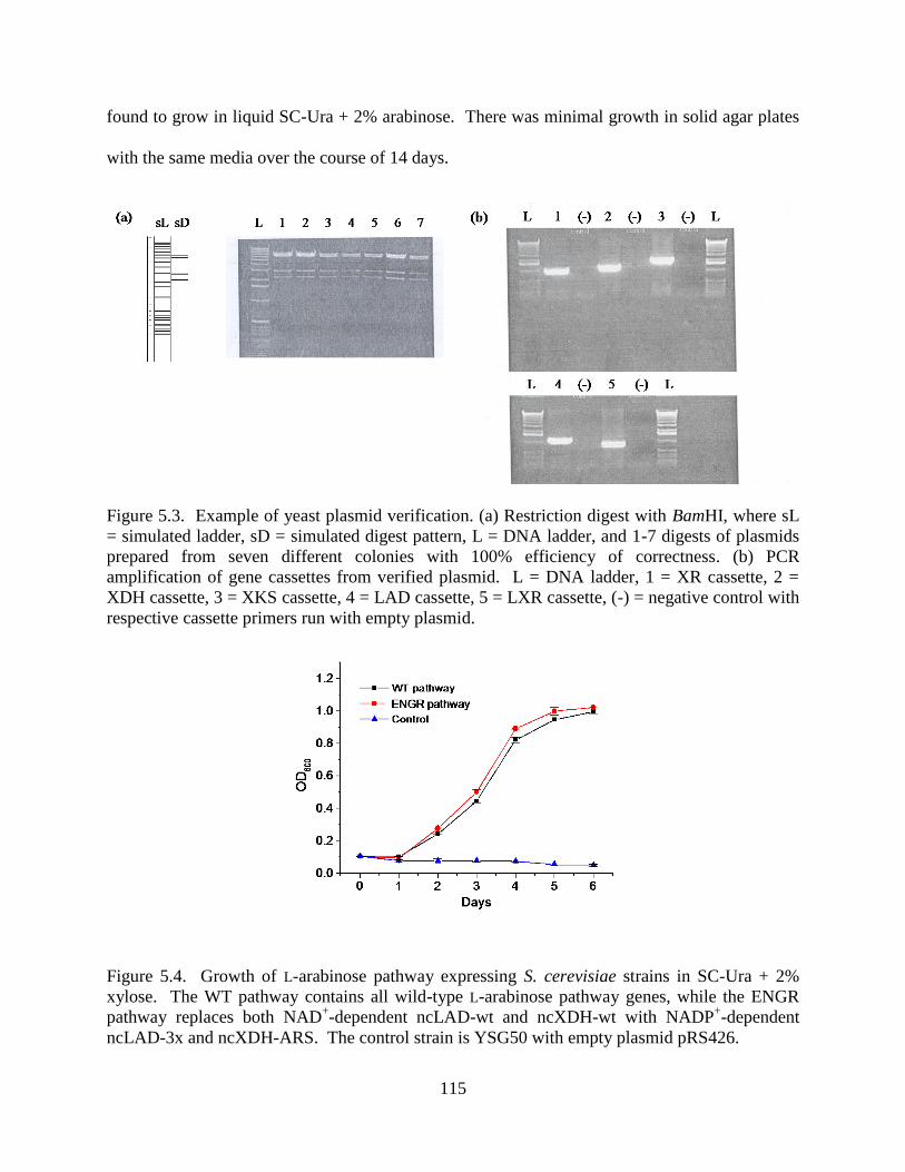
115
found to grow in liquid SC-Ura + 2% arabinose. There was minimal growth in solid agar plates
with the same media over the course of 14 days.
Figure 5.3. Example of yeast plasmid verification. (a) Restriction digest with BamHI, where sL
= simulated ladder, sD = simulated digest pattern, L = DNA ladder, and 1-7 digests of plasmids
prepared from seven different colonies with 100% efficiency of correctness. (b) PCR
amplification of gene cassettes from verified plasmid. L = DNA ladder, 1 = XR cassette, 2 =
XDH cassette, 3 = XKS cassette, 4 = LAD cassette, 5 = LXR cassette, (-) = negative control with
respective cassette primers run with empty plasmid.
Figure 5.4. Growth of L-arabinose pathway expressing S. cerevisiae strains in SC-Ura + 2%
xylose. The WT pathway contains all wild-type L-arabinose pathway genes, while the ENGR
pathway replaces both NAD+-dependent ncLAD-wt and ncXDH-wt with NADP
+-dependent
ncLAD-3x and ncXDH-ARS. The control strain is YSG50 with empty plasmid pRS426.

116
5.2.3. E. coli Complementation Experiments
Due to the inability to get a functional L-arabinose pathway in S. cerevisiae, E. coli was chosen
as a suitable host for the development of a selection method to determine (1) whether the fungal
pathway would be able to complement the bacterial pathway and circumvent the issues
associated with S. cerevisiae such as pentose transport, gene expression and protein solubility,
potential acidic intracellular pH conditions, and transcriptional regulation, and (2) whether the
cofactor balanced engineered pathway would result in improved growth and/or ethanol
production. Table 5.3 lists the strains used for xylose complementation experiments in E. coli
MG1655. The deletion of xylA by cat insertion was confirmed to abolish the ability of HZE60 to
grow in minimal media supplemented with xylose. In order to confirm that complementation
was possible by expression of the endogenous genes under the xylAp promoter in a plasmid,
constructs with either xylA or xylA and xylB together were transformed into HZE60 and selected
in xylose minimal media. As shown in Figure 5.5, over-expression of xylA alone was able to
complement growth in xylose minimal media. When both xylA and xylB were over-expressed,
the strain grew faster, which suggests that the deletion of XylA activity with the cat insertion
resulted in a polar effect that subsequently decreased XylB expression as well in HZE60.
Table 5.3. Strains constructed for D-xylose growth complementation experiments
MG1655 F-
- ilvG
- rfb-50 rph-1 (ATCC 700926)
HZE60 MG1655 xylA::cat (obtained from Tasha Desai)
HZE66 MG1655 xylA::cat pTrc99a
HZE67 MG1655 xylA::cat pTrc99a-xylAp-xylA
HZE68 MG1655 xylA::cat pTrc99a-xylAp-xylAB
HZE101 MG1655 xylA::cat pTrc99a-xylAp-ncXDH(wt)-ncXR-xylB
HZE102 MG1655 xylA::cat pTrc99a-xylAp-ncXDH(ARS)-ncXR-xylB
HZE109 MG1655 xylA::cat pTrc99a-xylAp-ncXDH(wt)-ncXR-psXKS
HZE110 MG1655 xylA::cat pTrc99a-xylAp-ncXDH(ARS)-ncXR-psXKS
HZE123 MG1655 xylA::cat pBLMA-xylAp-ncXDH(wt)-ncXR-xylB
HZE124 MG1655 xylA::cat pBLMA-xylAp-ncXDH(ARS)-ncXR-xylB

117
To determine whether XR and XDH together could complement XylA activity, two constructs
were tested, containing XR and either wild-type NAD+-dependent XDH or engineered NADP
+-
dependent XDH. In Figure 5.5, the wild-type pathway under the control of xylAp with either
xylB (HZE101) or psXKS (HZE109) was confirmed to complement growth, whereas the
engineered pathways (HZE102 and HZE110) did not complement growth in minimal media with
xylose. This is contradictory to what was seen in S. cerevisiae. However, when plated on M9
media with 5% xylose, slow grow was noticable with the engineered pathway under the control
of xylAp (HZE110, Figure 5.6).
Figure 5.5. Growth complementation on M9 + 0.4% xylose at 37 °C (See Table 5.3 for legend).

118
Figure 5.6. Growth complementation on M9 + 5% arabinose at 30 °C. (See Table 5.3 for
legend).
Table 5.4 lists the strains used for arabinose complementation experiments in E. coli MG1655.
Again, the same control strategy was applied by knocking out endogenous araBAD, which
resulted in the lack of cell growth in minimal media supplemented with L-arabinose, and
expression of araBAD in the knockout strain showing complementation was achieved.
Table 5.4. Strains constructed for L-arabinose growth complementation experiments
HZE62 MG1655araBAD (obtained from Tasha Desai)
HZE71 MG1655 araBAD pTrc99a
HZE72 MG1655 araBAD pTrc99a araBp-araBAD
HZE103 MG1655 araBAD pTrc99a + pRSFDuet
HZE104 MG1655 araBAD pTrc99a araBp-ncLAD(wt)-anLXR-ncXR + pRSFDuet araBp-
ncXDH(wt)-xylB HZE105 MG1655 araBAD pTrc99a araBp-ncLAD(3x)-anLXR-ncXR + pRSFDuet araBp-
ncXDH(ARS)-xylB
However, when the wild-type (HZE104) or engineered (HZE105) fungal arabinose pathways
were expressed in the araBAD-deletion strain, neither construct could complement cell growth
on L-arabinose media (data not shown).

119
5.3. Discussion
As mentioned earlier, the wild-type L-arabinose pathway has been introduced into S. cerevisiae
previously by Richard et al. although with little resultant ethanol production from L-arabinose.17
With the first four enzymes of the L-arabinose pathway already cloned, the final enzyme, D-
xylulokinase from Pichia stipitis (psXKS) was first cloned from P. stipitis cDNA into E. coli
prior to introduction into S. cerevisiae. Over-expression of xylulokinase in S. cerevisiae has
been reported to enhance specific ethanol production when grown on D-xylose25
. Introduction of
the L-arabinose pathway into S. cerevisiae was accomplished by a new homologous
recombination-based DNA assembly method developed by Zengyi Shao and Hua Zhao in the
Zhao laboratory.26
The full-length pathway placed in pRS426 was comprised of the following gene cassettes (―p‖
indicates promoter, ―t‖ indicates terminator sequence): ADH1p-ncXR-ADH1t, GPDp-ncLAD-
PYK1t, HXT7p-anLXR-HXT7t, PGK1p-ncXDH-CYC1t, and PYK1p-psXKS-ADH2t. Colonies
were selected on synthetic complete media lacking uracil (SC-Ura) with either D-xylose or L-
arabinose as the sole carbon source to confirm the pathway was functional. Colonies formed
readily on D-xylose after about four days, albeit slower than when compared to the typical two
day incubation for D-glucose containing media. The engineered pathway strain (containing the
NADP-dependent dehydrogenases ncLAD-SRN and ncXDH-ARS) also grew on D-xylose, and
had similar growth rate compared to the wild-type pathway. An interesting, although
unfortunate, observation was that while colonies were able to form on L-arabinose plates after
~10-14 days, subsequent inoculation of these colonies into liquid SC-Ura with L-arabinose as the
sole carbon source were unable to grow for reasons yet to be determined. Colonies that grew up

120
on both pentose sugar plates were checked by colony PCR and restriction digestion analysis to
confirm the pathway had been assembled properly.
It remains unknown why S. cerevisiae grew so poorly in L-arabinose media. Cofactor
imbalance, one of the key areas of research in the proposed project, is thought to be one reason.
However, transport of pentose sugars has also been brought to attention27-29
, and this is currently
being investigated by Jing Du and Sijin Li in the Zhao group. Recently, strong evidence has
shown support for the need of improvement of the LAD enzyme. Two natural L-arabinose-
utilizing yeasts, Candida arabinofermentans PYCC 5603T and Pichia guilliermondii PYCC
3012 were compared to gain more insights into the limitations of L-arabinose metabolism. 13
C
NMR was used to trace the fate of L-[2-13
C]arabinose through branching metabolic pathways,
and intracellular accumulation of arabinitol appeared in both strains, indicating a bottleneck at
the level of LAD which was also NAD+-dependent in these strains
30. It is hypothesized that an
improved ncLAD enzyme, along with cofactor reversal for the dehydrogenase enzymes for
NADP+-dependence, can shed light on the utilization of pentose sugars for ethanol production.
However, the results from expressing the fungal L-arabinose pathway in S. cerevisiae suggest
that the cofactor imbalance alone is not the answer to obtaining a strain that can grow readily on
L-arabinose.
As shown in Section 5.2.4, the L-arabinose pathways were also expressed in E. coli as a model
organism. E. coli can grow readily in either xylose or arabinose containing media, which
suggests it does not suffer from the possible limitations that S. cerevisiae has in pentose
utilization. Preliminary results have shown that the wild-type and, to a lesser extent, the
engineered fungal xylose pathway under the control of xylAp can complement growth in M9 +
5% xylose media at 30 °C.

121
5.4. Conclusions
The development of a selection method based on the complementation of E. coli pentose
utilization genes with those of fungal origin can help to determine the bottlenecks associated
with the initial pathway enzymes and conclude whether engineering a cofactor balanced pathway
helps in pentose utilization. The XDH from N. crassa was cloned, characterized, and engineered
for NADP+-dependence, and the five genes that convert L-arabinose to D-xylulose-5-phosphate
for entrance into the pentose phosphate pathway were introduced into S. cerevisiae.
Unfortuntely, only growth on xylose was attained, with minimal growth on solid media
containing arabinose only. Due to potential issues involved with transport, expression, etc., the
pathways were tested in E. coli with resultant growth only achievable on xylose and not
arabinose. Further optimization of the pathway expression is still required in order for the
arabinose pathway to be functional.
5.5. Materials and Methods
5.5.1. Materials
The Neurospora crassa genomic sequence and LAD protein sequences were accessed from the
National Center for Biotechnology Information (NCBI, www.ncbi.nlm.nih.gov). Neurospora
crassa 10333 was obtained from the American Type Culture Collection (ATCC). Escherichia
coli BL21(DE3) and pET-28a(+) expression vector were obtained from Novagen (Madison, WI).
SuperScript One-Step RT-PCR with Platinum
Taq kit was obtained from Invitrogen
(Carlsbad, CA). PCR grade dNTPs were obtained from Roche Applied Sciences (Indianapolis,
IN). Phusion High-Fidelity DNA Polymerase, shrimp alkaline phosphatase, and DNA-
modifying enzymes DNase I, NdeI, SacI, and T4 DNA ligase and their appropriate buffers were

122
purchased from New England Biolabs (NEB) (Beverly, MA). Xylitol, -D-
thiogalactopyranoside (IPTG), and NADP+ were purchased from Sigma (St. Louis, MO). NAD
+
was a gift from Jülich Fine Chemicals (Jülich, Germany). Other required salts and reagents were
purchased from Fisher (Pittsburgh, PA) or Sigma-Aldrich. The QIAprep spin plasmid mini-prep
kit, QIAquick gel purification kit, RNeasy midiprep kit, and QIAquick PCR purification kit were
purchased from Qiagen (Valencia, CA). Oligonucleotide primers were obtained from Integrated
DNA Technologies (Coralville, IA). SDS-PAGE gel materials, electrophoresis equipment, and
protein size markers were from Bio-Rad (Hercules, CA).
5.5.2. RT-PCR and Cloning
N. crassa 10333 was grown on rich potato media at 30 °C for 24 h and induced with 150 mM D-
xylose for 2 hr. Total ribonucleic acid (RNA) was purified from collected cells (RNeasy
purification kit, Qiagen) and treated with DNase I to remove residual genomic DNA. RT-PCR
was performed using SuperScript™ One-Step RT-PCR with Platinum® Taq (Invitrogen)
following the manufacturer‘s guidelines. A control reaction consisted of the same protocol
except for the SuperScript enzyme mix was heated to 95 ºC for ten minutes to thermally
inactivate the reverse transcriptase enzyme and the reverse transcription thermocycler step was
omitted. The primers used for the RT-PCR were: forward 5′- GTA GCT ACG TCA CAT ATG
CTA CCG ACG GCA AGT CT -3′ and reverse 5′- AGC TGA TAG CGA GCT CTT AGC AGC
AGC CAG AAC C -3′. The forward primer contained an NdeI restriction site (shown in bold),
while the reverse primer contained a SacI restriction site (shown in bold) and stop codon
(italicized). The resulting RT-PCR product was isolated by a QIAquick agarose gel purification
kit. The product was digested with NdeI and SacI restriction enzymes and purified again by
agarose gel electrophoresis. It was then ligated into pET-28a(+) which had been previously

123
prepared by NdeI and SacI digestion, dephosphorylation by shrimp alkaline phosphatase, and gel
purification. The ligation mixture was used to transform E. coli DH5 by electroporation.
Positive clones were selected on Luria–Bertani (LB) solid media with ampicillin at 37 °C
overnight. All colonies were then removed from the plates and grown to saturation in 5 mL
liquid LB from which the plasmids were purified using a QIAprep spin plasmid miniprep kit,
which were used to transform E. coli BL21(DE3) by heat shock. Positive clones were selected
on LB solid media with ampicillin, picked individually, and assayed for XDH activity by the cell
lysate assay described below. Plasmids were sequenced using the BigDye® Terminator
sequencing method and an ABI PRISM 3700 sequencer (Applied Biosystems, Foster City, CA).
Sequencing results determined at the Biotechnology Center of the University of Illinois.
5.5.3. Cell Lysate Assay
E. coli BL21 (DE3) harboring the pGEX-4T-3-derived vector was grown to a maximum optical
density at 600 nm (OD600) at 37 °C with shaking at 250 rpm. Fifty microliters was used to
inoculate a new culture, which was grown at 37 °C with shaking at 250 rpm until an OD600 of
~0.6 was reached. The cultures were then induced with 0.3 mM IPTG and shaken at 250 rpm at
25 °C for 4 h. One milliliter of cells was harvested by centrifugation and lysed by resuspension
in 1 mL of 1 mg/mL lysozyme/50 mM Tris-HCl (pH 8.0). The cells were frozen at −80 °C and
thawed at room temperature. The resulting lysate was vortexed thoroughly and centrifuged to
remove cell debris. Ten microliters of the lysate was used in a kinetic assay described below in
the Enzyme Kinetics Section with 200 mM xylitol and 2 mM NAD+ as the substrates in 50 mM
Tris (pH 8.0).

124
5.5.4. Enzyme Kinetics
Initial rates were determined by measuring the initial rate of reaction as observed by the increase
of absorbance at 340 nm using a Varian Cary 100 Bio UV-visible spectrophotometer (Varian,
Palo Alto, CA) at 25 °C in 50 mM Tris-HCl (pH 8.0). N. crassa XDH and XDH-ARS kinetic
measurements were taken with saturation of xylitol and varying cofactor NAD+ or NADP
+
concentrations or vice versa. The data were used to calculate the kinetic constants by fitting the
Michaelis-Menten equation using Origin 8.0. The data represent averages of assays performed
in duplicate or triplicate on two separate occasions.
5.5.5. pH Profile
Activity was measured at pH values between 3.5 and 8.0 under saturating concentrations of
NAD+ (2 mM) and xylitol (200 mM) in a universal buffer (50 mM morpholineethanesulfonic
acid (MES) / 50 mM Tris / 50 mM glycine). Ionic strength of the universal buffer was taken into
consideration according to previously established buffer compositions31
.
5.5.6. Pathway Construction (S. cerevisiae) (Notebook #5, p. 101)
S. cerevisiae YSG50 (MAT ade2-1 ade322 ura3-1 his3-11,15 trp1-1 leu2-3,112 and can1-
100) was used as the host for DNA assembly. pRS426 plasmid (New England Biolabs, Beverly,
MA) was linearized with BamHI and XhoI restriction enzymes. To prepare individual gene
expression cassettes, yeast promoters (ADH1p, 1500 bp; PGK1p, 750 bp; PYK1p, 1000 bp;
GPDp, 655 bp, and HXT7p, 395 bp), genes (ncXR, ncXDH, psXKS, ncLAD, and anLXR), and
yeast terminators (ADH1t, 327 bp; CYC1t, 250 bp, ADH2t, PYK1t, 400 bp; HXT7t, 400 bp)
were amplified from corresponding plasmids in the Zhao laboratory stock library. Each
individual gene cassette was assembled by overlap extension PCR (OE-PCR)32
. Following
electrophoresis, the OE-PCR product was gel purified from a 1.0% agarose gel using Qiagen Gel

125
Purification Kit (Valencia, CA). Each individual gene expression cassette (~300 ng) was mixed
with linearized pRS426 and transformed into S. cerevisiae.
5.5.7. Yeast Transformation (S. cerevisiae)
Yeast transformation was performed by electroporation. YPAD medium (50 mL) was inoculated
with a 0.5 mL overnight culture of S. cerevisiae YSG50 and incubated at 30 °C with shaking at
250 rpm for 4-5 hours until OD600 reached 0.8-1.0. Yeast cells were harvested by centrifugation
at 4 °C and 4000 rpm for 5 min. The supernatant was decanted and the cell pellet was washed
with 50 mL cold sterile water, followed by another wash with cold 1 M sorbitol and finally
resuspended in 250 L cold 1 M sorbitol. An aliquot of 50 L of yeast cells together with DNA
mixture containing linearized pRS426 vector and five gene cassettes, and electroporated in a 0.2
cm cuvette at 1.5 kV. The transformed cells were immediately mixed with 1 mL room
temperature YPAD medium and shaken at 30 °C for 1 hour. Cells were harvested by
centrifugation, washed with room temperature 1 M sorbitol several times to remove remaining
media, and finally resuspended in 1 mL 1 M sorbitol. Aliquots of 30-50 L were spread on SC-
Ura plates for the selection of the transformants, and the plates were incubated at 30 °C for 2-4
days until colonies appeared. Colonies were randomly picked into SC-Ura liquid media and
grown for 1 day, after which the yeast plasmid was isolated using Zymoprep II Yeast plasmid
Miniprep kit (Zymo Research, CA), and confirmed by restriction digestion analysis and PCR.
5.5.8. Pathway Construction (E. coli) (Notebook #4, p. 151)
Xylose and arabinose pathway pathway operons were amplified by overlap extension PCR (OE-
PCR). Xylose pathway operons consisted of XDH (wild-type XDH-wt or engineered XDH-
ARS), linker BA (see Chapter 4), XR, linker AD (see Chapter 4), and xylB under the xylose-
inducible xylAp promoter. Following electrophoresis, xylose pathway operons were digested

126
with NsiI and EcoRI and ligated into pTrc99a vector and transformed into DH5. Plasmids were
purified and verified by plasmid purification and digestion with respective flanking restriction
enzymes and PCR amplification of genes, yielding plasmids pRPS415-wt and pRPS415-eng that
were transformed into xylA-mutant E. coli HZE60. L-Arabinose pathways consisted of two
operons. The first operon contained the xylose pathway (wild-type or engineered) as described
above except that xylAp was replaced with the arabinose-inducible araBp promoter. Operons
were digested with NsiI and EcoRI, and ligated into pTrc99a. The second operon contained
LAD (wild-type LAD-wt or engineered LAD-3x), linker BA (see Chapter 4), and LXR under the
araBp promoter. These operons were digested with XbaI and EcoRI and ligated into pRSFDuet.
Ligations were transformed into DH5, constructs were verified and plasmids were co-
transformed into araBAD-mutant E. coli HZE62.
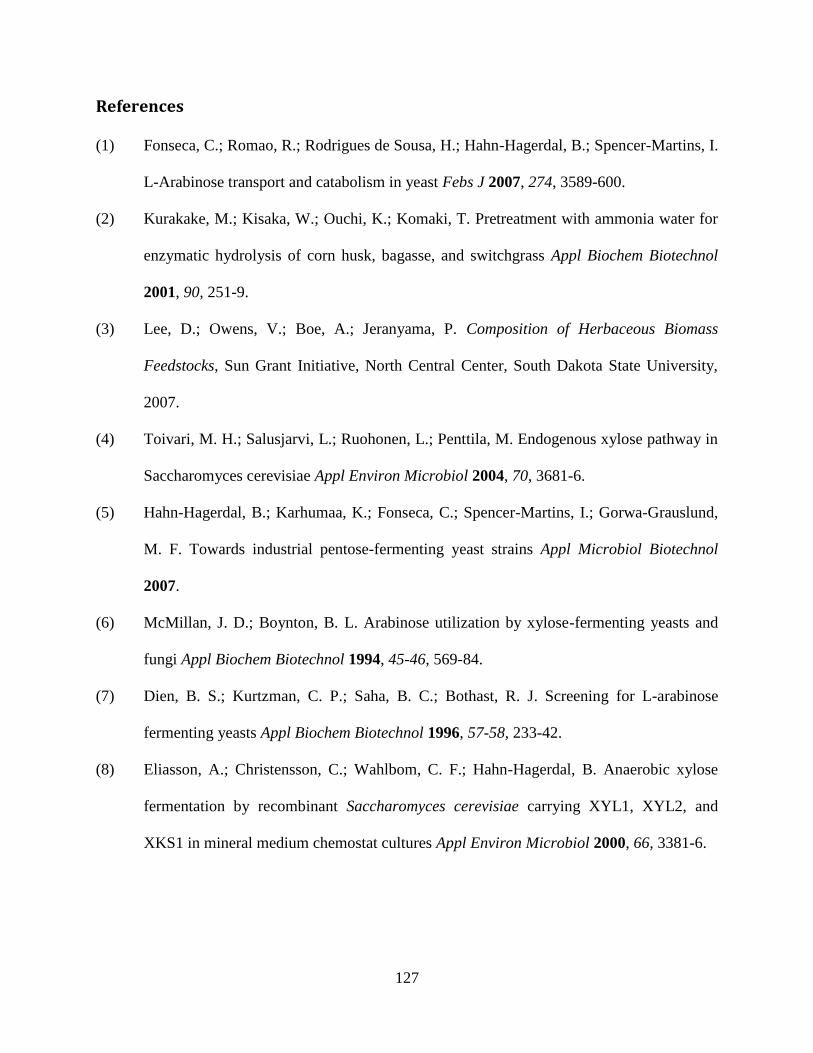
127
References
(1) Fonseca, C.; Romao, R.; Rodrigues de Sousa, H.; Hahn-Hagerdal, B.; Spencer-Martins, I.
L-Arabinose transport and catabolism in yeast Febs J 2007, 274, 3589-600.
(2) Kurakake, M.; Kisaka, W.; Ouchi, K.; Komaki, T. Pretreatment with ammonia water for
enzymatic hydrolysis of corn husk, bagasse, and switchgrass Appl Biochem Biotechnol
2001, 90, 251-9.
(3) Lee, D.; Owens, V.; Boe, A.; Jeranyama, P. Composition of Herbaceous Biomass
Feedstocks, Sun Grant Initiative, North Central Center, South Dakota State University,
2007.
(4) Toivari, M. H.; Salusjarvi, L.; Ruohonen, L.; Penttila, M. Endogenous xylose pathway in
Saccharomyces cerevisiae Appl Environ Microbiol 2004, 70, 3681-6.
(5) Hahn-Hagerdal, B.; Karhumaa, K.; Fonseca, C.; Spencer-Martins, I.; Gorwa-Grauslund,
M. F. Towards industrial pentose-fermenting yeast strains Appl Microbiol Biotechnol
2007.
(6) McMillan, J. D.; Boynton, B. L. Arabinose utilization by xylose-fermenting yeasts and
fungi Appl Biochem Biotechnol 1994, 45-46, 569-84.
(7) Dien, B. S.; Kurtzman, C. P.; Saha, B. C.; Bothast, R. J. Screening for L-arabinose
fermenting yeasts Appl Biochem Biotechnol 1996, 57-58, 233-42.
(8) Eliasson, A.; Christensson, C.; Wahlbom, C. F.; Hahn-Hagerdal, B. Anaerobic xylose
fermentation by recombinant Saccharomyces cerevisiae carrying XYL1, XYL2, and
XKS1 in mineral medium chemostat cultures Appl Environ Microbiol 2000, 66, 3381-6.

128
(9) Verho, R.; Londesborough, J.; Penttila, M.; Richard, P. Engineering redox cofactor
regeneration for improved pentose fermentation in Saccharomyces cerevisiae Appl
Environ Microbiol 2003, 69, 5892-7.
(10) Jeppsson, M.; Johansson, B.; Hahn-Hagerdal, B.; Gorwa-Grauslund, M. F. Reduced
oxidative pentose phosphate pathway flux in recombinant xylose-utilizing
Saccharomyces cerevisiae strains improves the ethanol yield from xylose Appl Environ
Microbiol 2002, 68, 1604-9.
(11) Petschacher, B.; Nidetzky, B. Engineering Candida tenuis Xylose reductase for improved
utilization of NADH: antagonistic effects of multiple side chain replacements and
performance of site-directed mutants under simulated in vivo conditions Appl Environ
Microbiol 2005, 71, 6390-3.
(12) Petschacher, B.; Nidetzky, B. Altering the coenzyme preference of xylose reductase to
favor utilization of NADH enhances ethanol yield from xylose in a metabolically
engineered strain of Saccharomyces cerevisiae Microb Cell Fact 2008, 7, 9.
(13) Watanabe, S.; Saleh, A. A.; Pack, S. P.; Annaluru, N.; Kodaki, T.; Makino, K. Ethanol
production from xylose by recombinant Saccharomyces cerevisiae expressing protein
engineered NADP+-dependent xylitol dehydrogenase J Biotechnol 2007, 130, 316-9.
(14) Watanabe, S.; Kodaki, T.; Makino, K. Complete reversal of coenzyme specificity of
xylitol dehydrogenase and increase of thermostability by the introduction of structural
zinc J Biol Chem 2005, 280, 10340-9.
(15) Becker, J.; Boles, E. A modified Saccharomyces cerevisiae strain that consumes L-
arabinose and produces ethanol Appl Environ Microbiol 2003, 69, 4144-50.

129
(16) Wisselink, H. W.; Toirkens, M. J.; del Rosario Franco Berriel, M.; Winkler, A. A.; van
Dijken, J. P.; Pronk, J. T.; van Maris, A. J. Engineering of Saccharomyces cerevisiae for
efficient anaerobic alcoholic fermentation of L-arabinose Appl Environ Microbiol 2007,
73, 4881-91.
(17) Richard, P.; Verho, R.; Putkonen, M.; Londesborough, J.; Penttila, M. Production of
ethanol from L-arabinose by Saccharomyces cerevisiae containing a fungal L-arabinose
pathway FEMS Yeast Res 2003, 3, 185-9.
(18) Bettiga, M.; Bengtsson, O.; Hahn-Hagerdal, B.; Gorwa-Grauslund, M. F. Arabinose and
xylose fermentation by recombinant Saccharomyces cerevisiae expressing a fungal
pentose utilization pathway Microb Cell Fact 2009, 8, 40.
(19) Karhumaa, K.; Wiedemann, B.; Hahn-Hagerdal, B.; Boles, E.; Gorwa-Grauslund, M. F.
Co-utilization of L-arabinose and D-xylose by laboratory and industrial Saccharomyces
cerevisiae strains Microb Cell Fact 2006, 5, 18.
(20) Witteveen, C. F. B.; Busink, R.; van de Vondervoort, P.; Dijkema, C.; Swart, K.; Visser,
J. L-arabinose and D-xylose catabolism in Aspergillus niger J Gen Microbiol 1989, 135,
2163-2171.
(21) Galagan, J. E.; Calvo, S. E.; Borkovich, K. A.; Selker, E. U.; Read, N. D.; Jaffe, D.;
FitzHugh, W.; Ma, L. J.; Smirnov, S.; Purcell, S.; Rehman, B.; Elkins, T.; Engels, R.;
Wang, S.; Nielsen, C. B.; Butler, J.; Endrizzi, M.; Qui, D.; Ianakiev, P.; Bell-Pedersen,
D.; Nelson, M. A.; Werner-Washburne, M.; Selitrennikoff, C. P.; Kinsey, J. A.; Braun, E.
L.; Zelter, A.; Schulte, U.; Kothe, G. O.; Jedd, G.; Mewes, W.; Staben, C.; Marcotte, E.;
Greenberg, D.; Roy, A.; Foley, K.; Naylor, J.; Stange-Thomann, N.; Barrett, R.; Gnerre,
S.; Kamal, M.; Kamvysselis, M.; Mauceli, E.; Bielke, C.; Rudd, S.; Frishman, D.;

130
Krystofova, S.; Rasmussen, C.; Metzenberg, R. L.; Perkins, D. D.; Kroken, S.; Cogoni,
C.; Macino, G.; Catcheside, D.; Li, W.; Pratt, R. J.; Osmani, S. A.; DeSouza, C. P.; Glass,
L.; Orbach, M. J.; Berglund, J. A.; Voelker, R.; Yarden, O.; Plamann, M.; Seiler, S.;
Dunlap, J.; Radford, A.; Aramayo, R.; Natvig, D. O.; Alex, L. A.; Mannhaupt, G.;
Ebbole, D. J.; Freitag, M.; Paulsen, I.; Sachs, M. S.; Lander, E. S.; Nusbaum, C.; Birren,
B. The genome sequence of the filamentous fungus Neurospora crassa Nature 2003, 422,
859-68.
(22) Bradford, M. M. A rapid and sensitive method for the quantitation of microgram
quantities of protein utilizing the principle of protein-dye binding Anal Biochem 1976,
72, 248-54.
(23) Diez-Gonzalez, F.; Russell, J. B. The ability of Escherichia coli O157:H7 to decrease its
intracellular pH and resist the toxicity of acetic acid Microbiology 1997, 143 ( Pt 4),
1175-80.
(24) Orij, R.; Postmus, J.; Ter Beek, A.; Brul, S.; Smits, G. J. In vivo measurement of
cytosolic and mitochondrial pH using a pH-sensitive GFP derivative in Saccharomyces
cerevisiae reveals a relation between intracellular pH and growth Microbiology 2009,
155, 268-78.
(25) Johansson, B.; Christensson, C.; Hobley, T.; Hahn-Hagerdal, B. Xylulokinase
overexpression in two strains of Saccharomyces cerevisiae also expressing xylose
reductase and xylitol dehydrogenase and its effect on fermentation of xylose and
lignocellulosic hydrolysate Appl Environ Microbiol 2001, 67, 4249-55.
(26) Shao, Z.; Zhao, H.; Zhao, H. DNA assembler, an in vivo genetic method for rapid
construction of biochemical pathways Nucleic Acids Res 2009, 37, e16.

131
(27) Jojima, T.; Omumasaba, C. A.; Inui, M.; Yukawa, H. Sugar transporters in efficient
utilization of mixed sugar substrates: current knowledge and outlook Appl Microbiol
Biotechnol 2009.
(28) Leandro, M. J.; Fonseca, C.; Goncalves, P. Hexose and pentose transport in
ascomycetous yeasts: an overview FEMS Yeast Res 2009, 9, 511-25.
(29) Jeffries, T. W.; Jin, Y. S. Metabolic engineering for improved fermentation of pentoses
by yeasts Appl Microbiol Biotechnol 2004, 63, 495-509.
(30) Fonseca, C.; Neves, A. R.; Antunes, A. M.; Noronha, J. P.; Hahn-Hagerdal, B.; Santos,
H.; Spencer-Martins, I. Use of in vivo 13C nuclear magnetic resonance spectroscopy to
elucidate L-arabinose metabolism in yeasts Appl Environ Microbiol 2008, 74, 1845-55.
(31) Ellis, K. J.; Morrison, J. F. Buffers of constant ionic strength for studying pH-dependent
processes Methods Enzymol 1982, 87, 405-26.
(32) Ho, S. N.; Hunt, H. D.; Horton, R. M.; Pullen, J. K.; Pease, L. R. Site-directed
mutagenesis by overlap extension using the polymerase chain reaction Gene 1989, 77,
51-9.