Embed Size (px)
Citation preview
www.Padasalai.Net www.TrbTnpsc.com
http://www.trbtnpsc.com/2018/06/latest-plus-one-11th-study-materials-tamil-medium-english-medium-new-syllabus-based.html

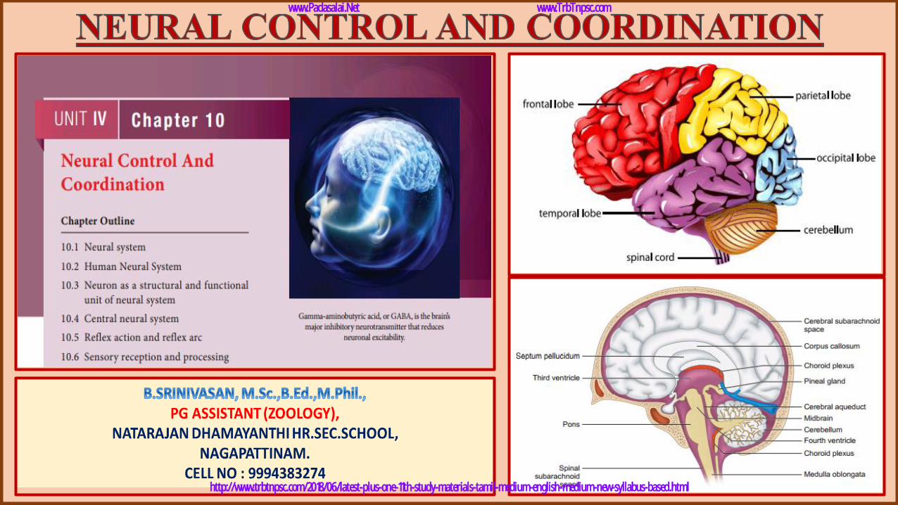
❖ Learning Objectives:
1. Understands the structure of neuron and neural system of human
beings.
2. Learns to differentiate the functions of sensory and motor neuron.
3. Understands the conduction of nerve impulses and learns the
importance of myelin sheathsaltatory conduction.
4. Outlines the role of synapse and neuromuscular junction.
5. Learns the structure and functionsof central neural system.
6. Understands the structure, sensory reception and processing in Photo,
Phono, Olfactory, Gustatory and Skin Receptors.
www.Padasalai.Net www.TrbTnpsc.com
http://www.trbtnpsc.com/2018/06/latest-plus-one-11th-study-materials-tamil-medium-english-medium-new-syllabus-based.html
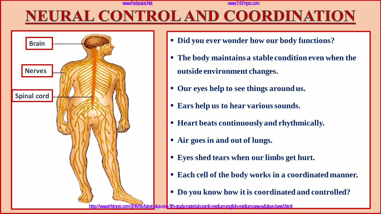
▪ Did you ever wonder how our body functions?
▪ The body maintains a stable condition even when the
outside environment changes.
▪ Our eyes help to see things around us.
▪ Ears help us to hear various sounds.
▪ Heart beats continuously and rhythmically.
▪ Air goes in and out of lungs.
▪ Eyes shed tears when our limbs get hurt.
▪ Each cell of the body works in a coordinated manner.
▪ Do you know how it is coordinated and controlled?
Brain
Nerves
Spinal cord
www.Padasalai.Net www.TrbTnpsc.com
http://www.trbtnpsc.com/2018/06/latest-plus-one-11th-study-materials-tamil-medium-english-medium-new-syllabus-based.html
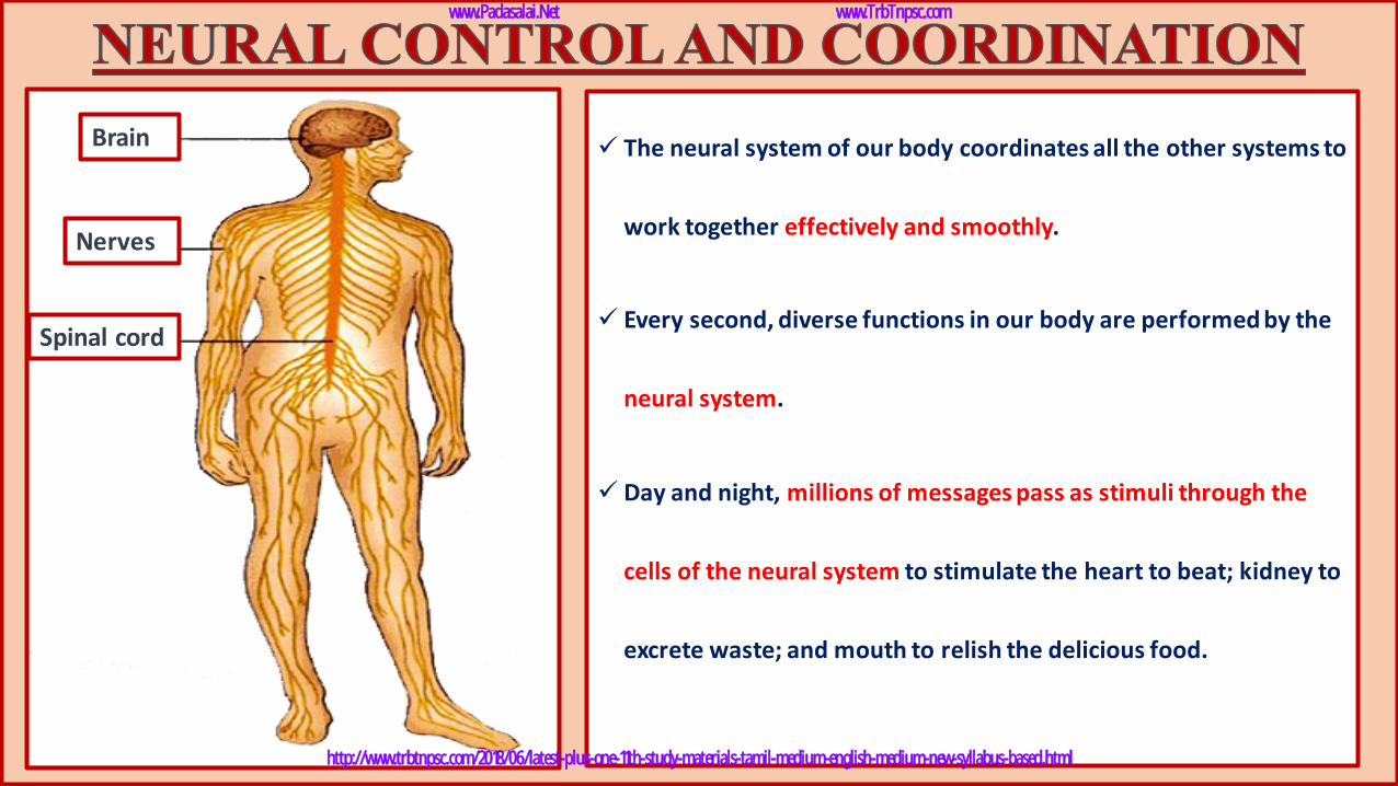
✓ The neural system of our body coordinates all the other systems to
work together effectively and smoothly.
✓ Every second, diverse functions in our body are performed by the
neural system.
✓ Day and night, millions of messages pass as stimuli through the
cells of the neural system to stimulate the heart to beat; kidney to
excrete waste; and mouth to relish the delicious food.
Brain
Nerves
Spinal cord
www.Padasalai.Net www.TrbTnpsc.com
http://www.trbtnpsc.com/2018/06/latest-plus-one-11th-study-materials-tamil-medium-english-medium-new-syllabus-based.html
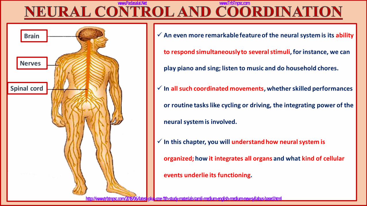
✓ An even more remarkable feature of the neural system is its ability
to respond simultaneously to several stimuli, for instance, we can
play piano and sing; listen to music and do household chores.
✓ In all such coordinated movements, whether skilled performances
or routine tasks like cycling or driving, the integrating power of the
neural system is involved.
✓ In this chapter, you will understand how neural system is
organized; how it integrates all organs and what kind of cellular
events underlie its functioning.
Brain
Nerves
Spinal cord
www.Padasalai.Net www.TrbTnpsc.com
http://www.trbtnpsc.com/2018/06/latest-plus-one-11th-study-materials-tamil-medium-english-medium-new-syllabus-based.html
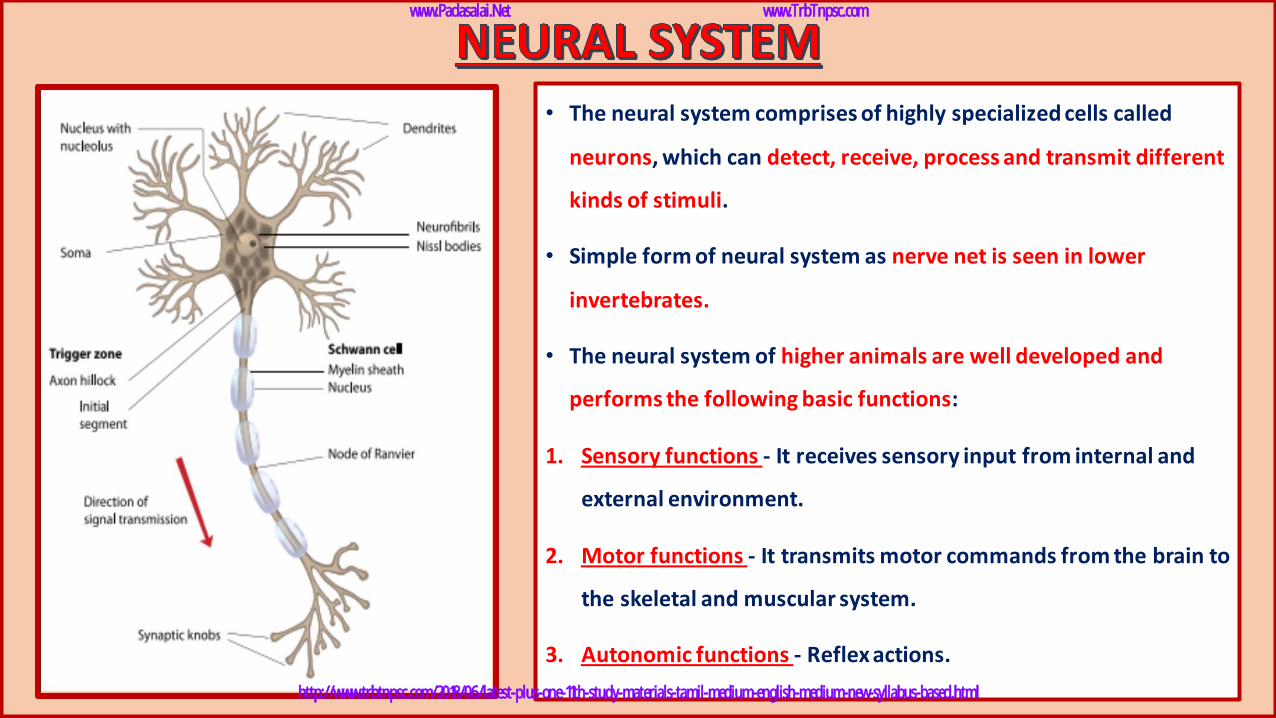
• The neural system comprises of highly specialized cells called
neurons, which can detect, receive, process and transmit different
kinds of stimuli.
• Simple form of neural system as nerve net is seen in lower
invertebrates.
• The neural system of higher animals are well developed and
performs the following basic functions:
1. Sensory functions - It receives sensory input from internal and
external environment.
2. Motor functions - It transmits motor commands from the brain to
the skeletal and muscular system.
3. Autonomic functions - Reflex actions.
www.Padasalai.Net www.TrbTnpsc.com
http://www.trbtnpsc.com/2018/06/latest-plus-one-11th-study-materials-tamil-medium-english-medium-new-syllabus-based.html
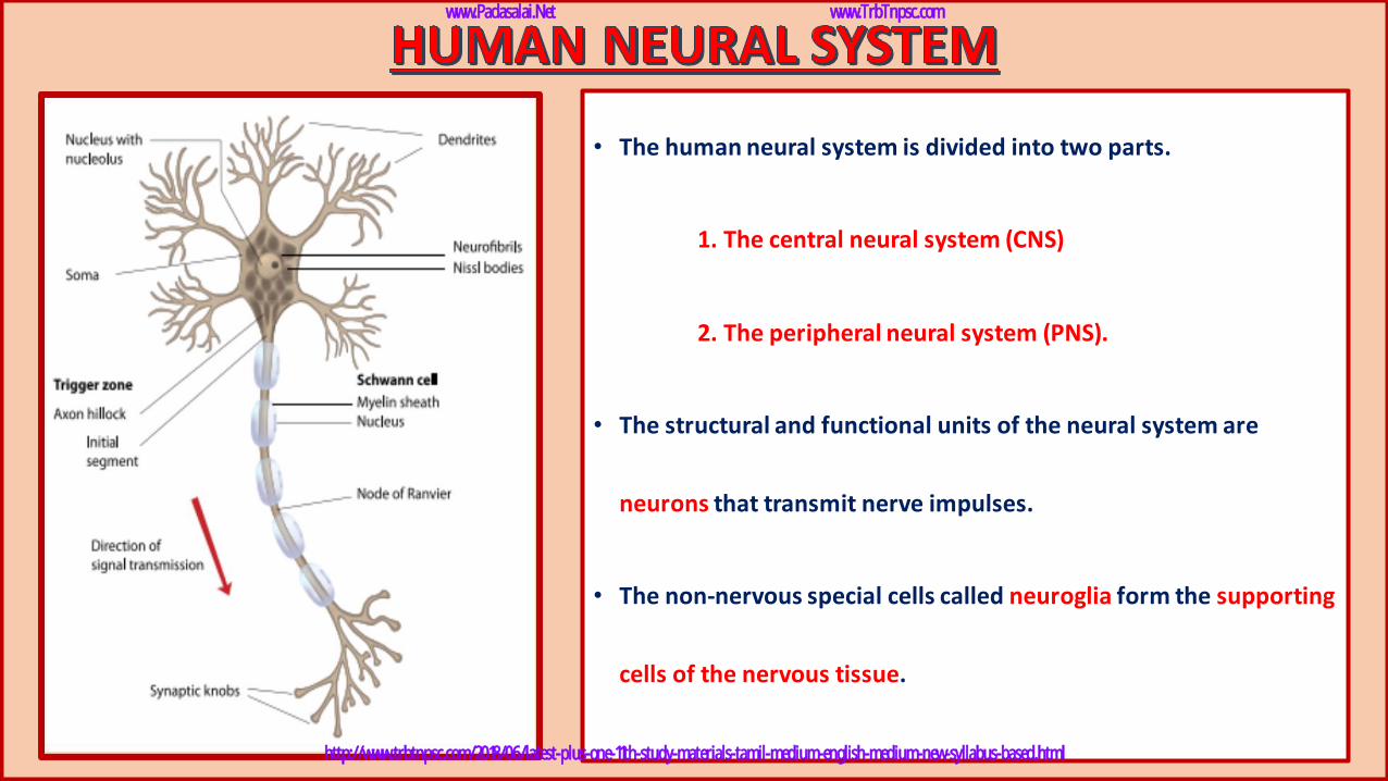
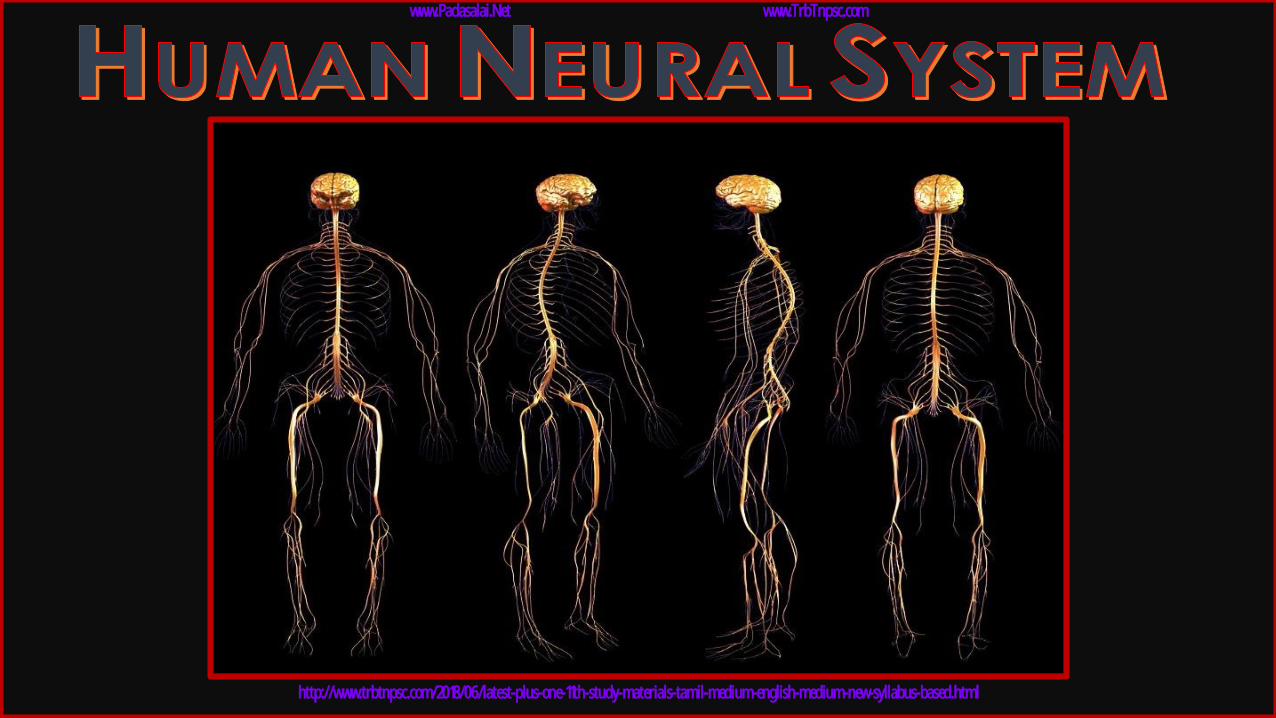
• The human neural system is divided into two parts.
1. The central neural system (CNS)
2. The peripheral neural system (PNS).
• The structural and functional units of the neural system are
neurons that transmit nerve impulses.
• The non-nervous special cells called neuroglia form the supporting
cells of the nervous tissue.
www.Padasalai.Net www.TrbTnpsc.com
http://www.trbtnpsc.com/2018/06/latest-plus-one-11th-study-materials-tamil-medium-english-medium-new-syllabus-based.html
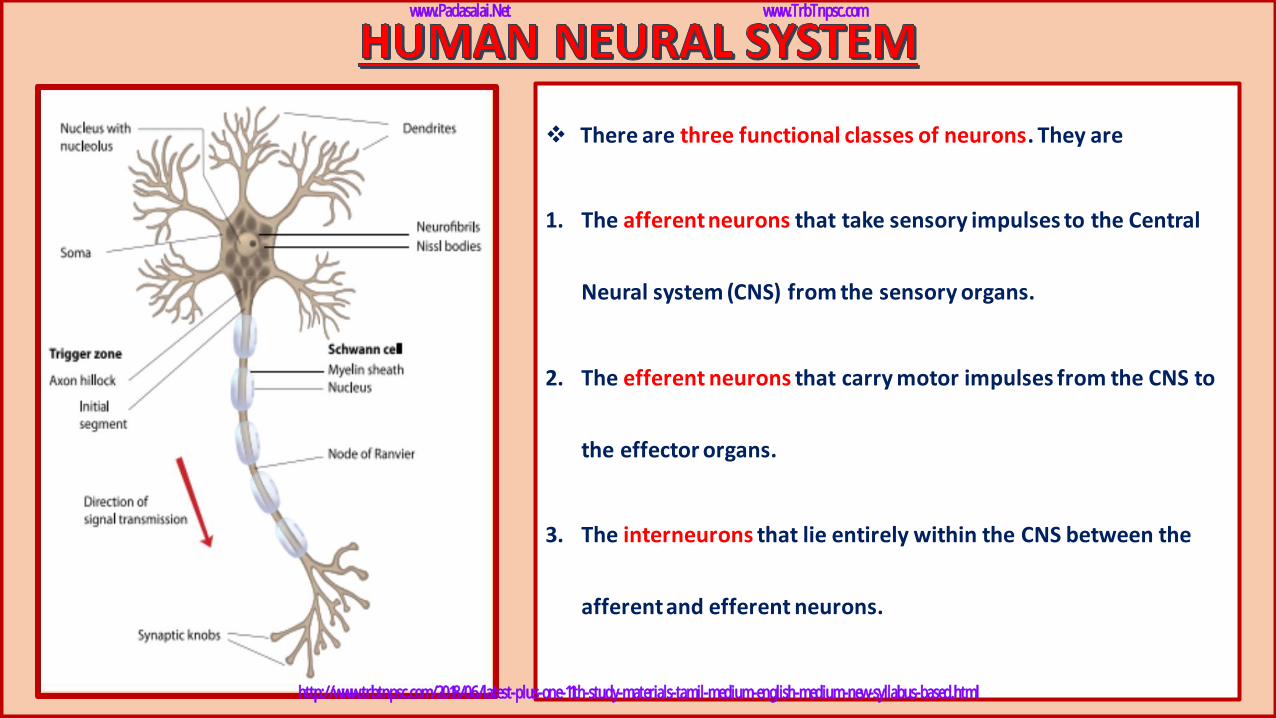
❖ There are three functional classes of neurons. They are
1. The afferent neurons that take sensory impulses to the Central
Neural system (CNS) from the sensory organs.
2. The efferent neurons that carry motor impulses from the CNS to
the effector organs.
3. The interneurons that lie entirely within the CNS between the
afferent and efferent neurons.
www.Padasalai.Net www.TrbTnpsc.com
http://www.trbtnpsc.com/2018/06/latest-plus-one-11th-study-materials-tamil-medium-english-medium-new-syllabus-based.html
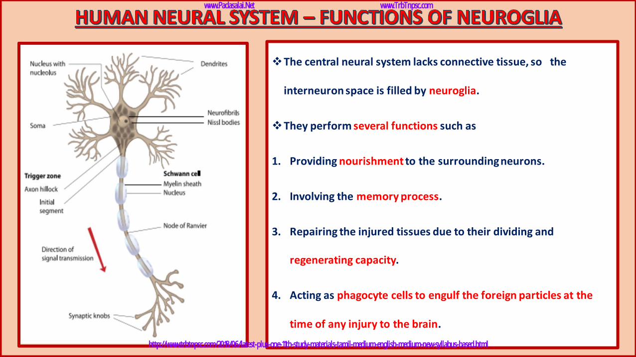
❖The central neural system lacks connective tissue, so the
interneuron space is filled by neuroglia.
❖They perform several functions such as
1. Providing nourishment to the surrounding neurons.
2. Involving the memory process.
3. Repairing the injured tissues due to their dividing and
regenerating capacity.
4. Acting as phagocyte cells to engulf the foreign particles at the
time of any injury to the brain.
www.Padasalai.Net www.TrbTnpsc.com
http://www.trbtnpsc.com/2018/06/latest-plus-one-11th-study-materials-tamil-medium-english-medium-new-syllabus-based.html
www.Padasalai.Net www.TrbTnpsc.com
http://www.trbtnpsc.com/2018/06/latest-plus-one-11th-study-materials-tamil-medium-english-medium-new-syllabus-based.html
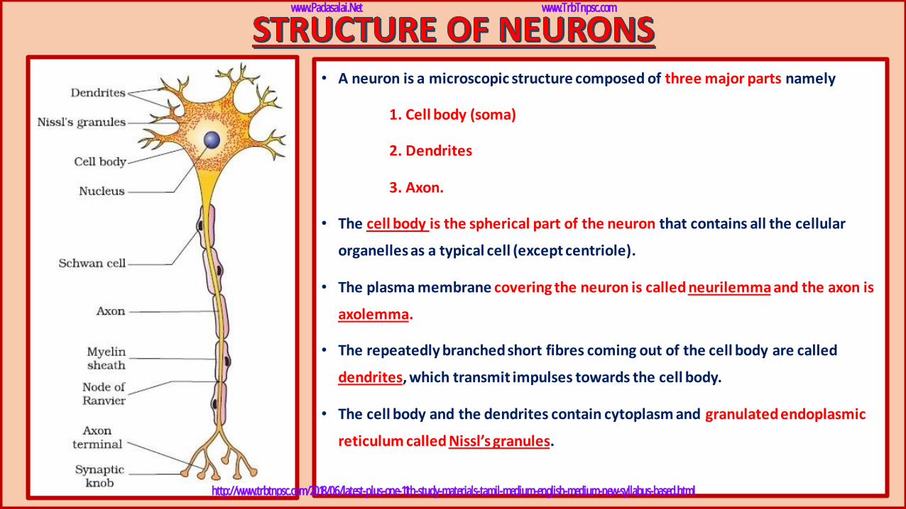
• A neuron is a microscopic structure composed of three major parts namely
1. Cell body (soma)
2. Dendrites
3. Axon.
• The cell body is the spherical part of the neuron that contains all the cellular
organelles as a typical cell (except centriole).
• The plasma membrane covering the neuron is called neurilemma and the axon is
axolemma.
• The repeatedly branched short fibres coming out of the cell body are called
dendrites, which transmit impulses towards the cell body.
• The cell body and the dendrites contain cytoplasm and granulated endoplasmic
reticulum called Nissl’s granules.
www.Padasalai.Net www.TrbTnpsc.com
http://www.trbtnpsc.com/2018/06/latest-plus-one-11th-study-materials-tamil-medium-english-medium-new-syllabus-based.html
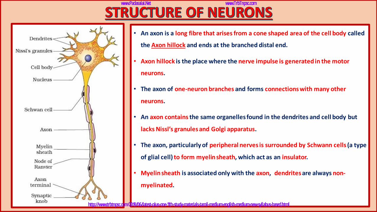
• An axon is a long fibre that arises from a cone shaped area of the cell body called
the Axon hillock and ends at the branched distal end.
• Axon hillock is the place where the nerve impulse is generated in the motor
neurons.
• The axon of one-neuron branches and forms connections with many other
neurons.
• An axon contains the same organelles found in the dendrites and cell body but
lacks Nissl’s granules and Golgi apparatus.
• The axon, particularly of peripheral nerves is surrounded by Schwann cells (a type
of glial cell) to form myelin sheath, which act as an insulator.
• Myelin sheath is associated only with the axon, dendrites are always non-
myelinated.
www.Padasalai.Net www.TrbTnpsc.com
http://www.trbtnpsc.com/2018/06/latest-plus-one-11th-study-materials-tamil-medium-english-medium-new-syllabus-based.html
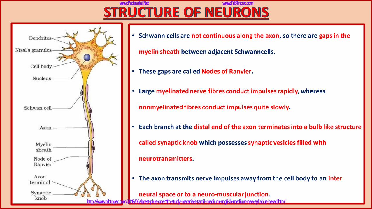
• Schwann cells are not continuous along the axon, so there are gaps in the
myelin sheath between adjacent Schwanncells.
• These gaps are called Nodes of Ranvier.
• Large myelinated nerve fibres conduct impulses rapidly, whereas
nonmyelinated fibres conduct impulses quite slowly.
• Each branch at the distal end of the axon terminates into a bulb like structure
called synaptic knob which possesses synaptic vesicles filled with
neurotransmitters.
• The axon transmits nerve impulses away from the cell body to an inter
neural space or to a neuro-muscular junction.
www.Padasalai.Net www.TrbTnpsc.com
http://www.trbtnpsc.com/2018/06/latest-plus-one-11th-study-materials-tamil-medium-english-medium-new-syllabus-based.html
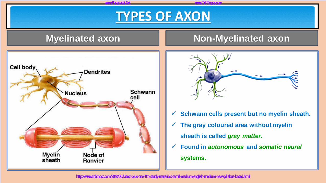
• It is enveloped with Schwann cells, which form a myelin sheath around the axon.
• Found in spinal and cranial nerves.
• White coloured area, formed of myelinated nerve fibres is called white matter.
• Gaps between two adjacent myelin sheaths are called nodes of Ranvier.
TYPES OF AXON
✓ Schwann cells present but no myelin sheath.
✓ The gray coloured area without myelin
sheath is called gray matter.
✓ Found in autonomous and somatic neural
systems.
Myelinated axon Non-Myelinated axon
www.Padasalai.Net www.TrbTnpsc.com
http://www.trbtnpsc.com/2018/06/latest-plus-one-11th-study-materials-tamil-medium-english-medium-new-syllabus-based.html
www.Padasalai.Net www.TrbTnpsc.com
http://www.trbtnpsc.com/2018/06/latest-plus-one-11th-study-materials-tamil-medium-english-medium-new-syllabus-based.html
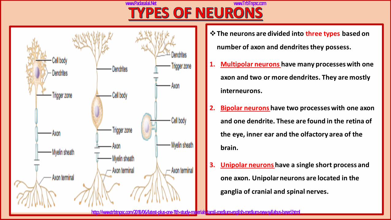
❖The neurons are divided into three types based on
number of axon and dendrites they possess.
1. Multipolar neurons have many processes with one
axon and two or more dendrites. They are mostly
interneurons.
2. Bipolar neurons have two processes with one axon
and one dendrite. These are found in the retina of
the eye, inner ear and the olfactory area of the
brain.
3. Unipolar neurons have a single short process and
one axon. Unipolar neurons are located in the
ganglia of cranial and spinal nerves.
www.Padasalai.Net www.TrbTnpsc.com
http://www.trbtnpsc.com/2018/06/latest-plus-one-11th-study-materials-tamil-medium-english-medium-new-syllabus-based.html
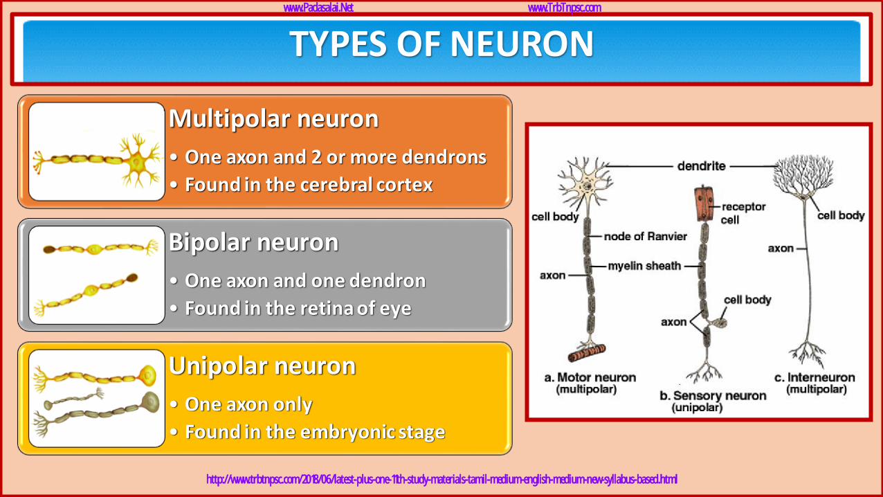
TYPES OF NEURON
Multipolar neuron
• One axon and 2 or more dendrons
• Found in the cerebral cortex
Bipolar neuron
• One axon and one dendron
• Found in the retina of eye
Unipolar neuron
• One axon only
• Found in the embryonic stage
www.Padasalai.Net www.TrbTnpsc.com
http://www.trbtnpsc.com/2018/06/latest-plus-one-11th-study-materials-tamil-medium-english-medium-new-syllabus-based.html
bankofbiology.blogspot.com
www.Padasalai.Net www.TrbTnpsc.com
http://www.trbtnpsc.com/2018/06/latest-plus-one-11th-study-materials-tamil-medium-english-medium-new-syllabus-based.html
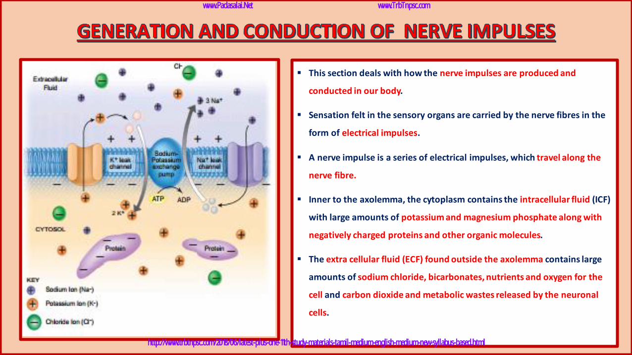
▪ This section deals with how the nerve impulses are produced and
conducted in our body.
▪ Sensation felt in the sensory organs are carried by the nerve fibres in the
form of electrical impulses.
▪ A nerve impulse is a series of electrical impulses, which travel along the
nerve fibre.
▪ Inner to the axolemma, the cytoplasm contains the intracellular fluid (ICF)
with large amounts of potassium and magnesium phosphate along with
negatively charged proteins and other organic molecules.
▪ The extra cellular fluid (ECF) found outside the axolemma contains large
amounts of sodium chloride, bicarbonates, nutrients and oxygen for the
cell and carbon dioxide and metabolic wastes released by the neuronal
cells.
www.Padasalai.Net www.TrbTnpsc.com
http://www.trbtnpsc.com/2018/06/latest-plus-one-11th-study-materials-tamil-medium-english-medium-new-syllabus-based.html
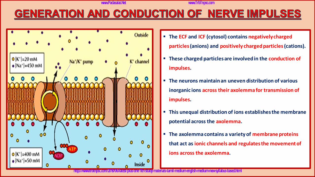
▪ The ECF and ICF (cytosol) contains negatively charged
particles (anions) and positively charged particles (cations).
▪ These charged particles are involved in the conduction of
impulses.
▪ The neurons maintain an uneven distribution of various
inorganic ions across their axolemma for transmission of
impulses.
▪ This unequal distribution of ions establishes the membrane
potential across the axolemma.
▪ The axolemma contains a variety of membrane proteins
that act as ionic channels and regulates the movement of
ions across the axolemma.
www.Padasalai.Net www.TrbTnpsc.com
http://www.trbtnpsc.com/2018/06/latest-plus-one-11th-study-materials-tamil-medium-english-medium-new-syllabus-based.html
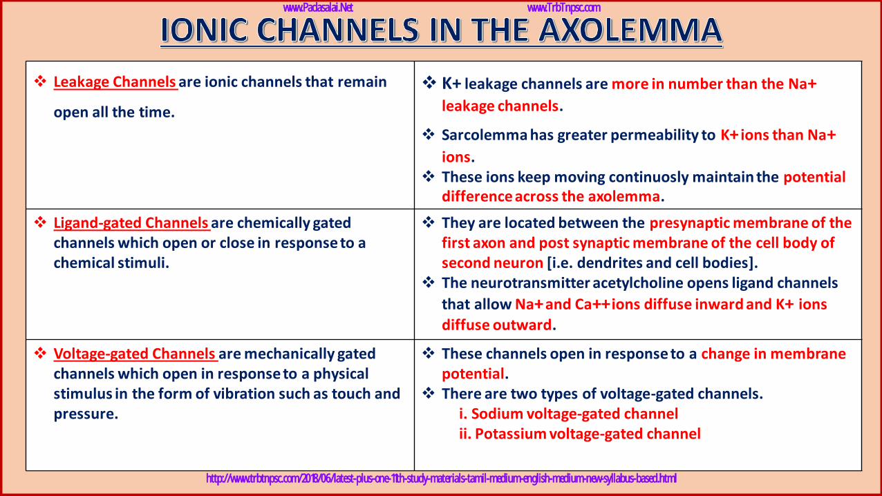
❖ Leakage Channels are ionic channels that remain
open all the time.
❖ K+ leakage channels are more in number than the Na+leakage channels.
❖ Sarcolemma has greater permeability to K+ ions than Na+ions.
❖ These ions keep moving continuosly maintain the potential difference across the axolemma.
❖ Ligand-gated Channels are chemically gated channels which open or close in response to a chemical stimuli.
❖ They are located between the presynaptic membrane of the first axon and post synaptic membrane of the cell body of second neuron [i.e. dendrites and cell bodies].
❖ The neurotransmitter acetylcholine opens ligand channels
that allow Na+and Ca++ions diffuse inward and K+ ions diffuse outward.
❖ Voltage-gated Channels are mechanically gated channels which open in response to a physical stimulus in the form of vibration such as touch and pressure.
❖ These channels open in response to a change in membrane potential.
❖ There are two types of voltage-gated channels.i. Sodium voltage-gated channelii. Potassium voltage-gated channel
www.Padasalai.Net www.TrbTnpsc.com
http://www.trbtnpsc.com/2018/06/latest-plus-one-11th-study-materials-tamil-medium-english-medium-new-syllabus-based.html
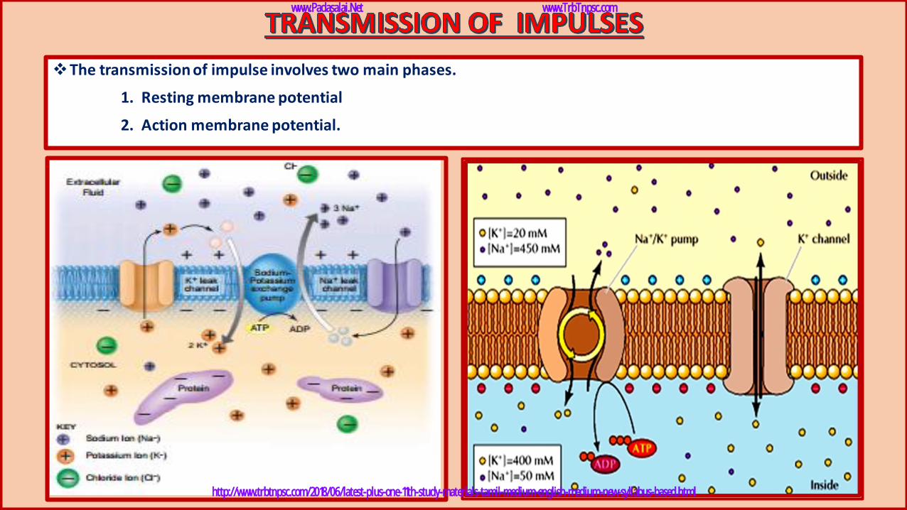
❖The transmission of impulse involves two main phases.
1. Resting membrane potential
2. Action membrane potential.
www.Padasalai.Net www.TrbTnpsc.com
http://www.trbtnpsc.com/2018/06/latest-plus-one-11th-study-materials-tamil-medium-english-medium-new-syllabus-based.html
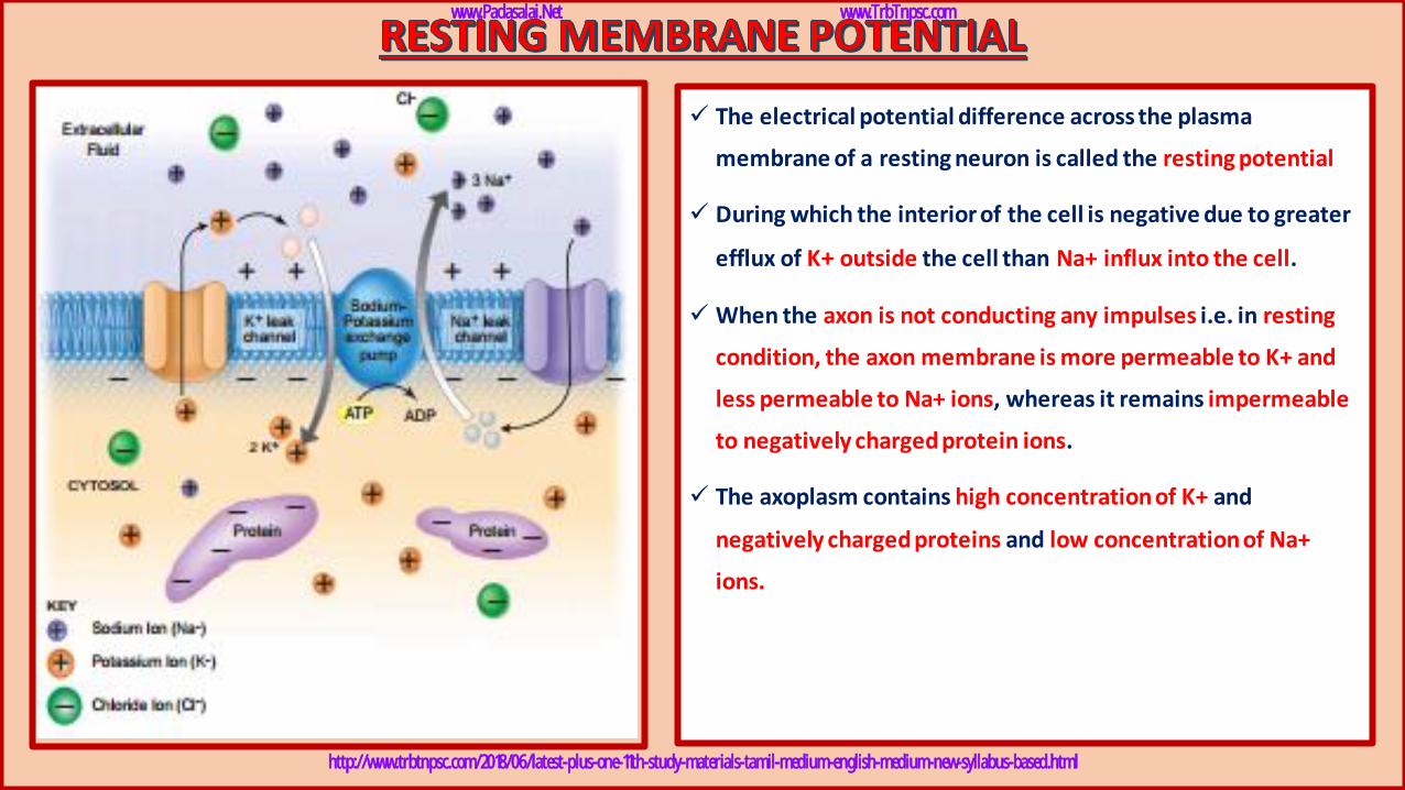
✓ The electrical potential difference across the plasma
membrane of a resting neuron is called the resting potential
✓ During which the interior of the cell is negative due to greater
efflux of K+ outside the cell than Na+ influx into the cell.
✓ When the axon is not conducting any impulses i.e. in resting
condition, the axon membrane is more permeable to K+ and
less permeable to Na+ ions, whereas it remains impermeable
to negatively charged protein ions.
✓ The axoplasm contains high concentration of K+ and
negatively charged proteins and low concentration of Na+
ions.
www.Padasalai.Net www.TrbTnpsc.com
http://www.trbtnpsc.com/2018/06/latest-plus-one-11th-study-materials-tamil-medium-english-medium-new-syllabus-based.html
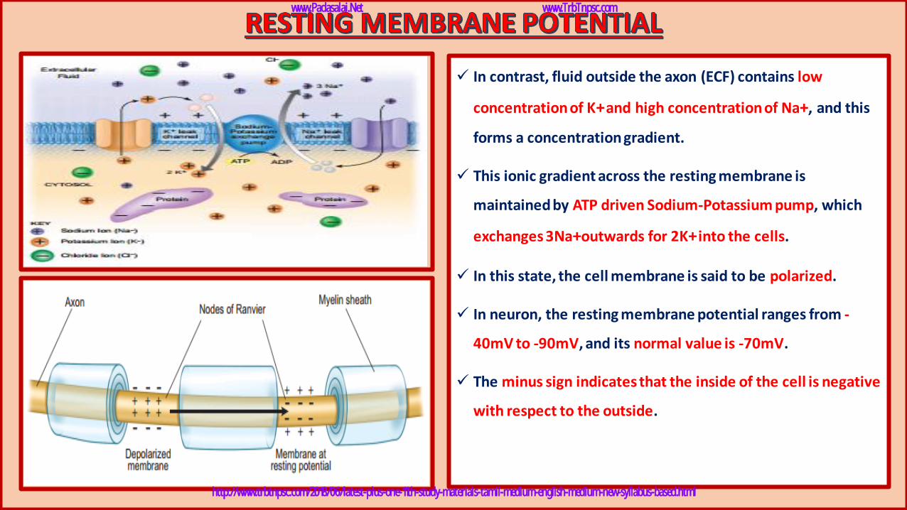
✓ In contrast, fluid outside the axon (ECF) contains low
concentration of K+and high concentration of Na+, and this
forms a concentration gradient.
✓ This ionic gradient across the resting membrane is
maintained by ATP driven Sodium-Potassium pump, which
exchanges 3Na+outwards for 2K+into the cells.
✓ In this state, the cell membrane is said to be polarized.
✓ In neuron, the resting membrane potential ranges from -
40mV to -90mV, and its normal value is -70mV.
✓ The minus sign indicates that the inside of the cell is negative
with respect to the outside.
www.Padasalai.Net www.TrbTnpsc.com
http://www.trbtnpsc.com/2018/06/latest-plus-one-11th-study-materials-tamil-medium-english-medium-new-syllabus-based.html
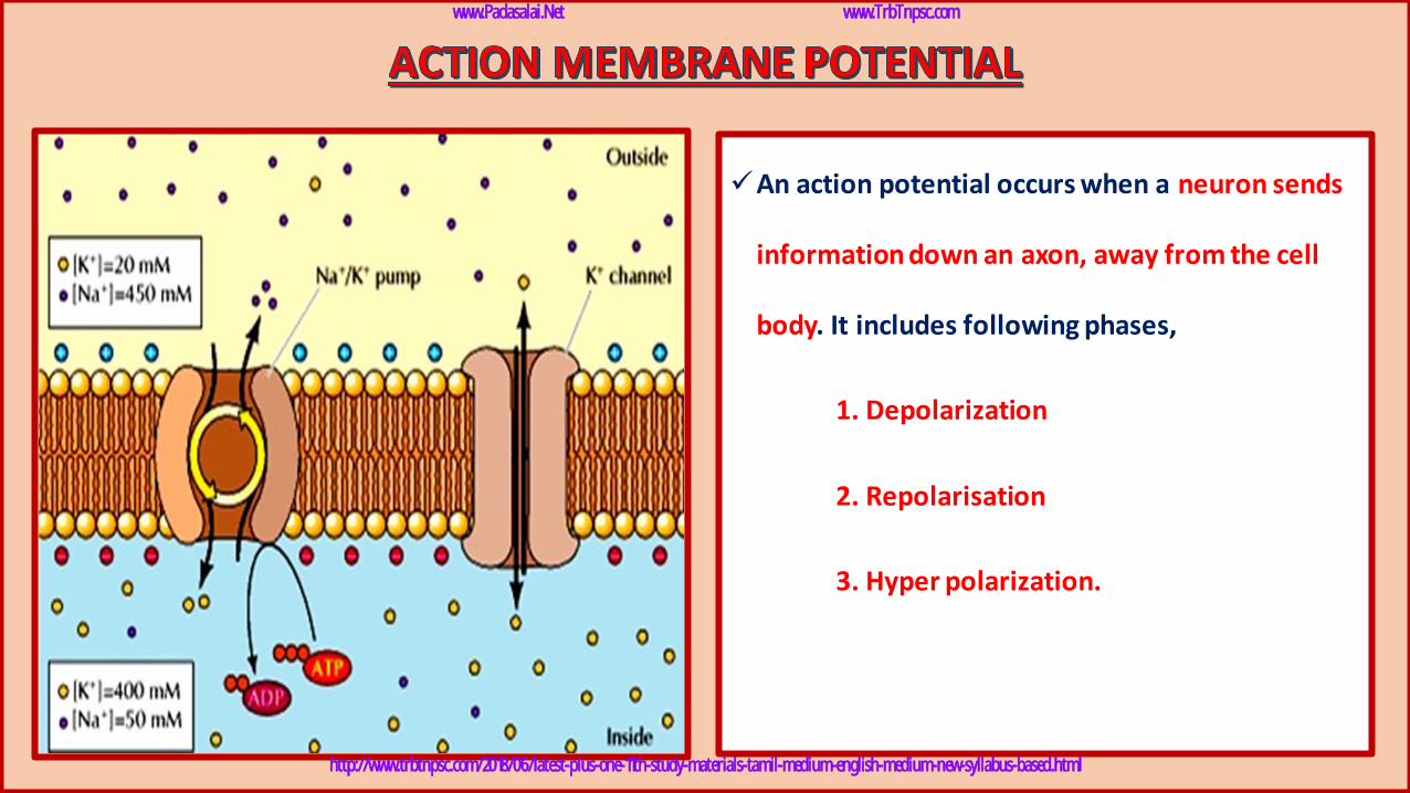
✓An action potential occurs when a neuron sends
information down an axon, away from the cell
body. It includes following phases,
1. Depolarization
2. Repolarisation
3. Hyper polarization.
www.Padasalai.Net www.TrbTnpsc.com
http://www.trbtnpsc.com/2018/06/latest-plus-one-11th-study-materials-tamil-medium-english-medium-new-syllabus-based.html

➢ When a nerve fibre is stimulated, sodium voltage-gate
opens and makes the axolemma permeable to Na+ ions,
meanwhile the potassium voltage gate closes.
➢ As a result, the rate of flow of Na+ ions into the axoplasm
exceeds the rate of flow of K+ ions to the outside fluid [ECF].
➢ Therefore, the axolemma becomes positively charged inside
and negatively charged outside.
➢ This reversal of electrical charge is called Depolarization.
www.Padasalai.Net www.TrbTnpsc.com
http://www.trbtnpsc.com/2018/06/latest-plus-one-11th-study-materials-tamil-medium-english-medium-new-syllabus-based.html
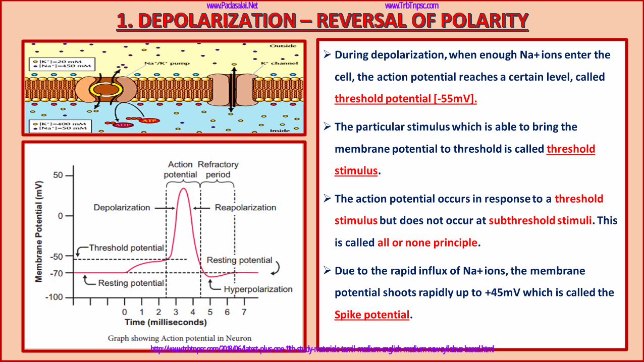
➢ During depolarization, when enough Na+ ions enter the
cell, the action potential reaches a certain level, called
threshold potential [-55mV].
➢ The particular stimulus which is able to bring the
membrane potential to threshold is called threshold
stimulus.
➢ The action potential occurs in response to a threshold
stimulus but does not occur at subthreshold stimuli. This
is called all or none principle.
➢ Due to the rapid influx of Na+ions, the membrane
potential shoots rapidly up to +45mV which is called the
Spike potential.
www.Padasalai.Net www.TrbTnpsc.com
http://www.trbtnpsc.com/2018/06/latest-plus-one-11th-study-materials-tamil-medium-english-medium-new-syllabus-based.html
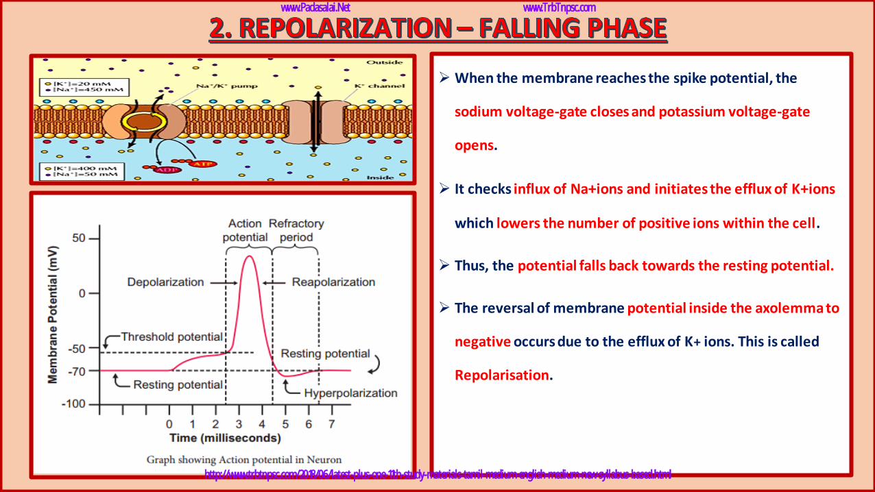
➢ When the membrane reaches the spike potential, the
sodium voltage-gate closes and potassium voltage-gate
opens.
➢ It checks influx of Na+ions and initiates the efflux of K+ions
which lowers the number of positive ions within the cell.
➢ Thus, the potential falls back towards the resting potential.
➢ The reversal of membrane potential inside the axolemma to
negative occurs due to the efflux of K+ ions. This is called
Repolarisation.
www.Padasalai.Net www.TrbTnpsc.com
http://www.trbtnpsc.com/2018/06/latest-plus-one-11th-study-materials-tamil-medium-english-medium-new-syllabus-based.html
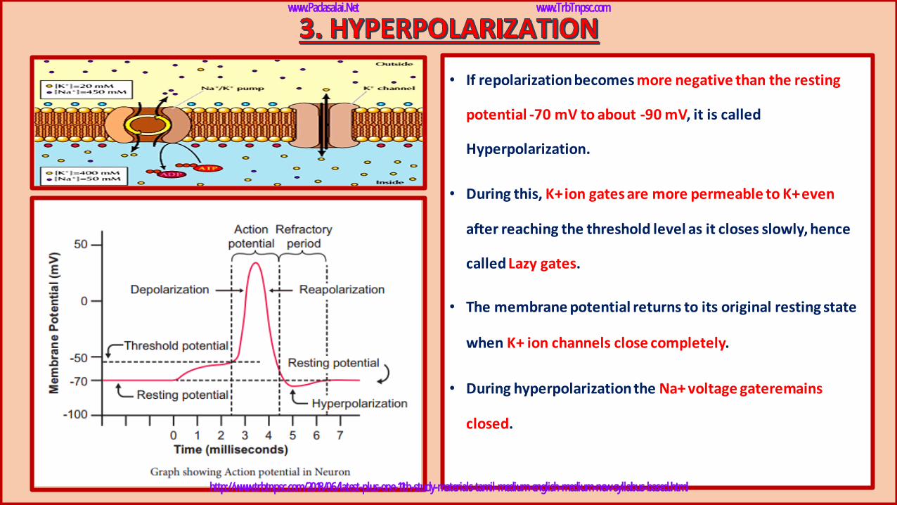
• If repolarization becomes more negative than the resting
potential -70 mV to about -90 mV, it is called
Hyperpolarization.
• During this, K+ ion gates are more permeable to K+even
after reaching the threshold level as it closes slowly, hence
called Lazy gates.
• The membrane potential returns to its original resting state
when K+ ion channels close completely.
• During hyperpolarization the Na+ voltage gateremains
closed.
www.Padasalai.Net www.TrbTnpsc.com
http://www.trbtnpsc.com/2018/06/latest-plus-one-11th-study-materials-tamil-medium-english-medium-new-syllabus-based.html
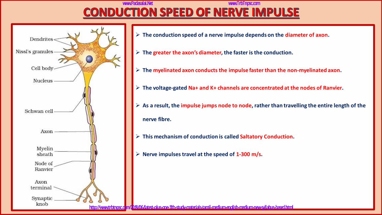
➢ The conduction speed of a nerve impulse depends on the diameter of axon.
➢ The greater the axon’s diameter, the faster is the conduction.
➢ The myelinated axon conducts the impulse faster than the non-myelinated axon.
➢ The voltage-gated Na+ and K+ channels are concentrated at the nodes of Ranvier.
➢ As a result, the impulse jumps node to node, rather than travelling the entire length of the
nerve fibre.
➢ This mechanism of conduction is called Saltatory Conduction.
➢ Nerve impulses travel at the speed of 1-300 m/s.
www.Padasalai.Net www.TrbTnpsc.com
http://www.trbtnpsc.com/2018/06/latest-plus-one-11th-study-materials-tamil-medium-english-medium-new-syllabus-based.html
www.Padasalai.Net www.TrbTnpsc.com
http://www.trbtnpsc.com/2018/06/latest-plus-one-11th-study-materials-tamil-medium-english-medium-new-syllabus-based.html
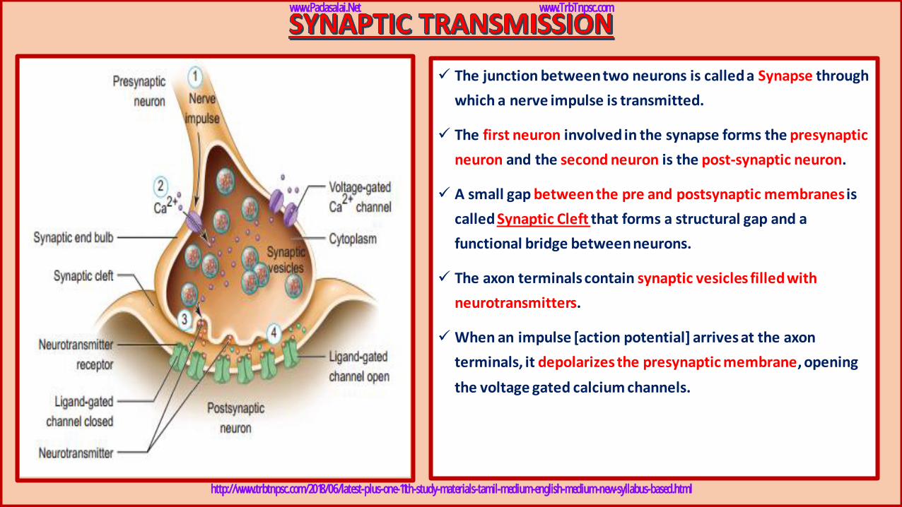
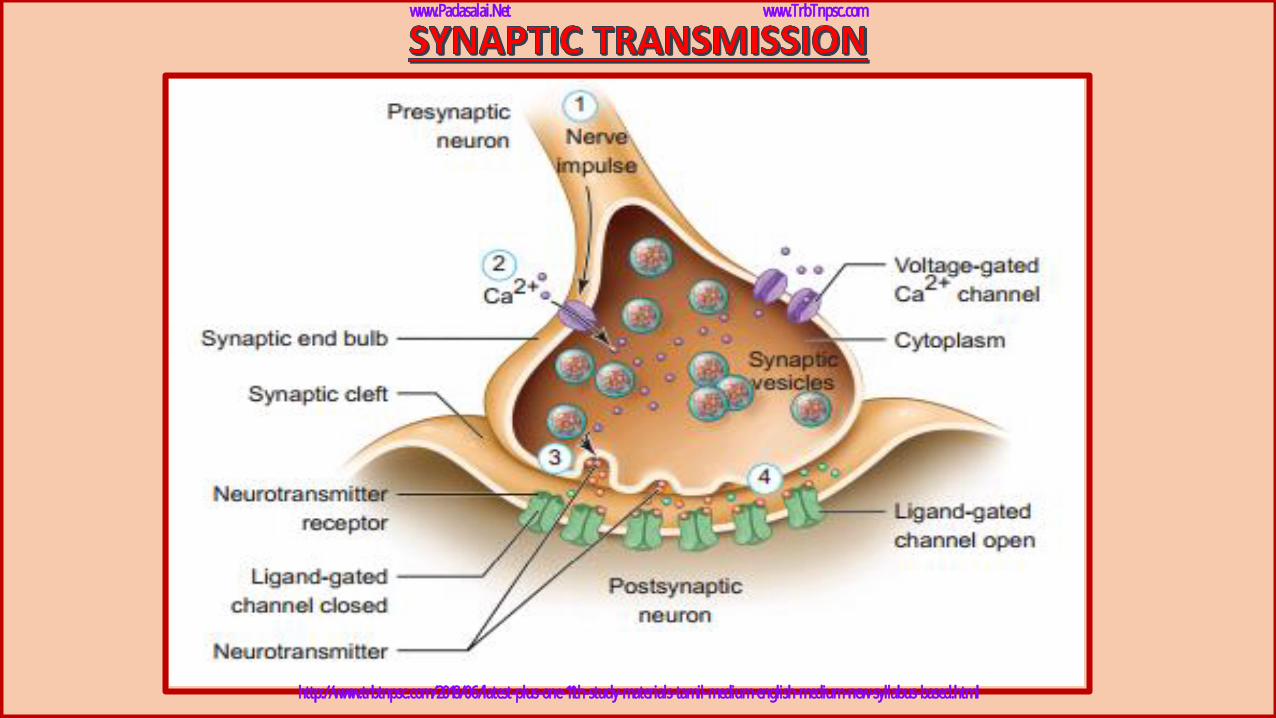
✓ The junction between two neurons is called a Synapse through
which a nerve impulse is transmitted.
✓ The first neuron involved in the synapse forms the presynaptic
neuron and the second neuron is the post-synaptic neuron.
✓ A small gap between the pre and postsynaptic membranesis
called Synaptic Cleft that forms a structural gap and a
functional bridge between neurons.
✓ The axon terminals contain synaptic vesicles filled with
neurotransmitters.
✓ When an impulse [action potential] arrives at the axon
terminals, it depolarizes the presynaptic membrane, opening
the voltage gated calcium channels.
www.Padasalai.Net www.TrbTnpsc.com
http://www.trbtnpsc.com/2018/06/latest-plus-one-11th-study-materials-tamil-medium-english-medium-new-syllabus-based.html
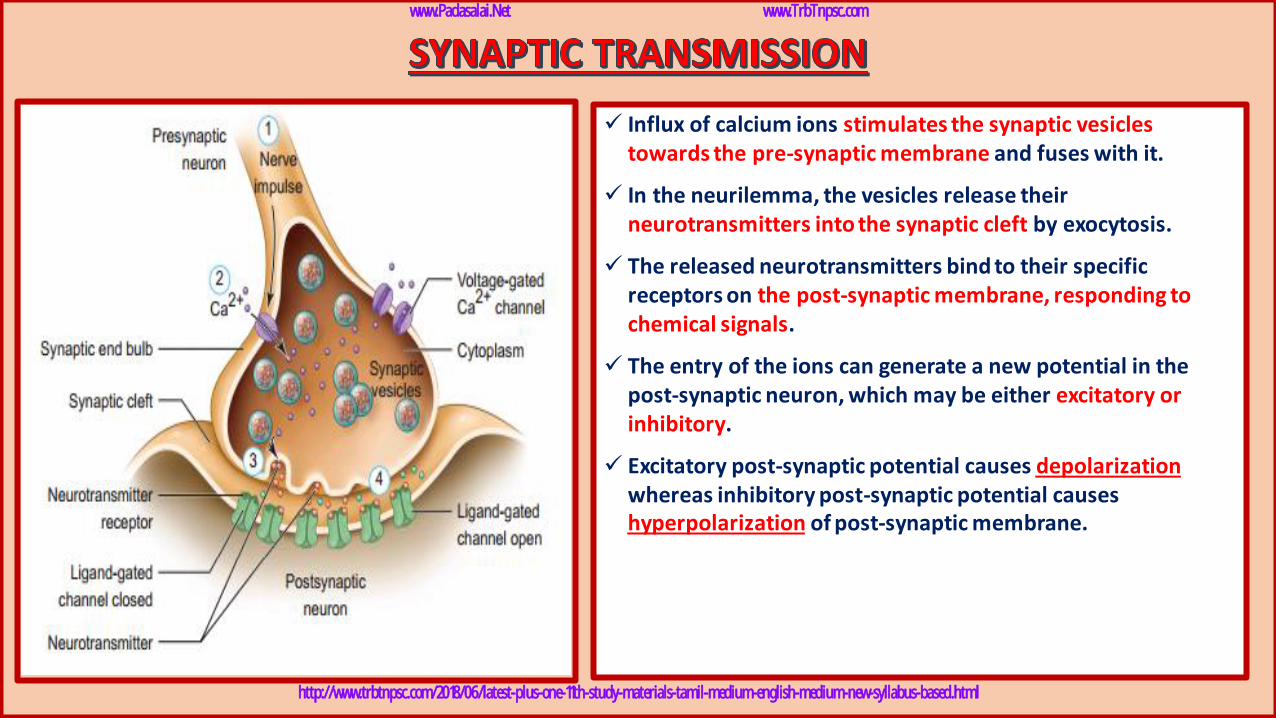
✓ Influx of calcium ions stimulates the synaptic vesicles towards the pre-synaptic membrane and fuses with it.
✓ In the neurilemma, the vesicles release their neurotransmitters into the synaptic cleft by exocytosis.
✓ The released neurotransmitters bind to their specific receptors on the post-synaptic membrane, responding to chemical signals.
✓ The entry of the ions can generate a new potential in the post-synaptic neuron, which may be either excitatory or inhibitory.
✓ Excitatory post-synaptic potential causes depolarizationwhereas inhibitory post-synaptic potential causes hyperpolarization of post-synaptic membrane.
www.Padasalai.Net www.TrbTnpsc.com
http://www.trbtnpsc.com/2018/06/latest-plus-one-11th-study-materials-tamil-medium-english-medium-new-syllabus-based.html
www.Padasalai.Net www.TrbTnpsc.com
http://www.trbtnpsc.com/2018/06/latest-plus-one-11th-study-materials-tamil-medium-english-medium-new-syllabus-based.html
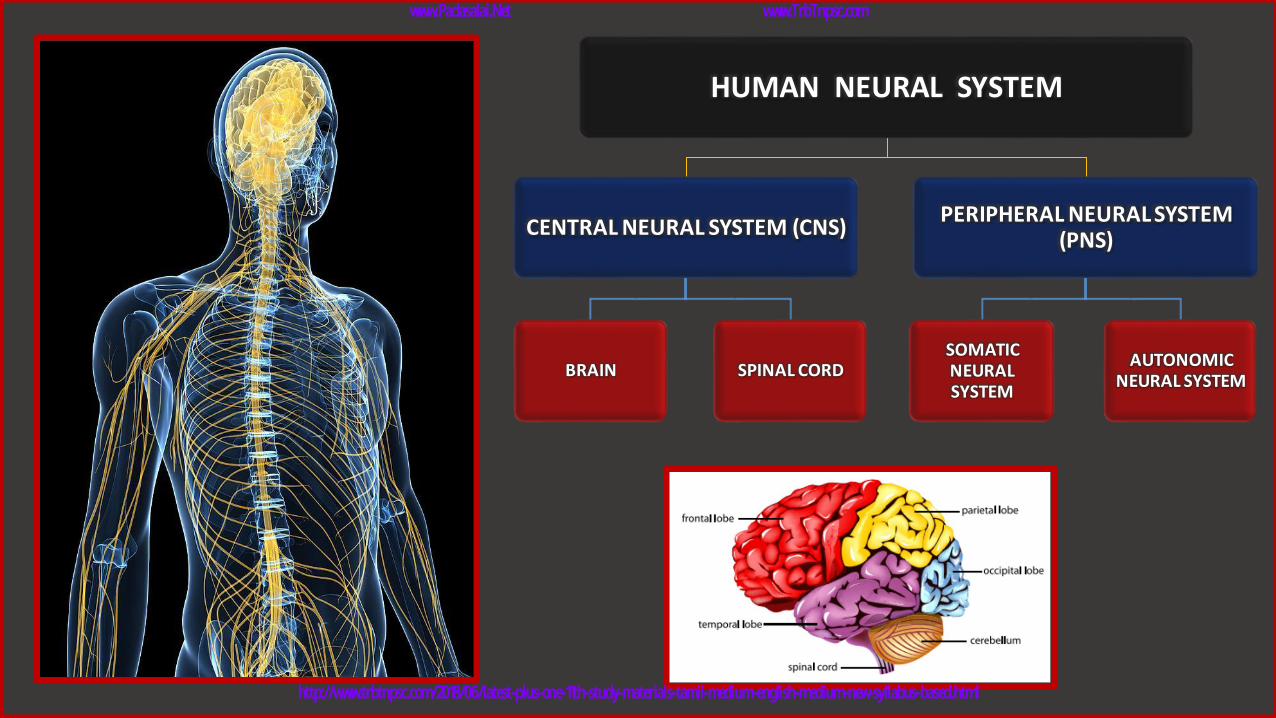
HUMAN NEURAL SYSTEM
CENTRAL NEURAL SYSTEM (CNS)
BRAIN SPINAL CORD
PERIPHERAL NEURAL SYSTEM (PNS)
SOMATIC NEURAL SYSTEM
AUTONOMIC NEURAL SYSTEM
www.Padasalai.Net www.TrbTnpsc.com
http://www.trbtnpsc.com/2018/06/latest-plus-one-11th-study-materials-tamil-medium-english-medium-new-syllabus-based.html
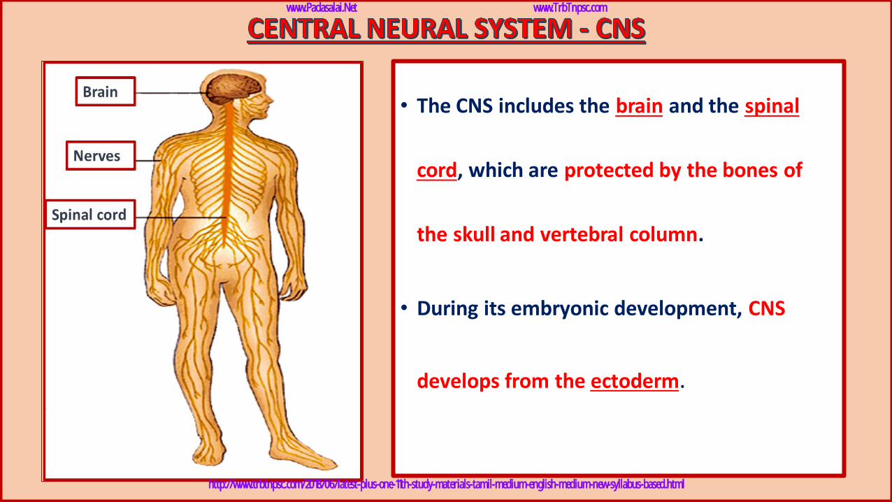
• The CNS includes the brain and the spinal
cord, which are protected by the bones of
the skull and vertebral column.
• During its embryonic development, CNS
develops from the ectoderm.
www.Padasalai.Net www.TrbTnpsc.com
http://www.trbtnpsc.com/2018/06/latest-plus-one-11th-study-materials-tamil-medium-english-medium-new-syllabus-based.html
www.Padasalai.Net www.TrbTnpsc.com
http://www.trbtnpsc.com/2018/06/latest-plus-one-11th-study-materials-tamil-medium-english-medium-new-syllabus-based.html
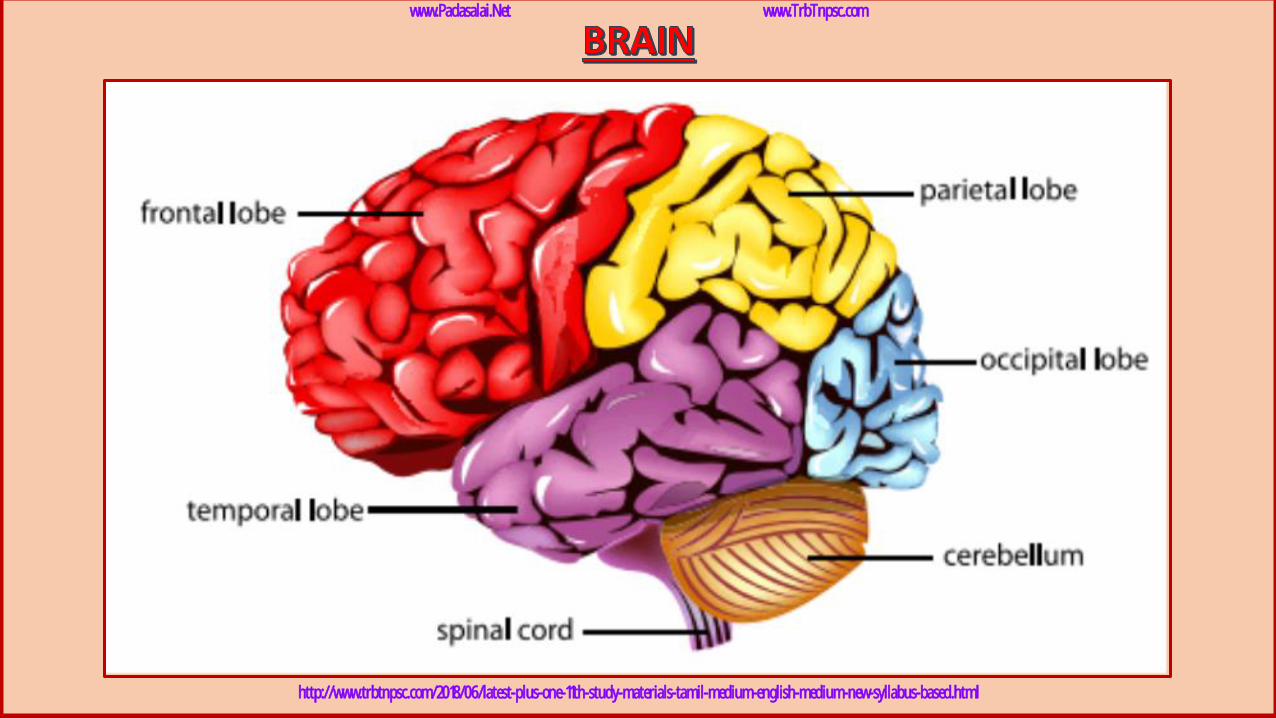
➢ The brain acts as the command and control system.
➢ It is the site of information processing.
➢ It is located in the cranial cavity and is covered by three cranial meninges.
1. The outer thick layer is Duramater which lines the inner surface
of the cranial cavity.
2. The median thin layer is Arachnoid mater which is separated from
the duramater by a narrow subdural space.
3. The innermost layer is Piamater which is closely adhered to the
brain but separated from the arachnoid mater by the subarachnoid
space.
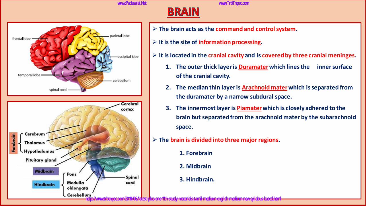
➢ The brain is divided into three major regions.
1. Forebrain
2. Midbrain
3. Hindbrain.
www.Padasalai.Net www.TrbTnpsc.com
http://www.trbtnpsc.com/2018/06/latest-plus-one-11th-study-materials-tamil-medium-english-medium-new-syllabus-based.html
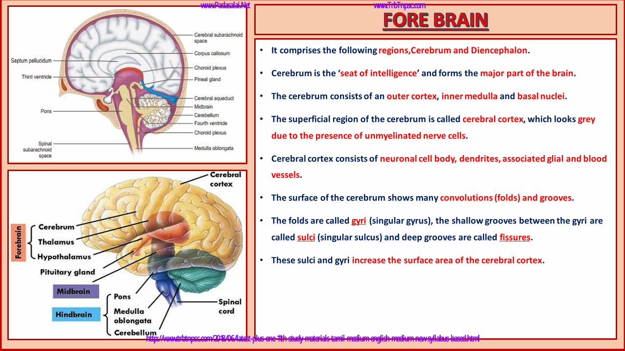
• It comprises the following regions,Cerebrum and Diencephalon.
• Cerebrum is the ‘seat of intelligence’ and forms the major part of the brain.
• The cerebrum consists of an outer cortex, inner medulla and basal nuclei.
• The superficial region of the cerebrum is called cerebral cortex, which looks grey
due to the presence of unmyelinated nerve cells.
• Cerebral cortex consists of neuronal cell body, dendrites, associated glial and blood
vessels.
• The surface of the cerebrum shows many convolutions (folds) and grooves.
• The folds are called gyri (singular gyrus), the shallow grooves between the gyri are
called sulci (singular sulcus) and deep grooves are called fissures.
• These sulci and gyri increase the surface area of the cerebral cortex.
www.Padasalai.Net www.TrbTnpsc.com
http://www.trbtnpsc.com/2018/06/latest-plus-one-11th-study-materials-tamil-medium-english-medium-new-syllabus-based.html
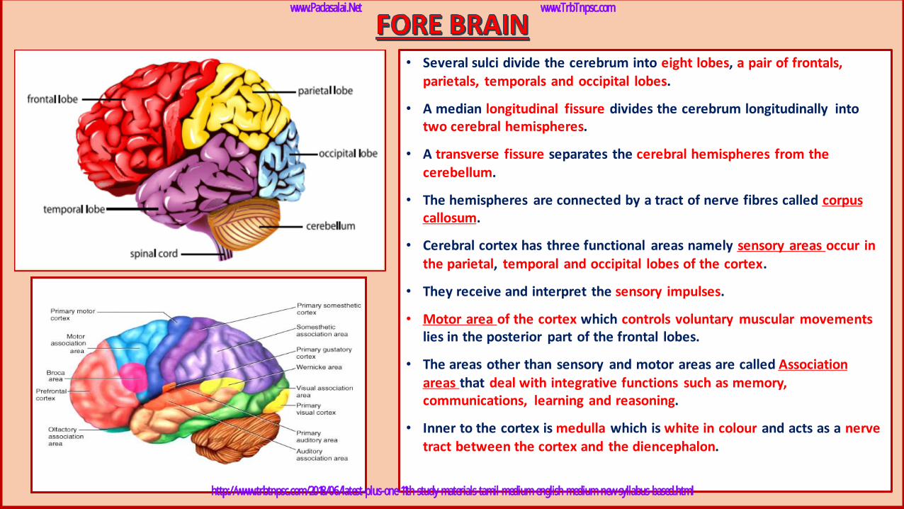
• Several sulci divide the cerebrum into eight lobes, a pair of frontals, parietals, temporals and occipital lobes.
• A median longitudinal fissure divides the cerebrum longitudinally into two cerebral hemispheres.
• A transverse fissure separates the cerebral hemispheres from the cerebellum.
• The hemispheres are connected by a tract of nerve fibres called corpus callosum.
• Cerebral cortex has three functional areas namely sensory areas occur in the parietal, temporal and occipital lobes of the cortex.
• They receive and interpret the sensory impulses.
• Motor area of the cortex which controls voluntary muscular movementslies in the posterior part of the frontal lobes.
• The areas other than sensory and motor areas are called Association areas that deal with integrative functions such as memory, communications, learning and reasoning.
• Inner to the cortex is medulla which is white in colour and acts as a nerve tract between the cortex and the diencephalon.
www.Padasalai.Net www.TrbTnpsc.com
http://www.trbtnpsc.com/2018/06/latest-plus-one-11th-study-materials-tamil-medium-english-medium-new-syllabus-based.html
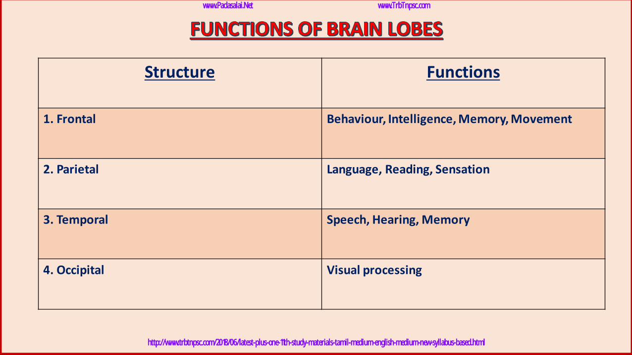
Structure Functions
1. Frontal Behaviour, Intelligence, Memory, Movement
2. Parietal Language, Reading, Sensation
3. Temporal Speech, Hearing, Memory
4. Occipital Visual processing
www.Padasalai.Net www.TrbTnpsc.com
http://www.trbtnpsc.com/2018/06/latest-plus-one-11th-study-materials-tamil-medium-english-medium-new-syllabus-based.html
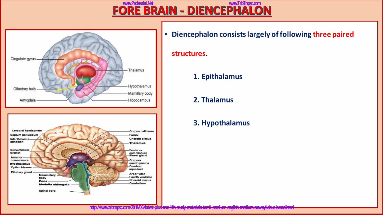
• Diencephalon consists largely of following three paired
structures.
1. Epithalamus
2. Thalamus
3. Hypothalamus
www.Padasalai.Net www.TrbTnpsc.com
http://www.trbtnpsc.com/2018/06/latest-plus-one-11th-study-materials-tamil-medium-english-medium-new-syllabus-based.html
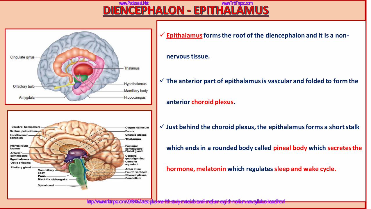
✓ Epithalamus forms the roof of the diencephalon and it is a non-
nervous tissue.
✓ The anterior part of epithalamus is vascular and folded to form the
anterior choroid plexus.
✓ Just behind the choroid plexus, the epithalamus forms a short stalk
which ends in a rounded body called pineal body which secretes the
hormone, melatonin which regulates sleep and wake cycle.
www.Padasalai.Net www.TrbTnpsc.com
http://www.trbtnpsc.com/2018/06/latest-plus-one-11th-study-materials-tamil-medium-english-medium-new-syllabus-based.html
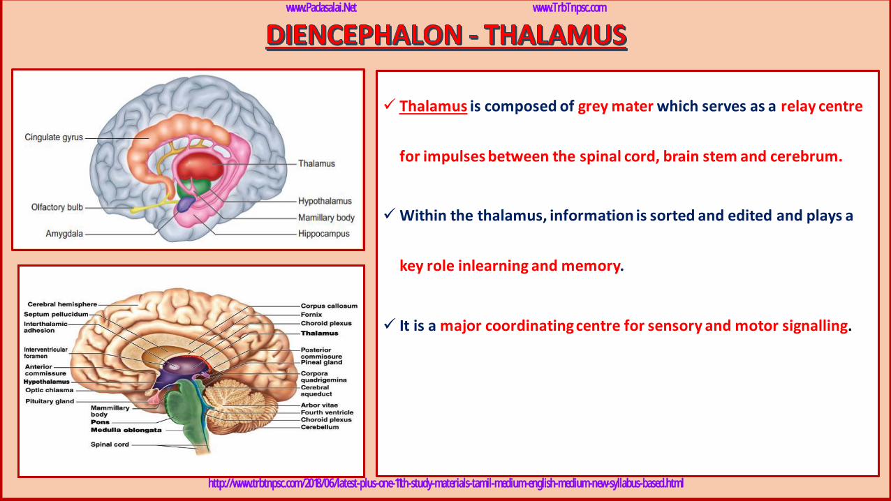
✓ Thalamus is composed of grey mater which serves as a relay centre
for impulses between the spinal cord, brain stem and cerebrum.
✓Within the thalamus, information is sorted and edited and plays a
key role inlearning and memory.
✓ It is a major coordinating centre for sensory and motor signalling.
www.Padasalai.Net www.TrbTnpsc.com
http://www.trbtnpsc.com/2018/06/latest-plus-one-11th-study-materials-tamil-medium-english-medium-new-syllabus-based.html
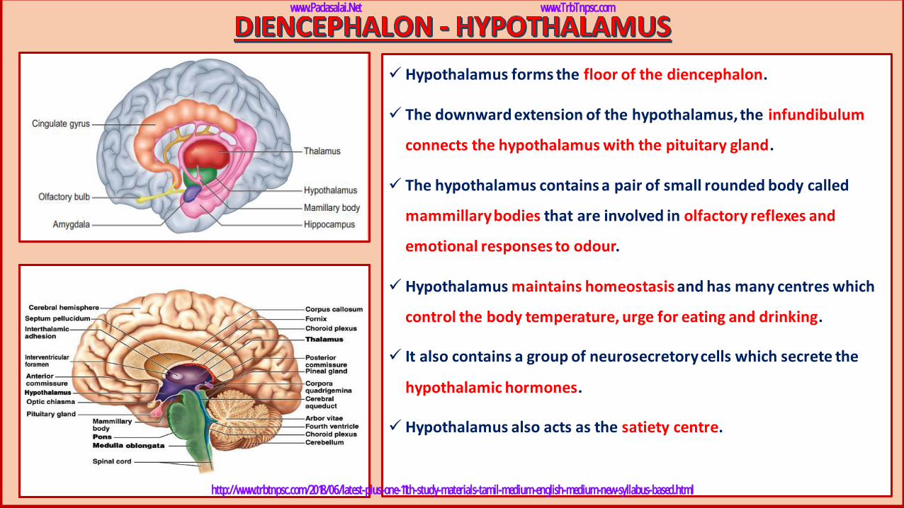
✓ Hypothalamus forms the floor of the diencephalon.
✓ The downward extension of the hypothalamus, the infundibulum
connects the hypothalamus with the pituitary gland.
✓ The hypothalamus contains a pair of small rounded body called
mammillary bodies that are involved in olfactory reflexes and
emotional responses to odour.
✓ Hypothalamus maintains homeostasis and has many centres which
control the body temperature, urge for eating and drinking.
✓ It also contains a group of neurosecretory cells which secrete the
hypothalamic hormones.
✓ Hypothalamus also acts as the satiety centre.
www.Padasalai.Net www.TrbTnpsc.com
http://www.trbtnpsc.com/2018/06/latest-plus-one-11th-study-materials-tamil-medium-english-medium-new-syllabus-based.html
www.Padasalai.Net www.TrbTnpsc.com
http://www.trbtnpsc.com/2018/06/latest-plus-one-11th-study-materials-tamil-medium-english-medium-new-syllabus-based.html

www.Padasalai.Net www.TrbTnpsc.com
http://www.trbtnpsc.com/2018/06/latest-plus-one-11th-study-materials-tamil-medium-english-medium-new-syllabus-based.html
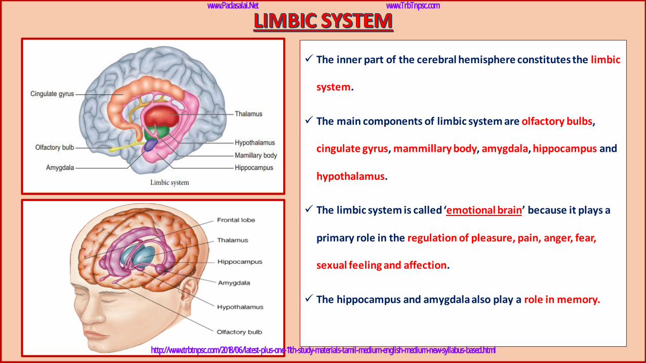
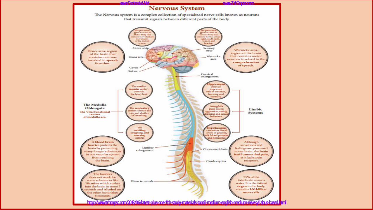
✓ The inner part of the cerebral hemisphere constitutes the limbic
system.
✓ The main components of limbic system are olfactory bulbs,
cingulate gyrus, mammillary body, amygdala, hippocampus and
hypothalamus.
✓ The limbic system is called ‘emotional brain’ because it plays a
primary role in the regulation of pleasure, pain, anger, fear,
sexual feeling and affection.
✓ The hippocampus and amygdala also play a role in memory.
www.Padasalai.Net www.TrbTnpsc.com
http://www.trbtnpsc.com/2018/06/latest-plus-one-11th-study-materials-tamil-medium-english-medium-new-syllabus-based.html
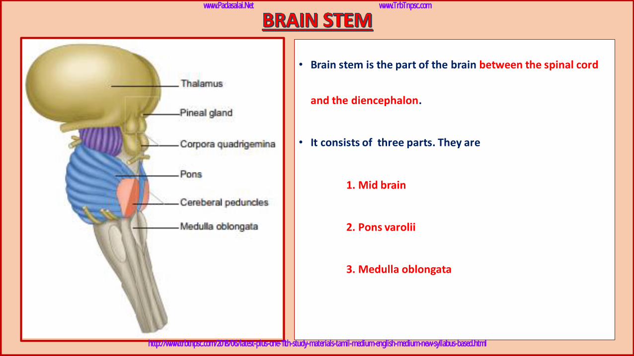
• Brain stem is the part of the brain between the spinal cord
and the diencephalon.
• It consists of three parts. They are
1. Mid brain
2. Pons varolii
3. Medulla oblongata
www.Padasalai.Net www.TrbTnpsc.com
http://www.trbtnpsc.com/2018/06/latest-plus-one-11th-study-materials-tamil-medium-english-medium-new-syllabus-based.html

✓ The mid brain is located between the diencephalon and the
pons.
✓ The lower portion of the midbrain consists of a pair of
longitudinal bands of nervous tissue called cerebral
peduncles which relay impulses back and forth between
cerebrum, cerebellum, pons and medulla.
✓ The dorsal portion of the midbrain consists of four rounded
bodies called corpora quadrigemina which acts as a reflex
centre for vision and hearing.
Cerebral
aqueduct
www.Padasalai.Net www.TrbTnpsc.com
http://www.trbtnpsc.com/2018/06/latest-plus-one-11th-study-materials-tamil-medium-english-medium-new-syllabus-based.html
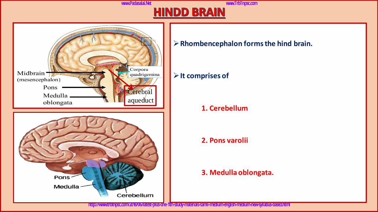
➢Rhombencephalon forms the hind brain.
➢ It comprises of
1. Cerebellum
2. Pons varolii
3. Medulla oblongata.
Cerebral
aqueduct
www.Padasalai.Net www.TrbTnpsc.com
http://www.trbtnpsc.com/2018/06/latest-plus-one-11th-study-materials-tamil-medium-english-medium-new-syllabus-based.html
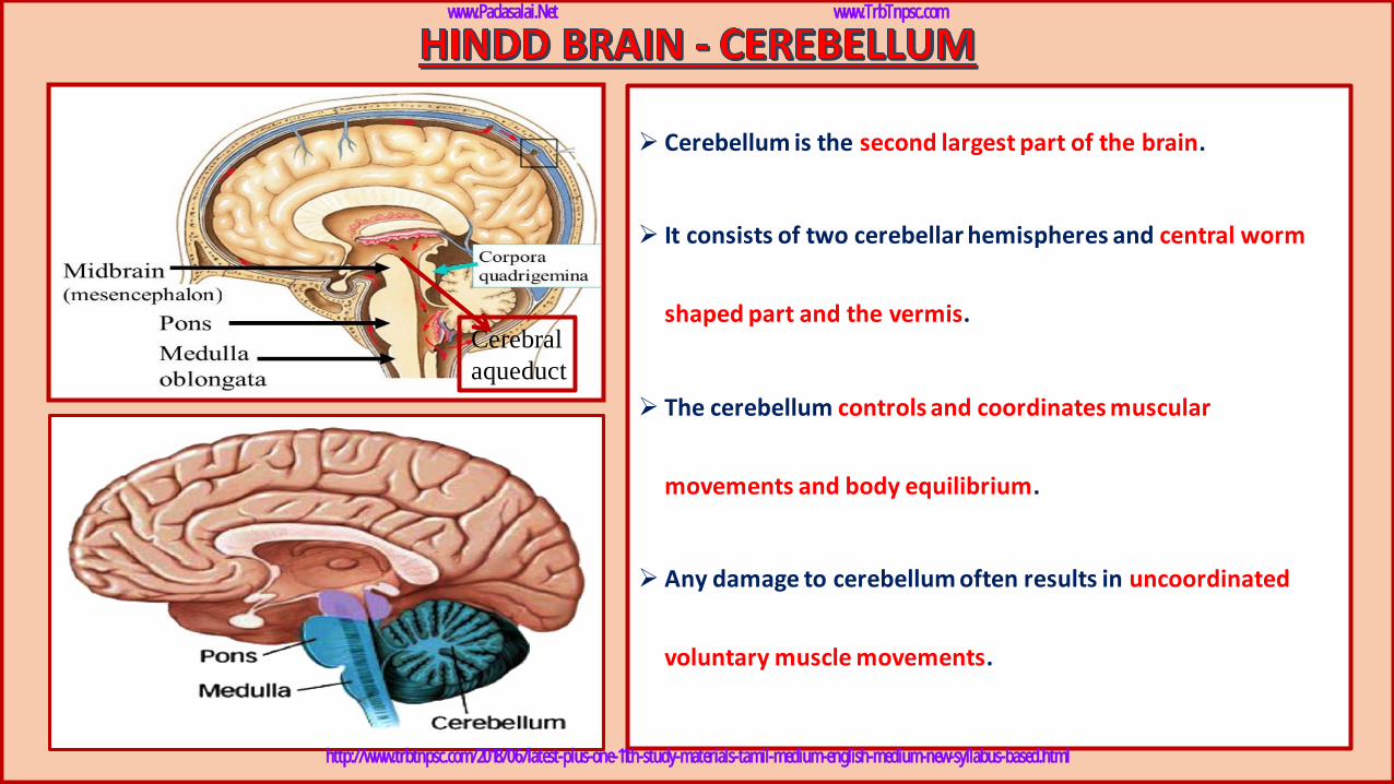
➢ Cerebellum is the second largest part of the brain.
➢ It consists of two cerebellar hemispheres and central worm
shaped part and the vermis.
➢ The cerebellum controls and coordinates muscular
movements and body equilibrium.
➢ Any damage to cerebellum often results in uncoordinated
voluntary muscle movements.
Cerebral
aqueduct
www.Padasalai.Net www.TrbTnpsc.com
http://www.trbtnpsc.com/2018/06/latest-plus-one-11th-study-materials-tamil-medium-english-medium-new-syllabus-based.html
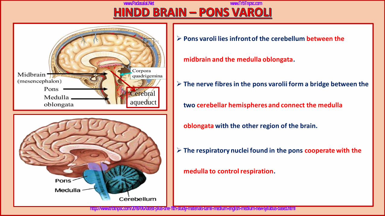
➢ Pons varoli lies infrontof the cerebellum between the
midbrain and the medulla oblongata.
➢ The nerve fibres in the pons varolii form a bridge between the
two cerebellar hemispheres and connect the medulla
oblongata with the other region of the brain.
➢ The respiratory nuclei found in the pons cooperate with the
medulla to control respiration.
Cerebral
aqueduct
www.Padasalai.Net www.TrbTnpsc.com
http://www.trbtnpsc.com/2018/06/latest-plus-one-11th-study-materials-tamil-medium-english-medium-new-syllabus-based.html
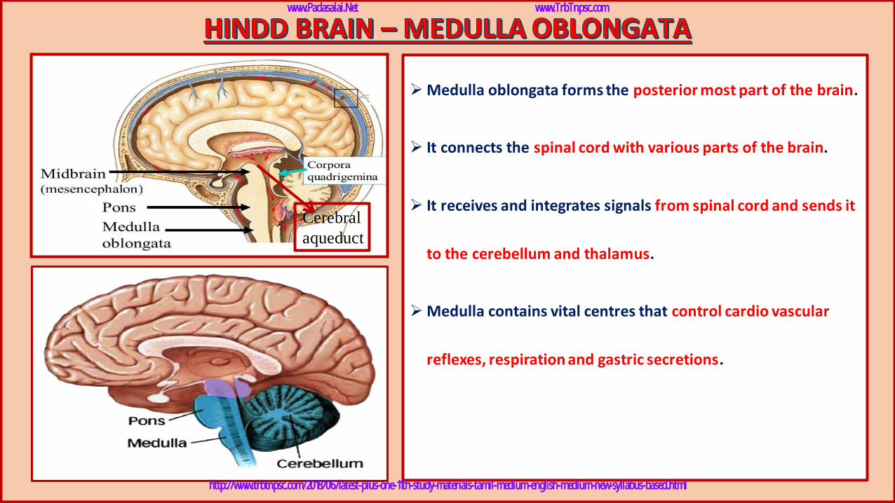
➢Medulla oblongata forms the posterior most part of the brain.
➢ It connects the spinal cord with various parts of the brain.
➢ It receives and integrates signals from spinal cord and sends it
to the cerebellum and thalamus.
➢Medulla contains vital centres that control cardio vascular
reflexes, respiration and gastric secretions.
Cerebral
aqueduct
www.Padasalai.Net www.TrbTnpsc.com
http://www.trbtnpsc.com/2018/06/latest-plus-one-11th-study-materials-tamil-medium-english-medium-new-syllabus-based.html
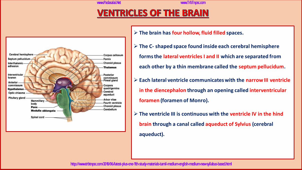
➢ The brain has four hollow, fluid filled spaces.
➢ The C- shaped space found inside each cerebral hemisphere
forms the lateral ventricles I and II which are separated from
each other by a thin membrane called the septum pellucidum.
➢ Each lateral ventricle communicates with the narrow III ventricle
in the diencephalon through an opening called interventricular
foramen (foramen of Monro).
➢ The ventricle III is continuous with the ventricle IV in the hind
brain through a canal called aqueduct of Sylvius (cerebral
aqueduct).
www.Padasalai.Net www.TrbTnpsc.com
http://www.trbtnpsc.com/2018/06/latest-plus-one-11th-study-materials-tamil-medium-english-medium-new-syllabus-based.html
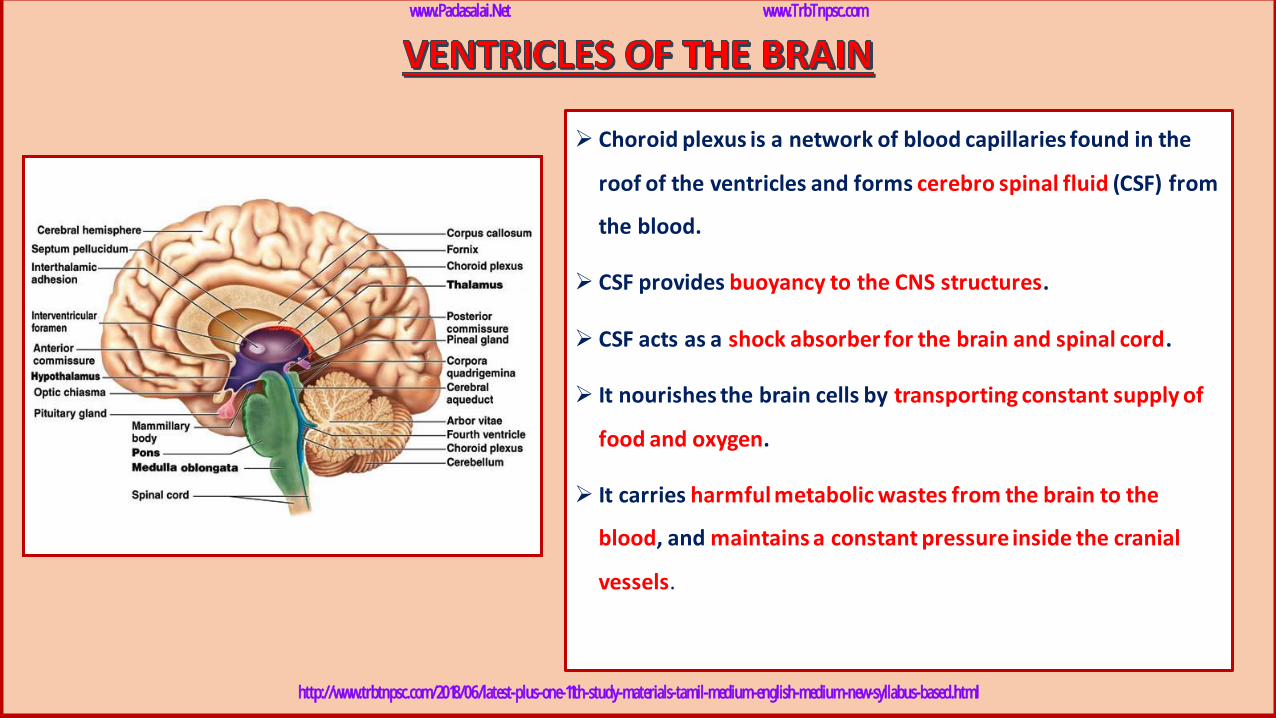
➢ Choroid plexus is a network of blood capillaries found in the
roof of the ventricles and forms cerebro spinal fluid (CSF) from
the blood.
➢ CSF provides buoyancy to the CNS structures.
➢ CSF acts as a shock absorber for the brain and spinal cord.
➢ It nourishes the brain cells by transporting constant supply of
food and oxygen.
➢ It carries harmful metabolic wastes from the brain to the
blood, and maintains a constant pressure inside the cranial
vessels.
www.Padasalai.Net www.TrbTnpsc.com
http://www.trbtnpsc.com/2018/06/latest-plus-one-11th-study-materials-tamil-medium-english-medium-new-syllabus-based.html
www.Padasalai.Net www.TrbTnpsc.com
http://www.trbtnpsc.com/2018/06/latest-plus-one-11th-study-materials-tamil-medium-english-medium-new-syllabus-based.html
www.Padasalai.Net www.TrbTnpsc.com
http://www.trbtnpsc.com/2018/06/latest-plus-one-11th-study-materials-tamil-medium-english-medium-new-syllabus-based.html
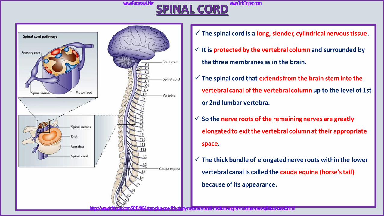
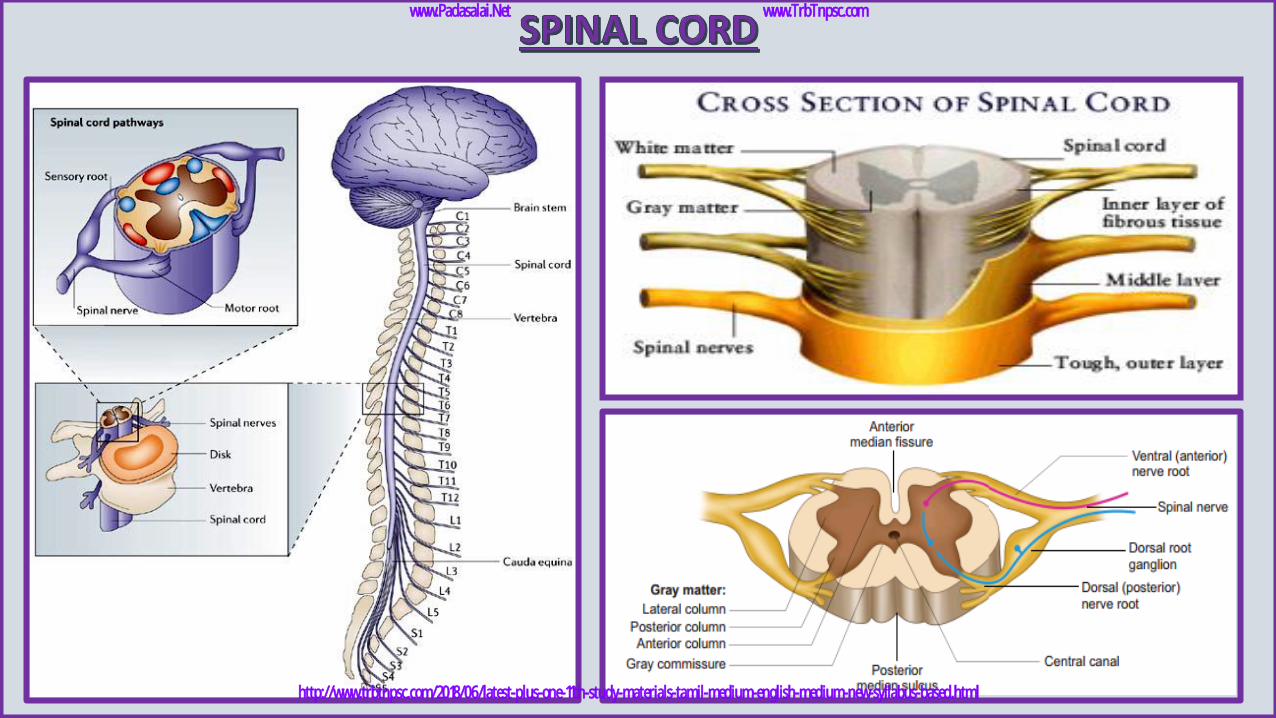
✓ The spinal cord is a long, slender, cylindrical nervous tissue.
✓ It is protected by the vertebral column and surrounded by
the three membranes as in the brain.
✓ The spinal cord that extends from the brain stem into the
vertebral canal of the vertebral column up to the level of 1st
or 2nd lumbar vertebra.
✓ So the nerve roots of the remaining nerves are greatly
elongated to exit the vertebral column at their appropriate
space.
✓ The thick bundle of elongated nerve roots within the lower
vertebral canal is called the cauda equina (horse’s tail)
because of its appearance.
www.Padasalai.Net www.TrbTnpsc.com
http://www.trbtnpsc.com/2018/06/latest-plus-one-11th-study-materials-tamil-medium-english-medium-new-syllabus-based.html
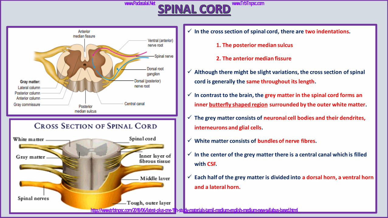
✓ In the cross section of spinal cord, there are two indentations.
1. The posterior median sulcus
2. The anterior median fissure
✓ Although there might be slight variations, the cross section of spinal
cord is generally the same throughout its length.
✓ In contrast to the brain, the grey matter in the spinal cord forms an
inner butterfly shaped region surrounded by the outer white matter.
✓ The grey matter consists of neuronal cell bodies and their dendrites,
interneurons and glial cells.
✓ White matter consists of bundles of nerve fibres.
✓ In the center of the grey matter there is a central canal which is filled
with CSF.
✓ Each half of the grey matter is divided into a dorsal horn, a ventral horn
and a lateral horn.
www.Padasalai.Net www.TrbTnpsc.com
http://www.trbtnpsc.com/2018/06/latest-plus-one-11th-study-materials-tamil-medium-english-medium-new-syllabus-based.html
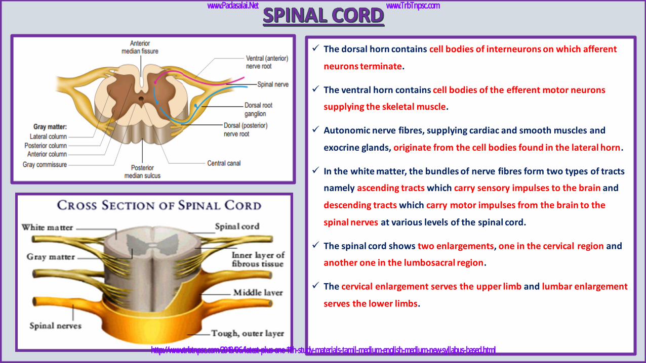
✓ The dorsal horn contains cell bodies of interneurons on which afferent
neurons terminate.
✓ The ventral horn contains cell bodies of the efferent motor neurons
supplying the skeletal muscle.
✓ Autonomic nerve fibres, supplying cardiac and smooth muscles and
exocrine glands, originate from the cell bodies found in the lateral horn.
✓ In the white matter, the bundles of nerve fibres form two types of tracts
namely ascending tracts which carry sensory impulses to the brain and
descending tracts which carry motor impulses from the brain to the
spinal nerves at various levels of the spinal cord.
✓ The spinal cord shows two enlargements, one in the cervical region and
another one in the lumbosacral region.
✓ The cervical enlargement serves the upper limb and lumbar enlargement
serves the lower limbs.
www.Padasalai.Net www.TrbTnpsc.com
http://www.trbtnpsc.com/2018/06/latest-plus-one-11th-study-materials-tamil-medium-english-medium-new-syllabus-based.html

www.Padasalai.Net www.TrbTnpsc.com
http://www.trbtnpsc.com/2018/06/latest-plus-one-11th-study-materials-tamil-medium-english-medium-new-syllabus-based.html
www.Padasalai.Net www.TrbTnpsc.com
http://www.trbtnpsc.com/2018/06/latest-plus-one-11th-study-materials-tamil-medium-english-medium-new-syllabus-based.html
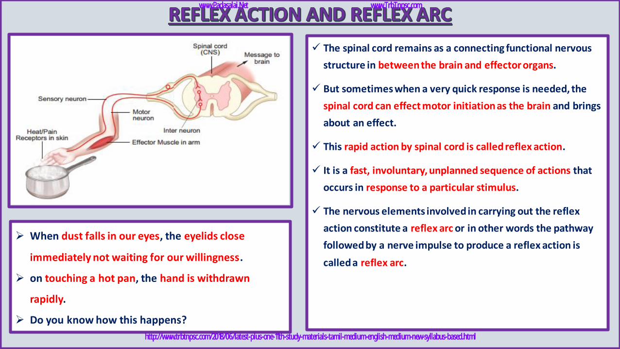
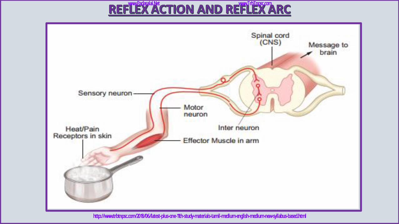
✓ The spinal cord remains as a connecting functional nervous
structure in between the brain and effector organs.
✓ But sometimes when a very quick response is needed, the
spinal cord can effect motor initiation as the brain and brings
about an effect.
✓ This rapid action by spinal cord is called reflex action.
✓ It is a fast, involuntary, unplanned sequence of actions that
occurs in response to a particular stimulus.
✓ The nervous elements involved in carrying out the reflex
action constitute a reflex arc or in other words the pathway
followed by a nerve impulse to produce a reflex action is
called a reflex arc.
➢ When dust falls in our eyes, the eyelids close
immediately not waiting for our willingness.
➢ on touching a hot pan, the hand is withdrawn
rapidly.
➢ Do you know how this happens?
www.Padasalai.Net www.TrbTnpsc.com
http://www.trbtnpsc.com/2018/06/latest-plus-one-11th-study-materials-tamil-medium-english-medium-new-syllabus-based.html
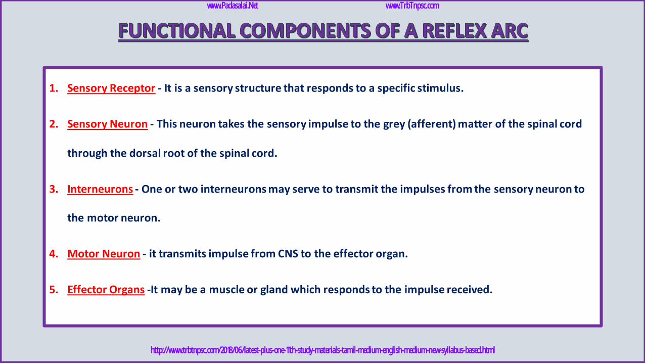
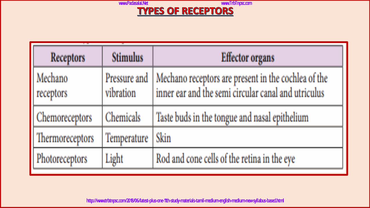
1. Sensory Receptor - It is a sensory structure that responds to a specific stimulus.
2. Sensory Neuron - This neuron takes the sensory impulse to the grey (afferent) matter of the spinal cord
through the dorsal root of the spinal cord.
3. Interneurons - One or two interneurons may serve to transmit the impulses from the sensory neuron to
the motor neuron.
4. Motor Neuron - it transmits impulse from CNS to the effector organ.
5. Effector Organs -It may be a muscle or gland which responds to the impulse received.
www.Padasalai.Net www.TrbTnpsc.com
http://www.trbtnpsc.com/2018/06/latest-plus-one-11th-study-materials-tamil-medium-english-medium-new-syllabus-based.html
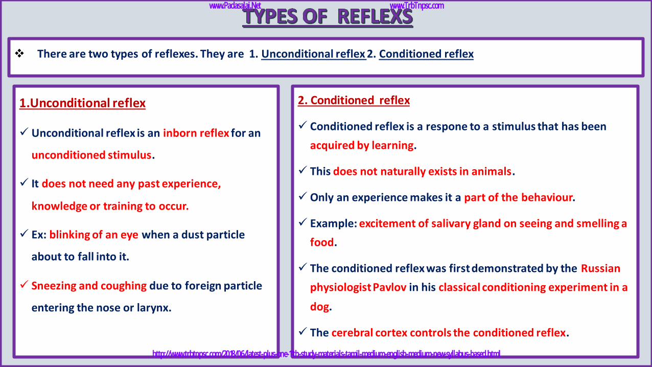
❖ There are two types of reflexes. They are 1. Unconditional reflex 2. Conditioned reflex
1.Unconditional reflex
✓ Unconditional reflex is an inborn reflex for an
unconditioned stimulus.
✓ It does not need any past experience,
knowledge or training to occur.
✓ Ex: blinking of an eye when a dust particle
about to fall into it.
✓ Sneezing and coughing due to foreign particle
entering the nose or larynx.
2. Conditioned reflex
✓ Conditioned reflex is a respone to a stimulus that has been
acquired by learning.
✓ This does not naturally exists in animals.
✓ Only an experience makes it a part of the behaviour.
✓ Example: excitement of salivary gland on seeing and smelling a
food.
✓ The conditioned reflex was first demonstrated by the Russian
physiologist Pavlov in his classical conditioning experiment in a
dog.
✓ The cerebral cortex controls the conditioned reflex.
www.Padasalai.Net www.TrbTnpsc.com
http://www.trbtnpsc.com/2018/06/latest-plus-one-11th-study-materials-tamil-medium-english-medium-new-syllabus-based.html
www.Padasalai.Net www.TrbTnpsc.com
http://www.trbtnpsc.com/2018/06/latest-plus-one-11th-study-materials-tamil-medium-english-medium-new-syllabus-based.html
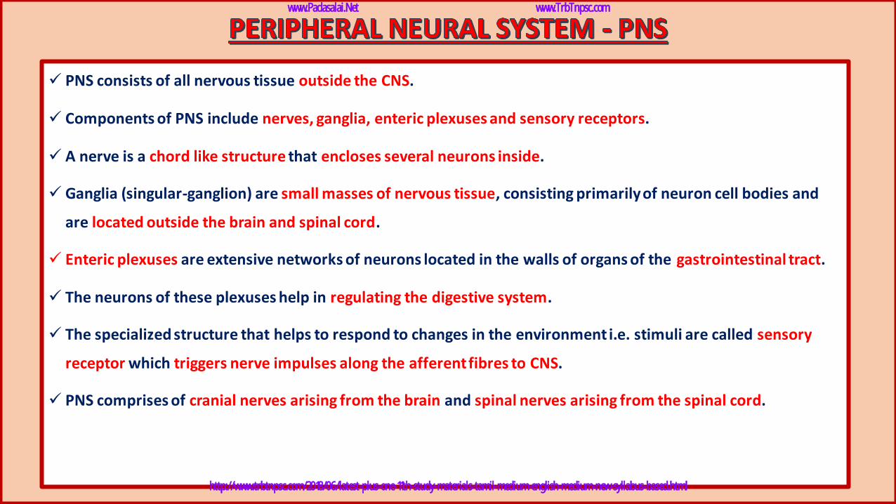
✓ PNS consists of all nervous tissue outside the CNS.
✓ Components of PNS include nerves, ganglia, enteric plexuses and sensory receptors.
✓ A nerve is a chord like structure that encloses several neurons inside.
✓ Ganglia (singular-ganglion) are small masses of nervous tissue, consisting primarily of neuron cell bodies and
are located outside the brain and spinal cord.
✓ Enteric plexuses are extensive networks of neurons located in the walls of organs of the gastrointestinal tract.
✓ The neurons of these plexuses help in regulating the digestive system.
✓ The specialized structure that helps to respond to changes in the environment i.e. stimuli are called sensory
receptor which triggers nerve impulses along the afferent fibres to CNS.
✓ PNS comprises of cranial nerves arising from the brain and spinal nerves arising from the spinal cord.
www.Padasalai.Net www.TrbTnpsc.com
http://www.trbtnpsc.com/2018/06/latest-plus-one-11th-study-materials-tamil-medium-english-medium-new-syllabus-based.html
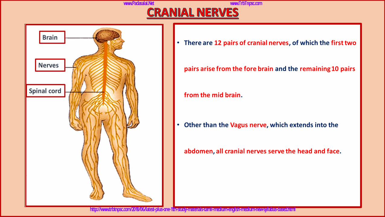
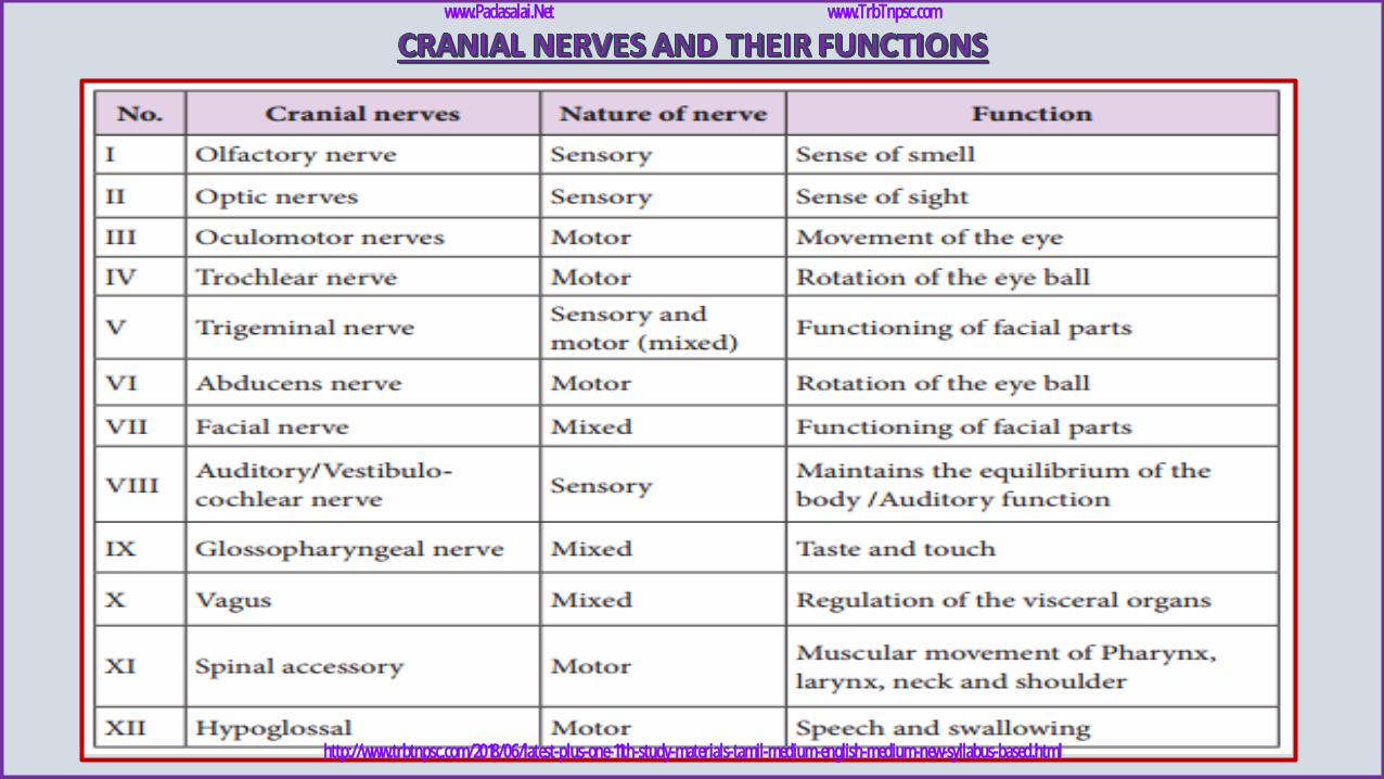
• There are 12 pairs of cranial nerves, of which the first two
pairs arise from the fore brain and the remaining 10 pairs
from the mid brain.
• Other than the Vagus nerve, which extends into the
abdomen, all cranial nerves serve the head and face.
www.Padasalai.Net www.TrbTnpsc.com
http://www.trbtnpsc.com/2018/06/latest-plus-one-11th-study-materials-tamil-medium-english-medium-new-syllabus-based.html
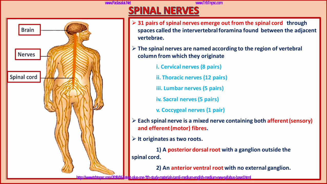
➢ 31 pairs of spinal nerves emerge out from the spinal cord through spaces called the intervertebral foramina found between the adjacent vertebrae.
➢ The spinal nerves are named according to the region of vertebral column from which they originate
i. Cervical nerves (8 pairs)
ii. Thoracic nerves (12 pairs)
iii. Lumbar nerves (5 pairs)
iv. Sacral nerves (5 pairs)
v. Coccygeal nerves (1 pair)
➢ Each spinal nerve is a mixed nerve containing both afferent (sensory) and efferent (motor) fibres.
➢ It originates as two roots.
1) A posterior dorsal root with a ganglion outside the spinal cord.
2) An anterior ventral root with no external ganglion.
www.Padasalai.Net www.TrbTnpsc.com
http://www.trbtnpsc.com/2018/06/latest-plus-one-11th-study-materials-tamil-medium-english-medium-new-syllabus-based.html
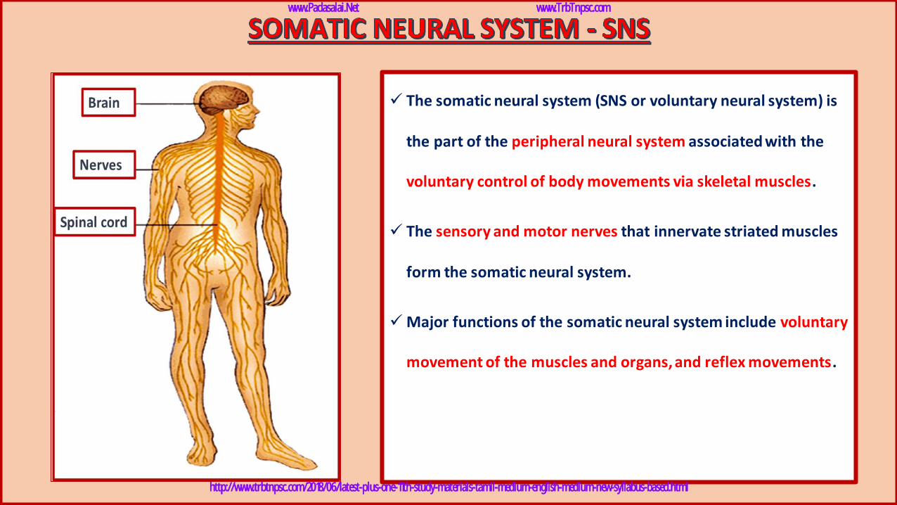
✓ The somatic neural system (SNS or voluntary neural system) is
the part of the peripheral neural system associated with the
voluntary control of body movements via skeletal muscles.
✓ The sensory and motor nerves that innervate striated muscles
form the somatic neural system.
✓Major functions of the somatic neural system include voluntary
movement of the muscles and organs, and reflex movements.
www.Padasalai.Net www.TrbTnpsc.com
http://www.trbtnpsc.com/2018/06/latest-plus-one-11th-study-materials-tamil-medium-english-medium-new-syllabus-based.html
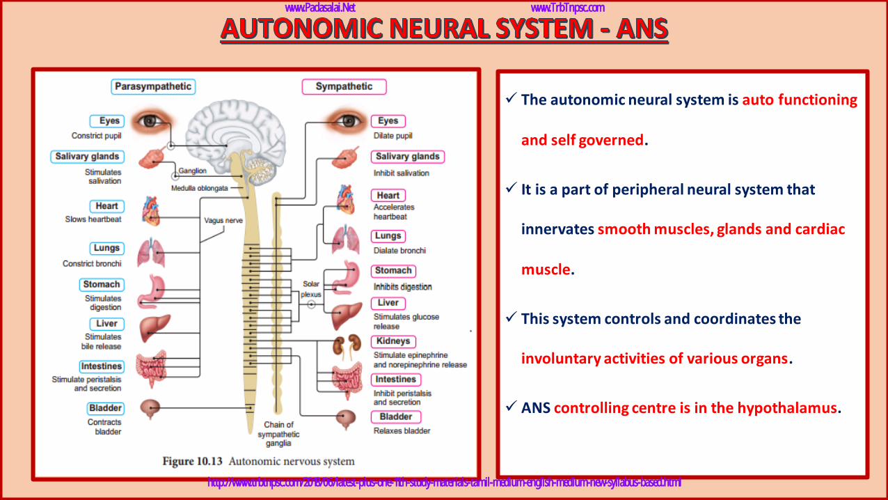
✓ The autonomic neural system is auto functioning
and self governed.
✓ It is a part of peripheral neural system that
innervates smooth muscles, glands and cardiac
muscle.
✓ This system controls and coordinates the
involuntary activities of various organs.
✓ ANS controlling centre is in the hypothalamus.
www.Padasalai.Net www.TrbTnpsc.com
http://www.trbtnpsc.com/2018/06/latest-plus-one-11th-study-materials-tamil-medium-english-medium-new-syllabus-based.html
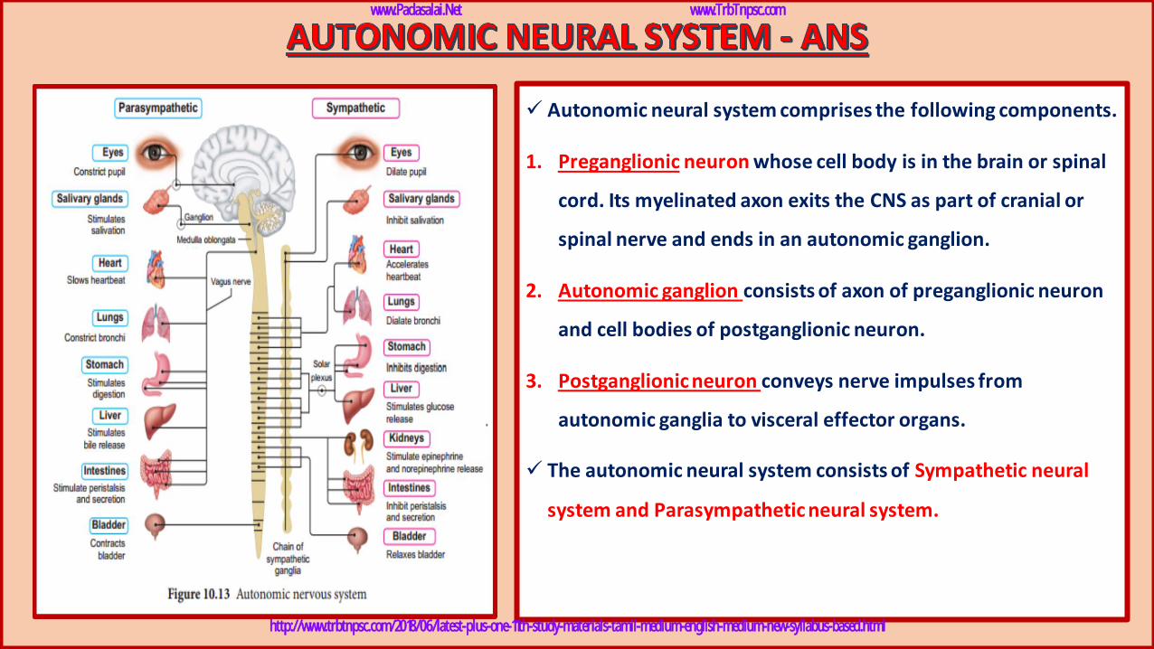
✓ Autonomic neural system comprises the following components.
1. Preganglionic neuron whose cell body is in the brain or spinal
cord. Its myelinated axon exits the CNS as part of cranial or
spinal nerve and ends in an autonomic ganglion.
2. Autonomic ganglion consists of axon of preganglionic neuron
and cell bodies of postganglionic neuron.
3. Postganglionic neuron conveys nerve impulses from
autonomic ganglia to visceral effector organs.
✓ The autonomic neural system consists of Sympathetic neural
system and Parasympathetic neural system.
www.Padasalai.Net www.TrbTnpsc.com
http://www.trbtnpsc.com/2018/06/latest-plus-one-11th-study-materials-tamil-medium-english-medium-new-syllabus-based.html
www.Padasalai.Net www.TrbTnpsc.com
http://www.trbtnpsc.com/2018/06/latest-plus-one-11th-study-materials-tamil-medium-english-medium-new-syllabus-based.html
www.Padasalai.Net www.TrbTnpsc.com
http://www.trbtnpsc.com/2018/06/latest-plus-one-11th-study-materials-tamil-medium-english-medium-new-syllabus-based.html
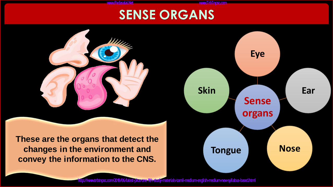
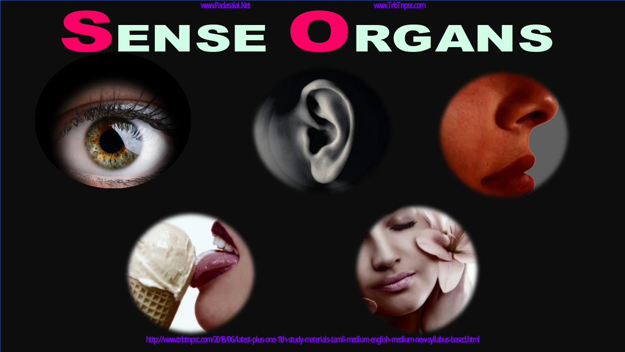
These are the organs that detect the
changes in the environment and
convey the information to the CNS.
SENSE ORGANS
Sense organs
Eye
Ear
Nose Tongue
Skin
www.Padasalai.Net www.TrbTnpsc.com
http://www.trbtnpsc.com/2018/06/latest-plus-one-11th-study-materials-tamil-medium-english-medium-new-syllabus-based.html
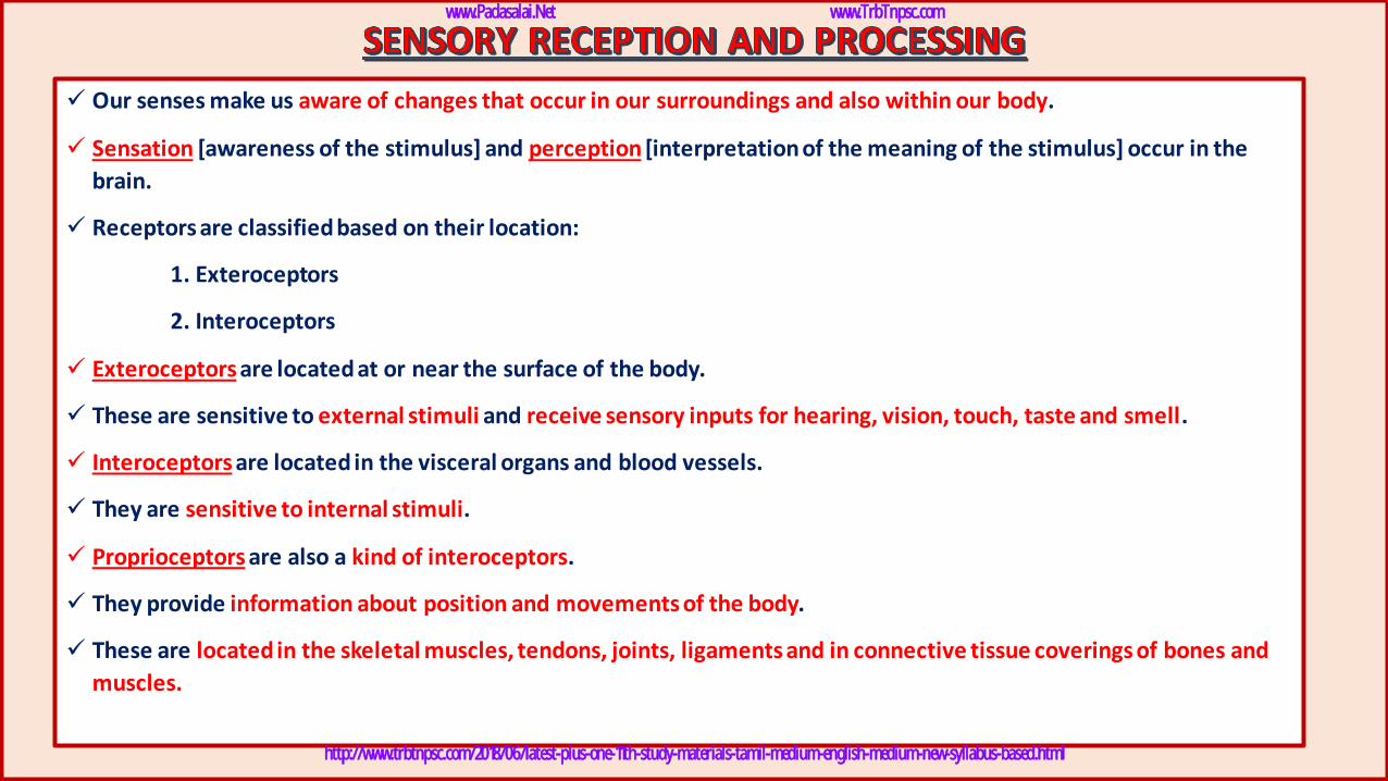
✓ Our senses make us aware of changes that occur in our surroundings and also within our body.
✓ Sensation [awareness of the stimulus] and perception [interpretation of the meaning of the stimulus] occur in the
brain.
✓ Receptors are classified based on their location:
1. Exteroceptors
2. Interoceptors
✓ Exteroceptors are located at or near the surface of the body.
✓ These are sensitive to external stimuli and receive sensory inputs for hearing, vision, touch, taste and smell.
✓ Interoceptors are located in the visceral organs and blood vessels.
✓ They are sensitive to internal stimuli.
✓ Proprioceptors are also a kind of interoceptors.
✓ They provide information about position and movements of the body.
✓ These are located in the skeletal muscles, tendons, joints, ligaments and in connective tissue coverings of bones and
muscles.
www.Padasalai.Net www.TrbTnpsc.com
http://www.trbtnpsc.com/2018/06/latest-plus-one-11th-study-materials-tamil-medium-english-medium-new-syllabus-based.html
www.Padasalai.Net www.TrbTnpsc.com
http://www.trbtnpsc.com/2018/06/latest-plus-one-11th-study-materials-tamil-medium-english-medium-new-syllabus-based.html
www.Padasalai.Net www.TrbTnpsc.com
http://www.trbtnpsc.com/2018/06/latest-plus-one-11th-study-materials-tamil-medium-english-medium-new-syllabus-based.html
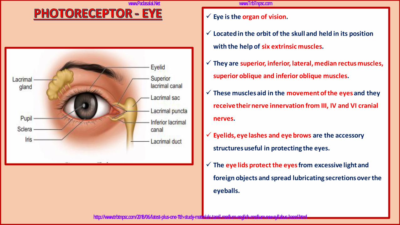
✓ Eye is the organ of vision.
✓ Located in the orbit of the skull and held in its position
with the help of six extrinsic muscles.
✓ They are superior, inferior, lateral, median rectus muscles,
superior oblique and inferior oblique muscles.
✓ These muscles aid in the movement of the eyes and they
receive their nerve innervation from III, IV and VI cranial
nerves.
✓ Eyelids, eye lashes and eye brows are the accessory
structures useful in protecting the eyes.
✓ The eye lids protect the eyes from excessive light and
foreign objects and spread lubricating secretions over the
eyeballs.
www.Padasalai.Net www.TrbTnpsc.com
http://www.trbtnpsc.com/2018/06/latest-plus-one-11th-study-materials-tamil-medium-english-medium-new-syllabus-based.html
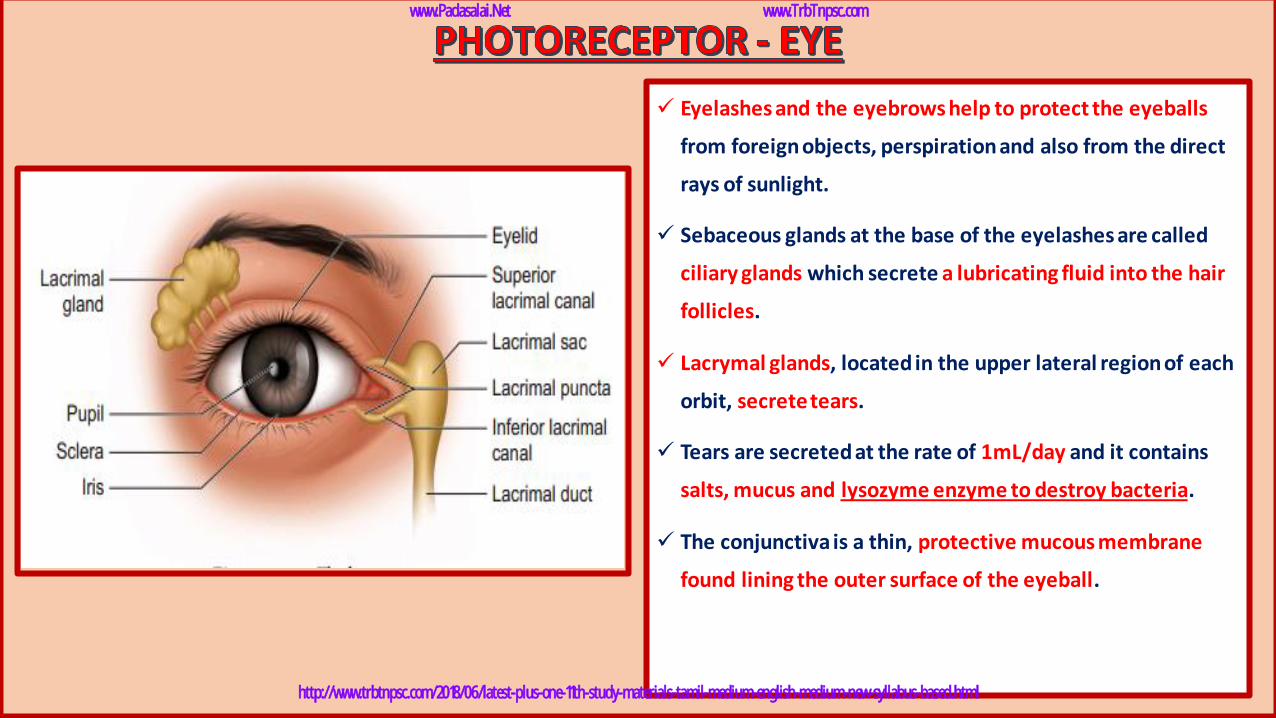
✓ Eyelashes and the eyebrows help to protect the eyeballs
from foreign objects, perspiration and also from the direct
rays of sunlight.
✓ Sebaceous glands at the base of the eyelashes are called
ciliary glands which secrete a lubricating fluid into the hair
follicles.
✓ Lacrymal glands, located in the upper lateral region of each
orbit, secrete tears.
✓ Tears are secreted at the rate of 1mL/day and it contains
salts, mucus and lysozyme enzyme to destroy bacteria.
✓ The conjunctiva is a thin, protective mucous membrane
found lining the outer surface of the eyeball.
www.Padasalai.Net www.TrbTnpsc.com
http://www.trbtnpsc.com/2018/06/latest-plus-one-11th-study-materials-tamil-medium-english-medium-new-syllabus-based.html
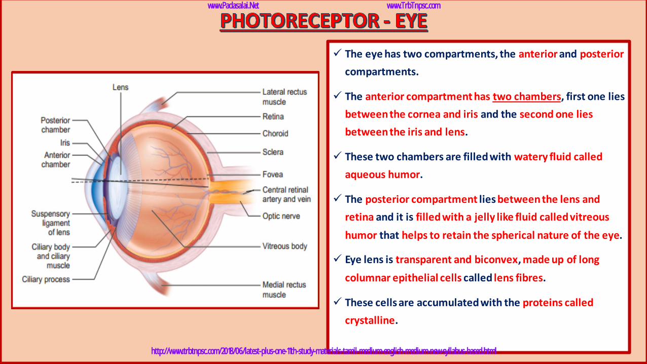
✓ The eye has two compartments, the anterior and posterior
compartments.
✓ The anterior compartment has two chambers, first one lies
between the cornea and iris and the second one lies
between the iris and lens.
✓ These two chambers are filled with watery fluid called
aqueous humor.
✓ The posterior compartment lies between the lens and
retina and it is filled with a jelly like fluid called vitreous
humor that helps to retain the spherical nature of the eye.
✓ Eye lens is transparent and biconvex, made up of long
columnar epithelial cells called lens fibres.
✓ These cells are accumulated with the proteins called
crystalline.
www.Padasalai.Net www.TrbTnpsc.com
http://www.trbtnpsc.com/2018/06/latest-plus-one-11th-study-materials-tamil-medium-english-medium-new-syllabus-based.html
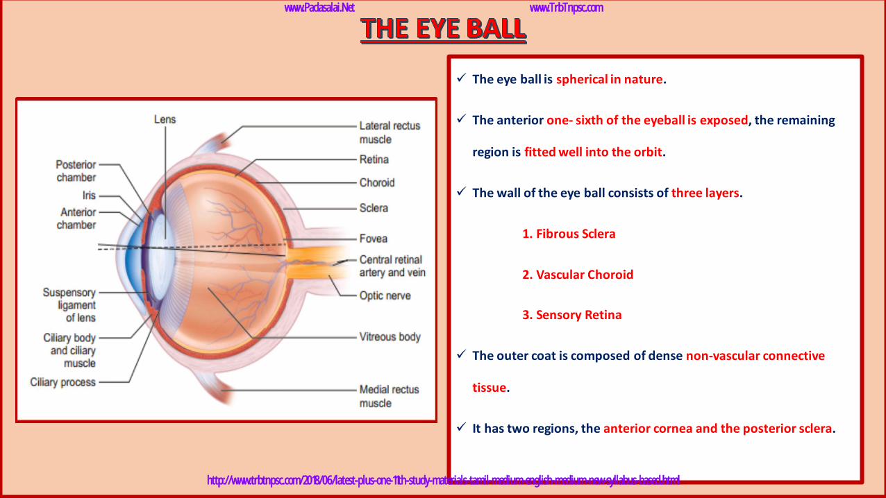
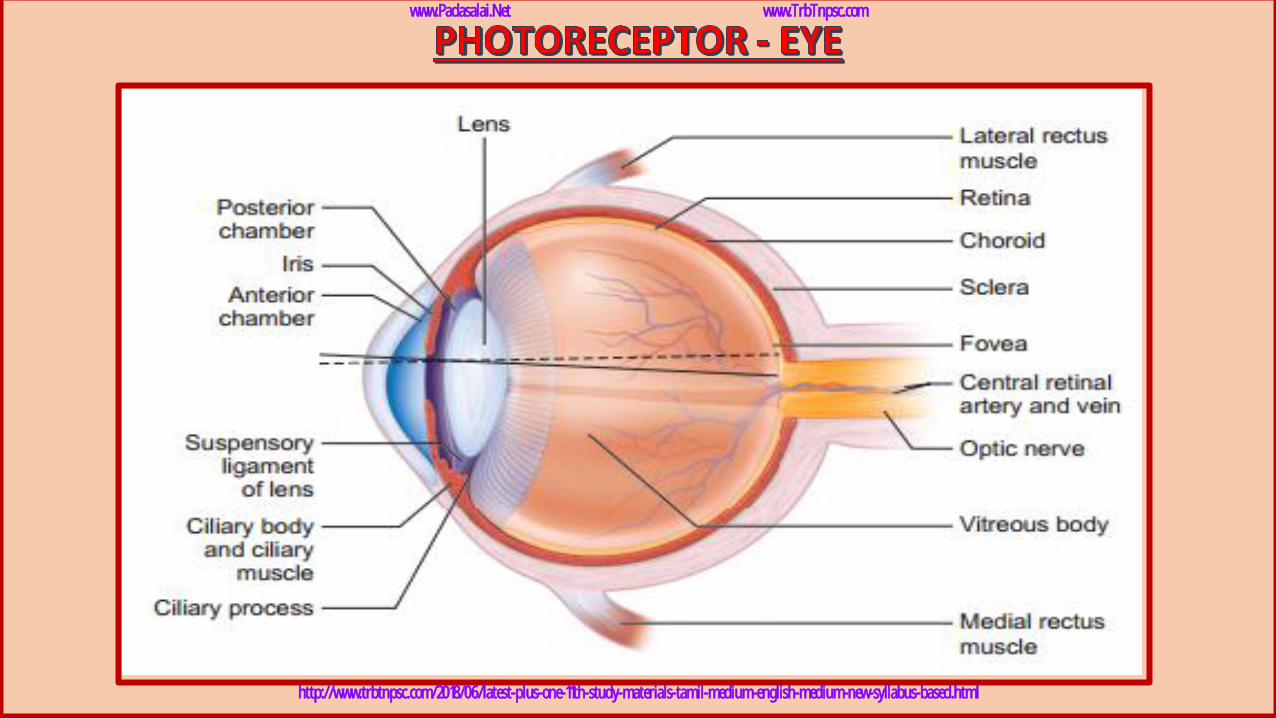
✓ The eye ball is spherical in nature.
✓ The anterior one- sixth of the eyeball is exposed, the remaining
region is fitted well into the orbit.
✓ The wall of the eye ball consists of three layers.
1. Fibrous Sclera
2. Vascular Choroid
3. Sensory Retina
✓ The outer coat is composed of dense non-vascular connective
tissue.
✓ It has two regions, the anterior cornea and the posterior sclera.
www.Padasalai.Net www.TrbTnpsc.com
http://www.trbtnpsc.com/2018/06/latest-plus-one-11th-study-materials-tamil-medium-english-medium-new-syllabus-based.html
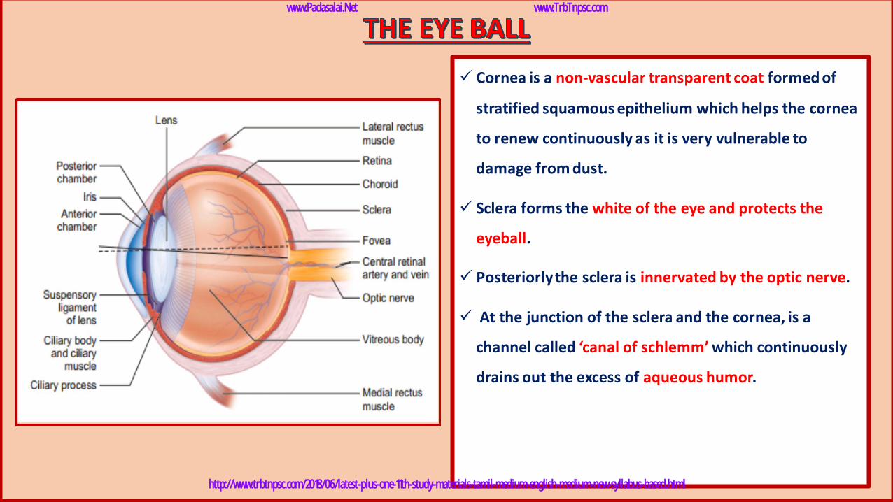
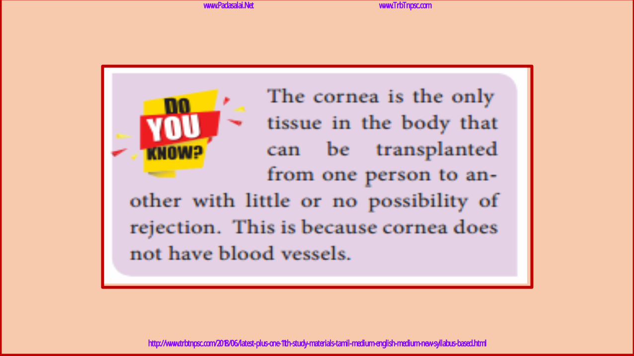
✓ Cornea is a non-vascular transparent coat formed of
stratified squamous epithelium which helps the cornea
to renew continuously as it is very vulnerable to
damage from dust.
✓ Sclera forms the white of the eye and protects the
eyeball.
✓ Posteriorly the sclera is innervated by the optic nerve.
✓ At the junction of the sclera and the cornea, is a
channel called ‘canal of schlemm’ which continuously
drains out the excess of aqueous humor.
www.Padasalai.Net www.TrbTnpsc.com
http://www.trbtnpsc.com/2018/06/latest-plus-one-11th-study-materials-tamil-medium-english-medium-new-syllabus-based.html
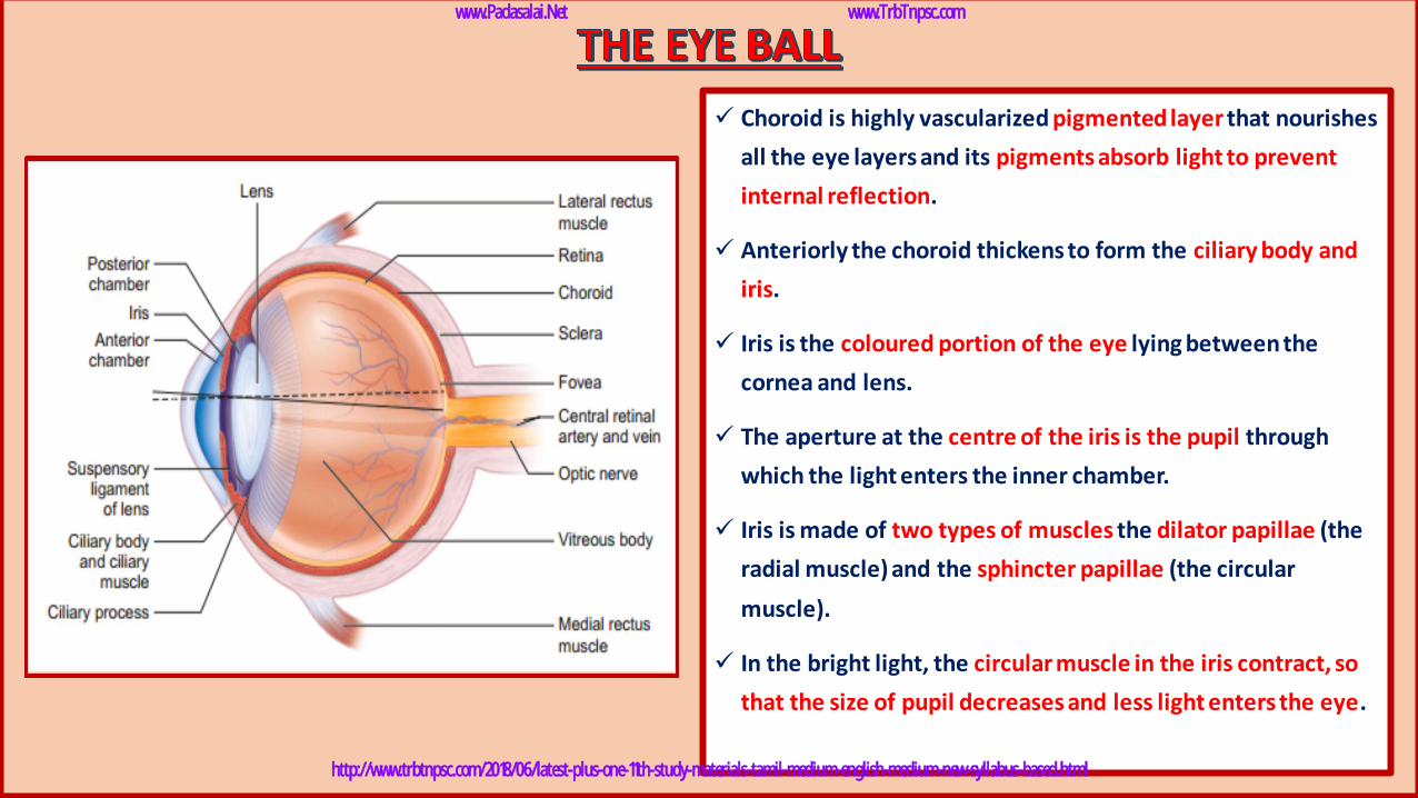
✓ Choroid is highly vascularized pigmented layer that nourishes
all the eye layers and its pigments absorb light to prevent
internal reflection.
✓ Anteriorly the choroid thickens to form the ciliary body and
iris.
✓ Iris is the coloured portion of the eye lying between the
cornea and lens.
✓ The aperture at the centre of the iris is the pupil through
which the light enters the inner chamber.
✓ Iris is made of two types of muscles the dilator papillae (the
radial muscle) and the sphincter papillae (the circular
muscle).
✓ In the bright light, the circular muscle in the iris contract, so
that the size of pupil decreases and less light enters the eye.
www.Padasalai.Net www.TrbTnpsc.com
http://www.trbtnpsc.com/2018/06/latest-plus-one-11th-study-materials-tamil-medium-english-medium-new-syllabus-based.html
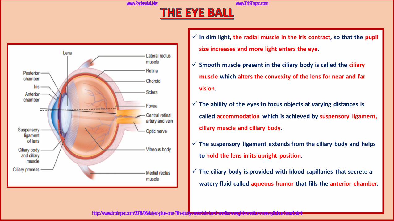
✓ In dim light, the radial muscle in the iris contract, so that the pupil
size increases and more light enters the eye.
✓ Smooth muscle present in the ciliary body is called the ciliary
muscle which alters the convexity of the lens for near and far
vision.
✓ The ability of the eyes to focus objects at varying distances is
called accommodation which is achieved by suspensory ligament,
ciliary muscle and ciliary body.
✓ The suspensory ligament extends from the ciliary body and helps
to hold the lens in its upright position.
✓ The ciliary body is provided with blood capillaries that secrete a
watery fluid called aqueous humor that fills the anterior chamber.
www.Padasalai.Net www.TrbTnpsc.com
http://www.trbtnpsc.com/2018/06/latest-plus-one-11th-study-materials-tamil-medium-english-medium-new-syllabus-based.html
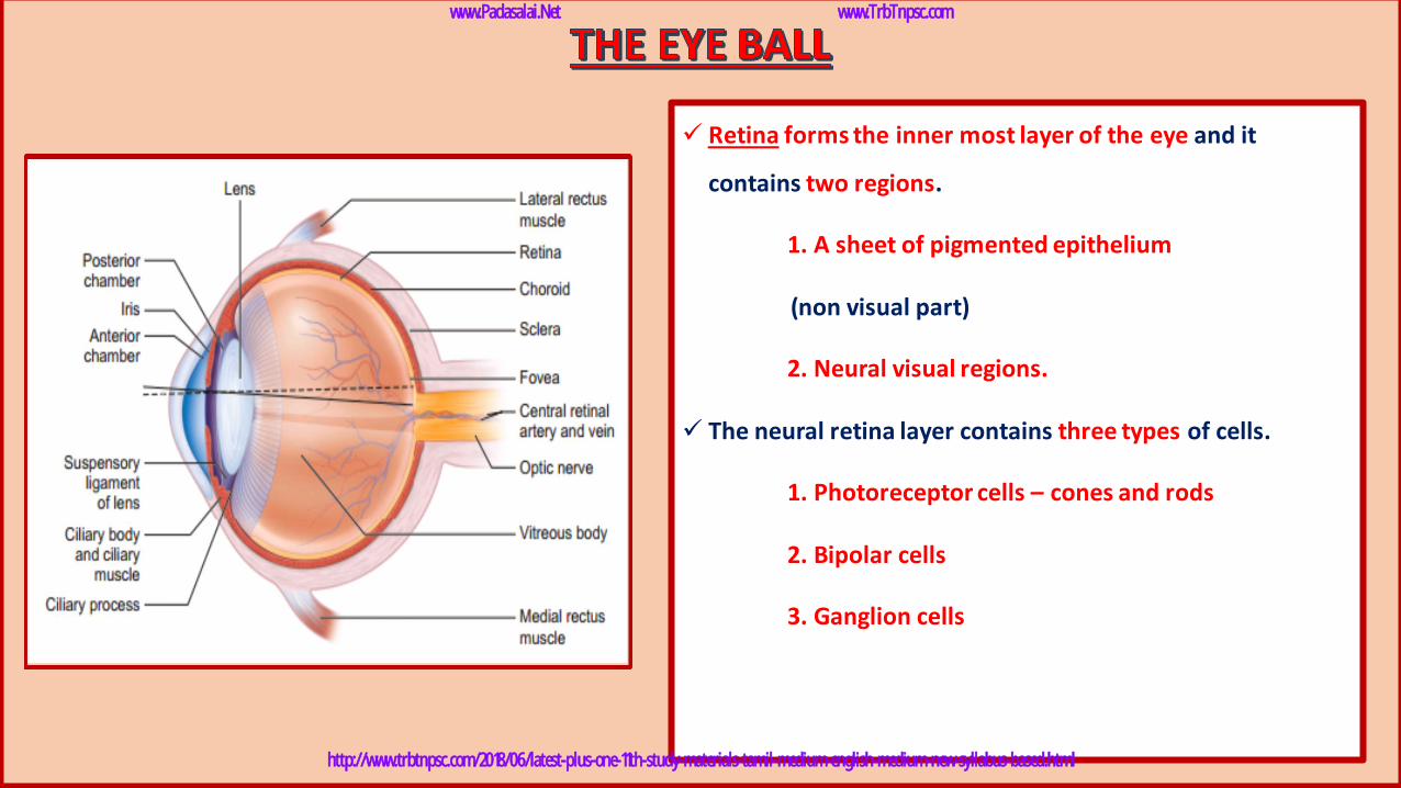
✓ Retina forms the inner most layer of the eye and it
contains two regions.
1. A sheet of pigmented epithelium
(non visual part)
2. Neural visual regions.
✓ The neural retina layer contains three types of cells.
1. Photoreceptor cells – cones and rods
2. Bipolar cells
3. Ganglion cells
www.Padasalai.Net www.TrbTnpsc.com
http://www.trbtnpsc.com/2018/06/latest-plus-one-11th-study-materials-tamil-medium-english-medium-new-syllabus-based.html
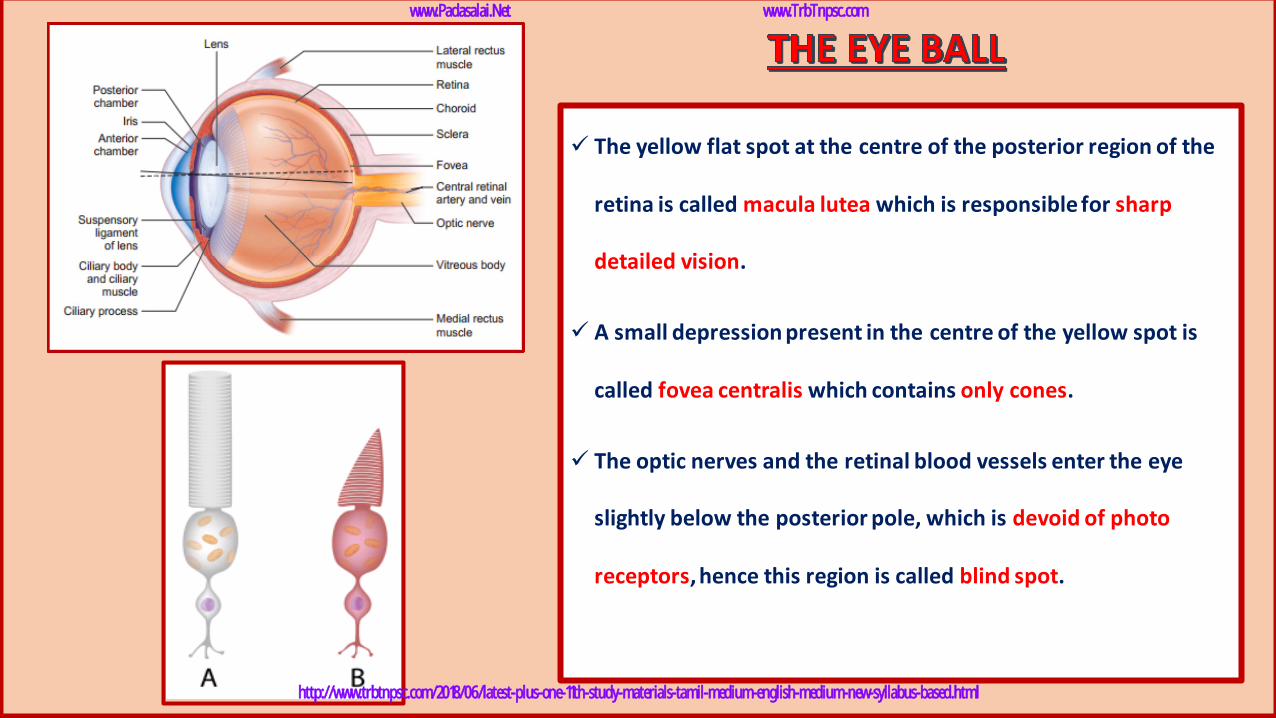
✓ The yellow flat spot at the centre of the posterior region of the
retina is called macula lutea which is responsible for sharp
detailed vision.
✓ A small depression present in the centre of the yellow spot is
called fovea centralis which contains only cones.
✓ The optic nerves and the retinal blood vessels enter the eye
slightly below the posterior pole, which is devoid of photo
receptors, hence this region is called blind spot.
www.Padasalai.Net www.TrbTnpsc.com
http://www.trbtnpsc.com/2018/06/latest-plus-one-11th-study-materials-tamil-medium-english-medium-new-syllabus-based.html

www.Padasalai.Net www.TrbTnpsc.com
http://www.trbtnpsc.com/2018/06/latest-plus-one-11th-study-materials-tamil-medium-english-medium-new-syllabus-based.html
www.Padasalai.Net www.TrbTnpsc.com
http://www.trbtnpsc.com/2018/06/latest-plus-one-11th-study-materials-tamil-medium-english-medium-new-syllabus-based.html
www.Padasalai.Net www.TrbTnpsc.com
http://www.trbtnpsc.com/2018/06/latest-plus-one-11th-study-materials-tamil-medium-english-medium-new-syllabus-based.html
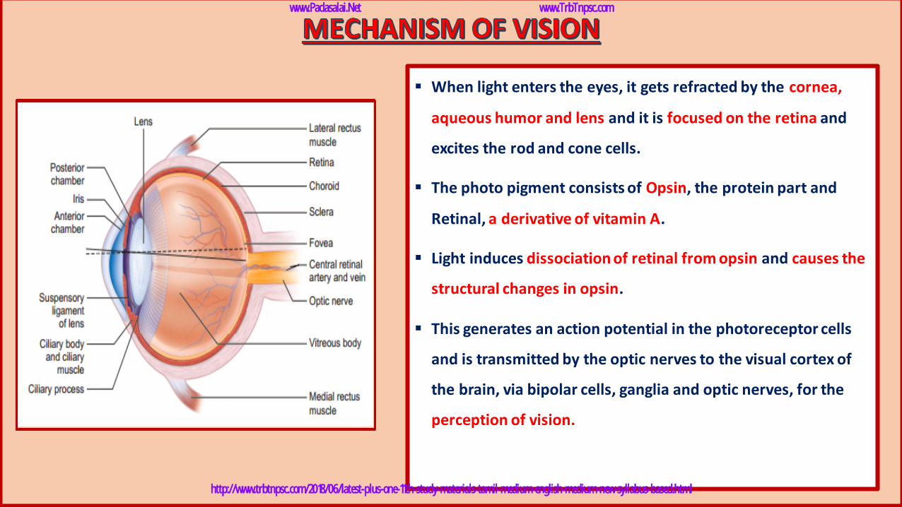
▪ When light enters the eyes, it gets refracted by the cornea,
aqueous humor and lens and it is focused on the retina and
excites the rod and cone cells.
▪ The photo pigment consists of Opsin, the protein part and
Retinal, a derivative of vitamin A.
▪ Light induces dissociation of retinal from opsin and causes the
structural changes in opsin.
▪ This generates an action potential in the photoreceptor cells
and is transmitted by the optic nerves to the visual cortex of
the brain, via bipolar cells, ganglia and optic nerves, for the
perception of vision.
www.Padasalai.Net www.TrbTnpsc.com
http://www.trbtnpsc.com/2018/06/latest-plus-one-11th-study-materials-tamil-medium-english-medium-new-syllabus-based.html
www.Padasalai.Net www.TrbTnpsc.com
http://www.trbtnpsc.com/2018/06/latest-plus-one-11th-study-materials-tamil-medium-english-medium-new-syllabus-based.html
www.Padasalai.Net www.TrbTnpsc.com
http://www.trbtnpsc.com/2018/06/latest-plus-one-11th-study-materials-tamil-medium-english-medium-new-syllabus-based.html

www.Padasalai.Net www.TrbTnpsc.com
http://www.trbtnpsc.com/2018/06/latest-plus-one-11th-study-materials-tamil-medium-english-medium-new-syllabus-based.html
www.Padasalai.Net www.TrbTnpsc.com
http://www.trbtnpsc.com/2018/06/latest-plus-one-11th-study-materials-tamil-medium-english-medium-new-syllabus-based.html
1. Myopia (near sightedness):
✓ The affected person can see the nearby objects but not
the distant objects.
✓ This condition may result due to an elongated eyeball or
thickened lens, so that the image of distant object is
formed in front of the yellow spot.
✓ This error can be corrected using concave lens that
diverge the entering light rays and focuses it on the
retina.
www.Padasalai.Net www.TrbTnpsc.com
http://www.trbtnpsc.com/2018/06/latest-plus-one-11th-study-materials-tamil-medium-english-medium-new-syllabus-based.html
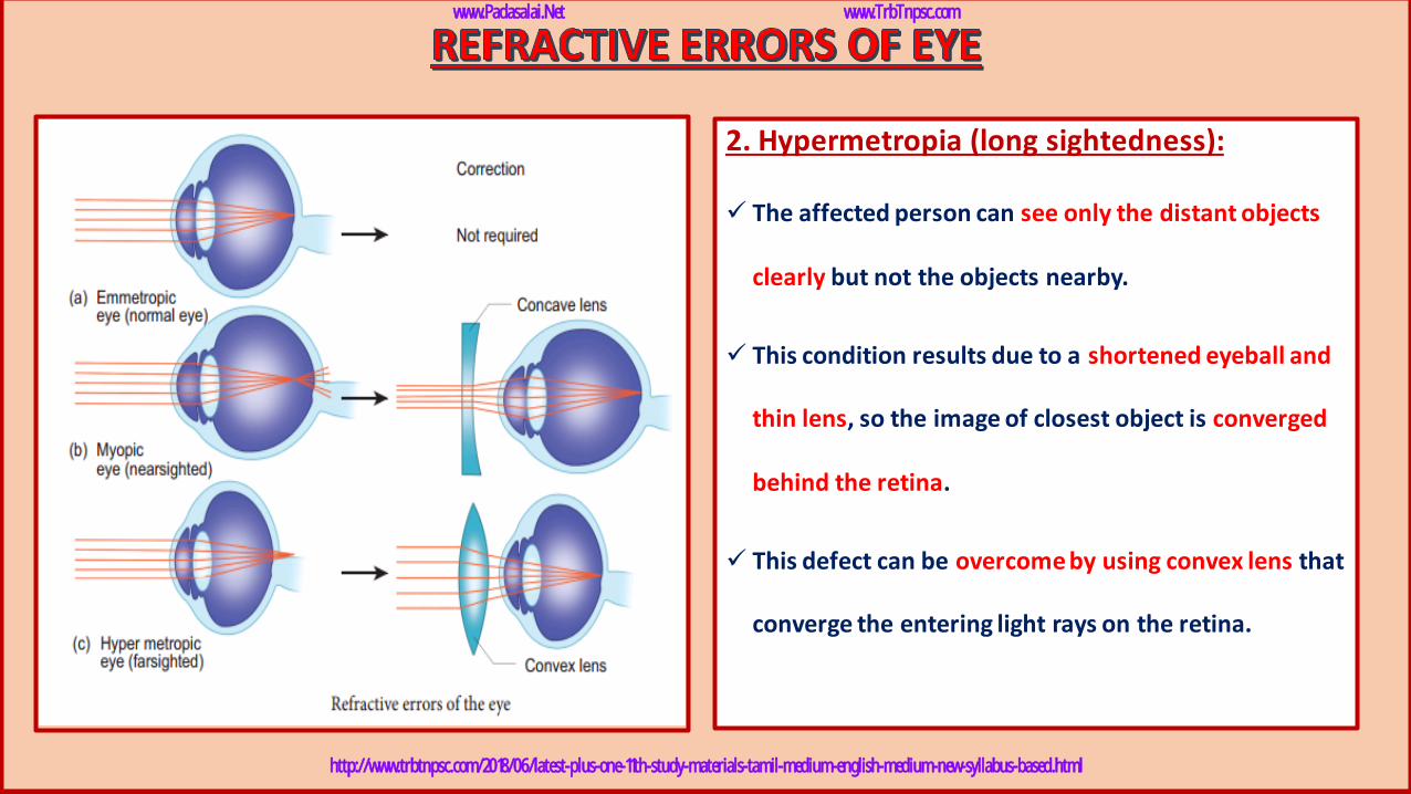
2. Hypermetropia (long sightedness):
✓ The affected person can see only the distant objects
clearly but not the objects nearby.
✓ This condition results due to a shortened eyeball and
thin lens, so the image of closest object is converged
behind the retina.
✓ This defect can be overcome by using convex lens that
converge the entering light rays on the retina.
www.Padasalai.Net www.TrbTnpsc.com
http://www.trbtnpsc.com/2018/06/latest-plus-one-11th-study-materials-tamil-medium-english-medium-new-syllabus-based.html
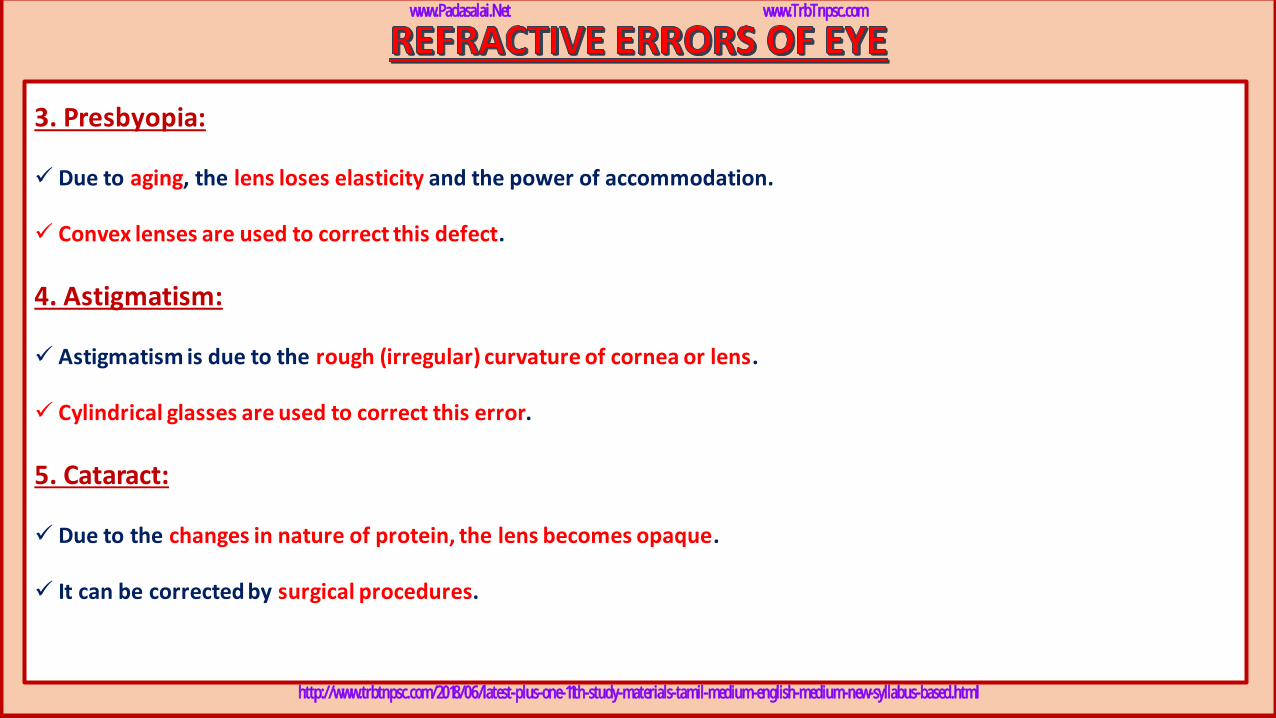
3. Presbyopia:
✓ Due to aging, the lens loses elasticity and the power of accommodation.
✓ Convex lenses are used to correct this defect.
4. Astigmatism:
✓ Astigmatism is due to the rough (irregular) curvature of cornea or lens.
✓ Cylindrical glasses are used to correct this error.
5. Cataract:
✓ Due to the changes in nature of protein, the lens becomes opaque.
✓ It can be corrected by surgical procedures.
www.Padasalai.Net www.TrbTnpsc.com
http://www.trbtnpsc.com/2018/06/latest-plus-one-11th-study-materials-tamil-medium-english-medium-new-syllabus-based.html
www.Padasalai.Net www.TrbTnpsc.com
http://www.trbtnpsc.com/2018/06/latest-plus-one-11th-study-materials-tamil-medium-english-medium-new-syllabus-based.html
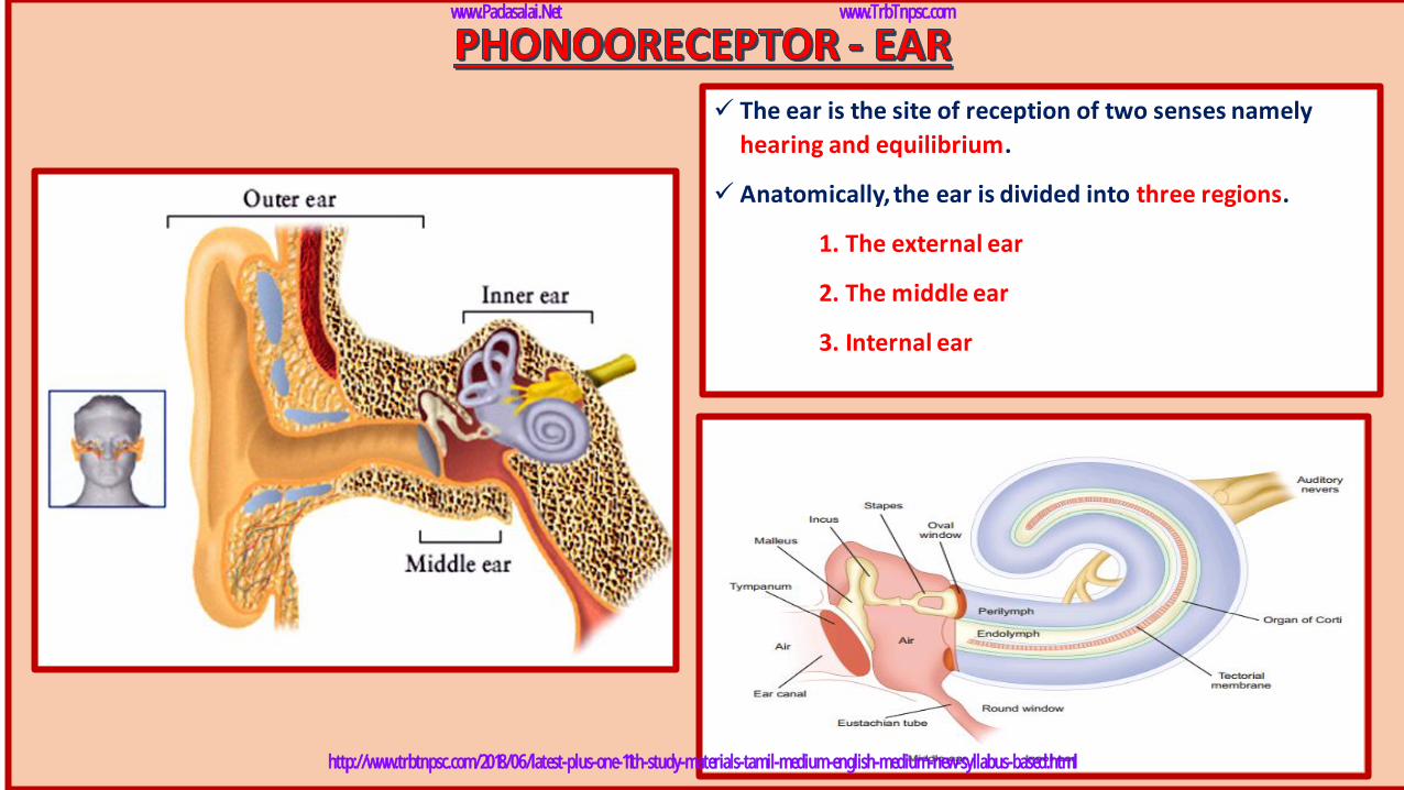
✓ The ear is the site of reception of two senses namely
hearing and equilibrium.
✓ Anatomically, the ear is divided into three regions.
1. The external ear
2. The middle ear
3. Internal ear
www.Padasalai.Net www.TrbTnpsc.com
http://www.trbtnpsc.com/2018/06/latest-plus-one-11th-study-materials-tamil-medium-english-medium-new-syllabus-based.html
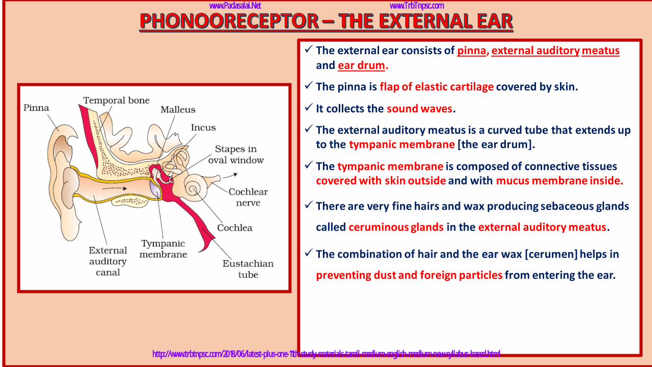
✓ The external ear consists of pinna, external auditory meatusand ear drum.
✓ The pinna is flap of elastic cartilage covered by skin.
✓ It collects the sound waves.
✓ The external auditory meatus is a curved tube that extends up to the tympanic membrane [the ear drum].
✓ The tympanic membrane is composed of connective tissues covered with skin outside and with mucus membrane inside.
✓ There are very fine hairs and wax producing sebaceous glands
called ceruminous glands in the external auditory meatus.
✓ The combination of hair and the ear wax [cerumen] helps in
preventing dust and foreign particles from entering the ear.
www.Padasalai.Net www.TrbTnpsc.com
http://www.trbtnpsc.com/2018/06/latest-plus-one-11th-study-materials-tamil-medium-english-medium-new-syllabus-based.html
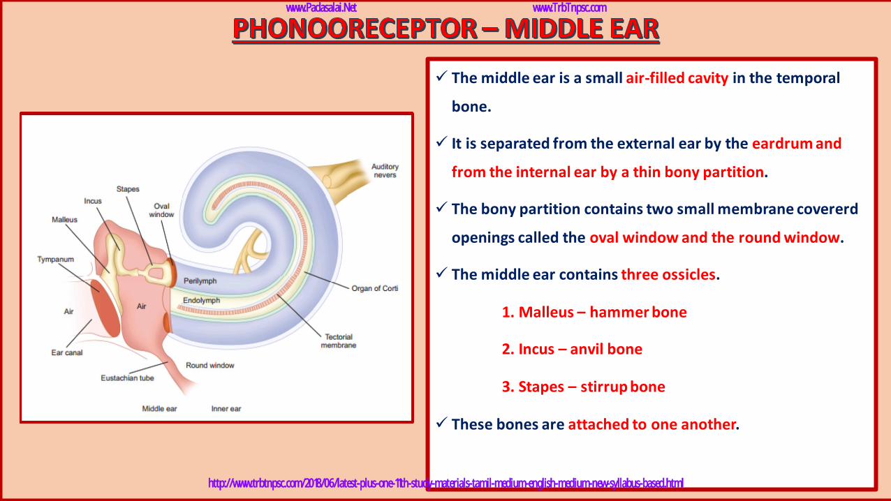
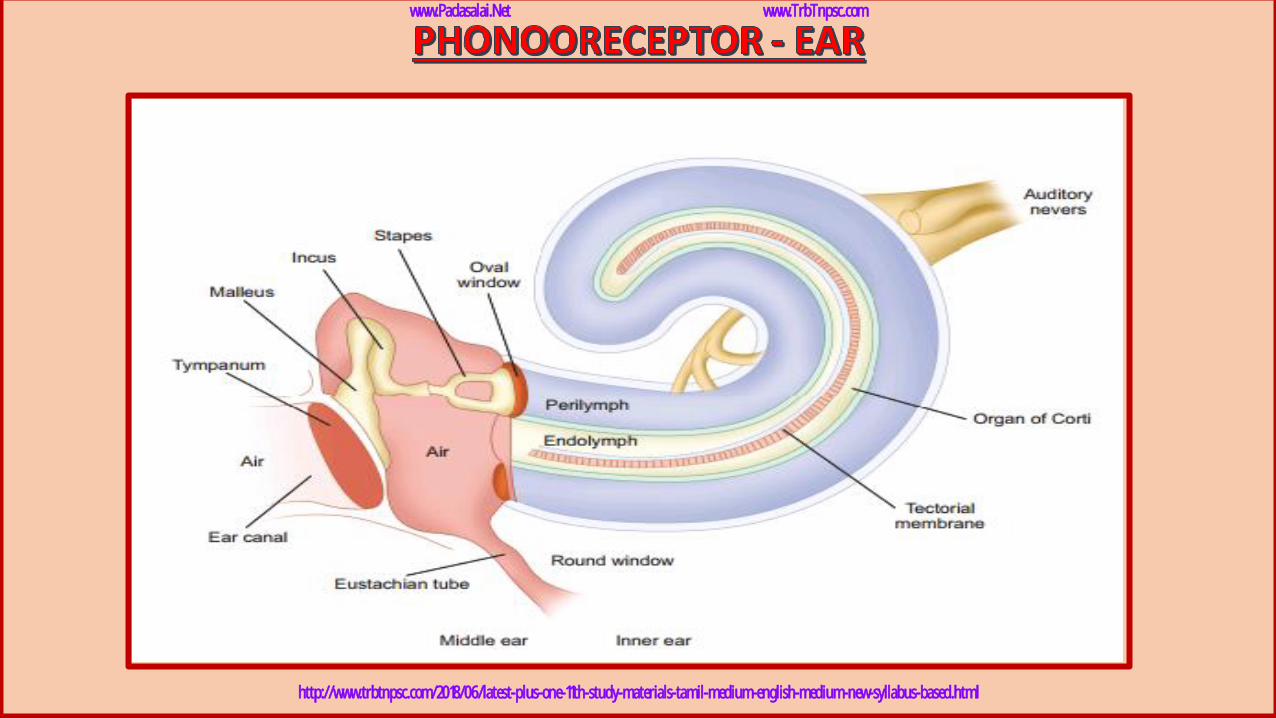
✓ The middle ear is a small air-filled cavity in the temporal
bone.
✓ It is separated from the external ear by the eardrum and
from the internal ear by a thin bony partition.
✓ The bony partition contains two small membrane covererd
openings called the oval window and the round window.
✓ The middle ear contains three ossicles.
1. Malleus – hammer bone
2. Incus – anvil bone
3. Stapes – stirrup bone
✓ These bones are attached to one another.
www.Padasalai.Net www.TrbTnpsc.com
http://www.trbtnpsc.com/2018/06/latest-plus-one-11th-study-materials-tamil-medium-english-medium-new-syllabus-based.html
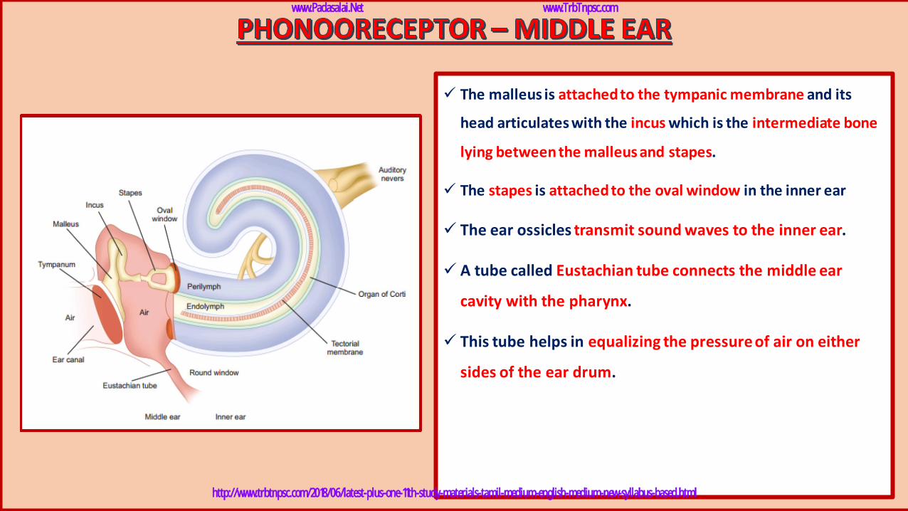
✓ The malleus is attached to the tympanic membrane and its
head articulates with the incus which is the intermediate bone
lying between the malleus and stapes.
✓ The stapes is attached to the oval window in the inner ear
✓ The ear ossicles transmit sound waves to the inner ear.
✓ A tube called Eustachian tube connects the middle ear
cavity with the pharynx.
✓ This tube helps in equalizing the pressure of air on either
sides of the ear drum.
www.Padasalai.Net www.TrbTnpsc.com
http://www.trbtnpsc.com/2018/06/latest-plus-one-11th-study-materials-tamil-medium-english-medium-new-syllabus-based.html
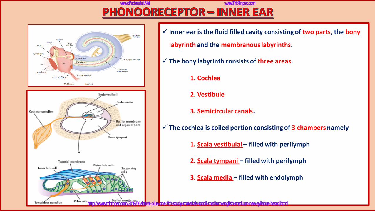
✓ Inner ear is the fluid filled cavity consisting of two parts, the bony
labyrinth and the membranous labyrinths.
✓ The bony labyrinth consists of three areas.
1. Cochlea
2. Vestibule
3. Semicircular canals.
✓ The cochlea is coiled portion consisting of 3 chambers namely
1. Scala vestibulai – filled with perilymph
2. Scala tympani – filled with perilymph
3. Scala media – filled with endolymph
www.Padasalai.Net www.TrbTnpsc.com
http://www.trbtnpsc.com/2018/06/latest-plus-one-11th-study-materials-tamil-medium-english-medium-new-syllabus-based.html
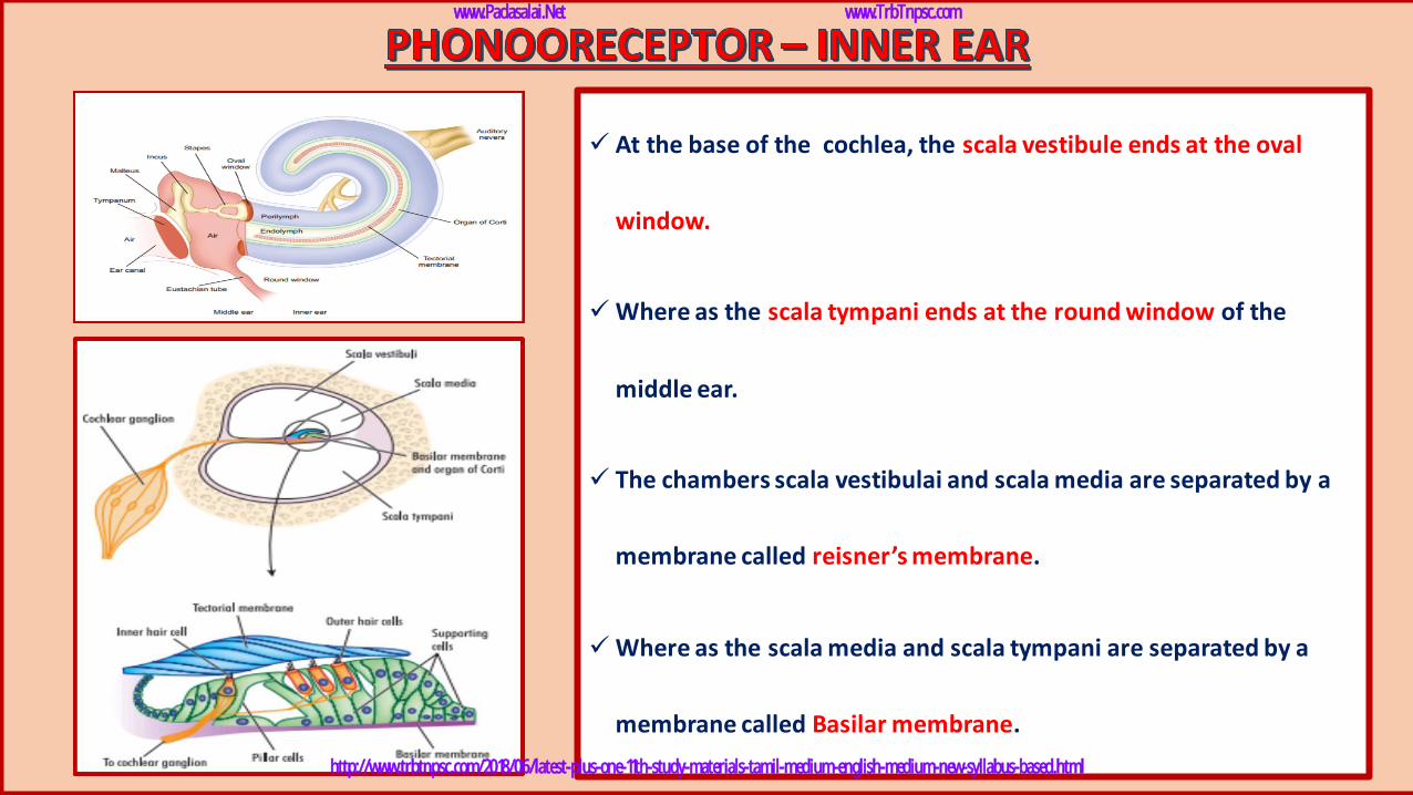
✓ At the base of the cochlea, the scala vestibule ends at the oval
window.
✓Where as the scala tympani ends at the round window of the
middle ear.
✓ The chambers scala vestibulai and scala media are separated by a
membrane called reisner’s membrane.
✓Where as the scala media and scala tympani are separated by a
membrane called Basilar membrane.
www.Padasalai.Net www.TrbTnpsc.com
http://www.trbtnpsc.com/2018/06/latest-plus-one-11th-study-materials-tamil-medium-english-medium-new-syllabus-based.html
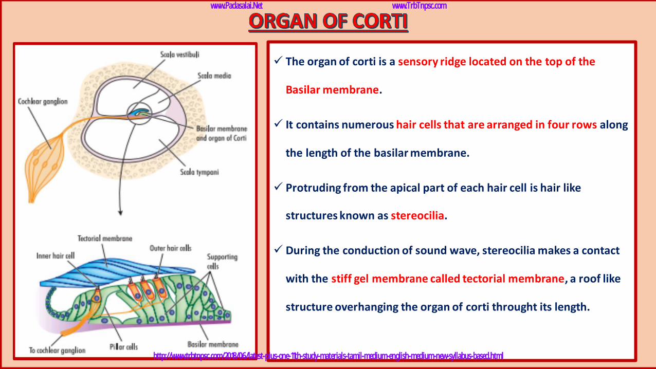
✓ The organ of corti is a sensory ridge located on the top of the
Basilar membrane.
✓ It contains numerous hair cells that are arranged in four rows along
the length of the basilar membrane.
✓ Protruding from the apical part of each hair cell is hair like
structures known as stereocilia.
✓ During the conduction of sound wave, stereocilia makes a contact
with the stiff gel membrane called tectorial membrane, a roof like
structure overhanging the organ of corti throught its length.
www.Padasalai.Net www.TrbTnpsc.com
http://www.trbtnpsc.com/2018/06/latest-plus-one-11th-study-materials-tamil-medium-english-medium-new-syllabus-based.html
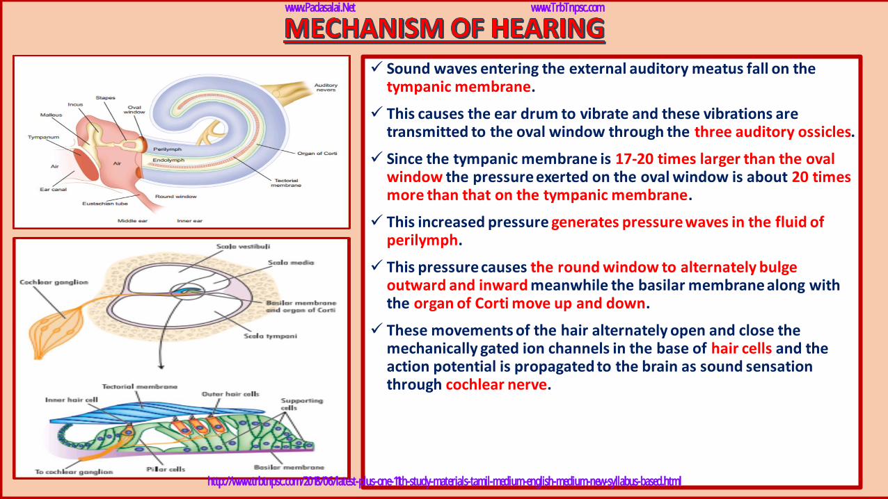
✓ Sound waves entering the external auditory meatus fall on the tympanic membrane.
✓ This causes the ear drum to vibrate and these vibrations are transmitted to the oval window through the three auditory ossicles.
✓ Since the tympanic membrane is 17-20 times larger than the oval window the pressure exerted on the oval window is about 20 times more than that on the tympanic membrane.
✓ This increased pressure generates pressure waves in the fluid of perilymph.
✓ This pressure causes the round window to alternately bulge outward and inward meanwhile the basilar membrane along with the organ of Corti move up and down.
✓ These movements of the hair alternately open and close the mechanically gated ion channels in the base of hair cells and the action potential is propagated to the brain as sound sensation through cochlear nerve.
www.Padasalai.Net www.TrbTnpsc.com
http://www.trbtnpsc.com/2018/06/latest-plus-one-11th-study-materials-tamil-medium-english-medium-new-syllabus-based.html
➢ Deafness may be temporary or permanent.
➢ It can be further classified into
1. Conductive deafness
2. Sensory-neural deafness.
➢ Possible causes for conductive deafness may be due to
i. the blockage of ear canal with earwax,
ii. Rupture of eardrum
iii. Middle ear infection with fluid accumulation
iv. Restriction of ossicular movement.
➢ In sensory-neural deafness, the defect may be in the organ of Corti or the auditory nerve or in the ascending
auditory pathways or auditory cortex.
www.Padasalai.Net www.TrbTnpsc.com
http://www.trbtnpsc.com/2018/06/latest-plus-one-11th-study-materials-tamil-medium-english-medium-new-syllabus-based.html
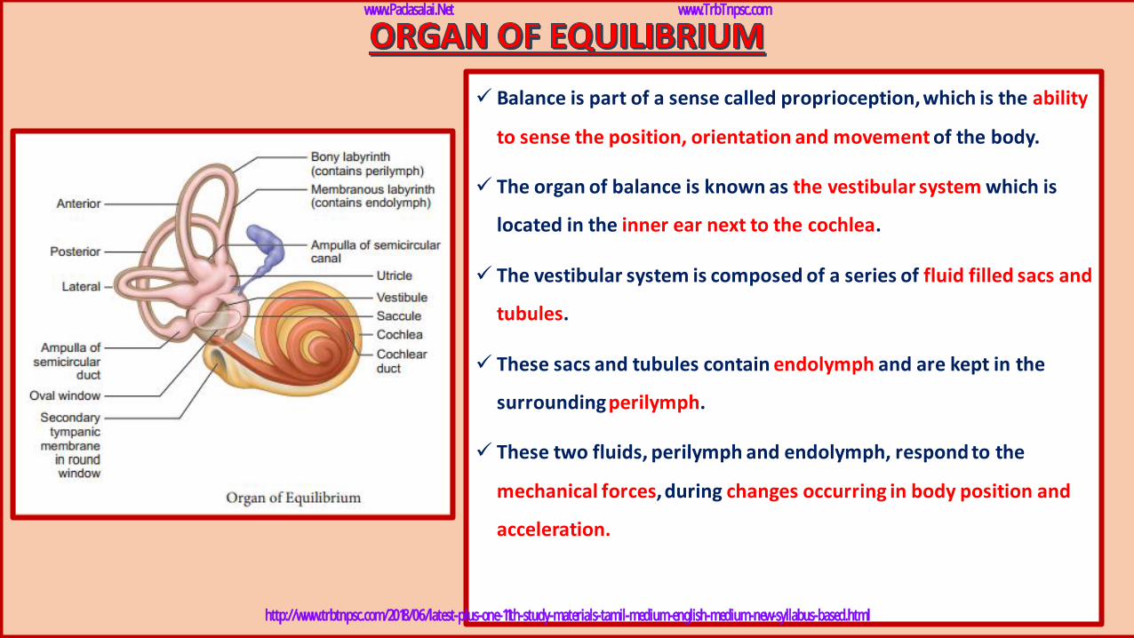
✓ Balance is part of a sense called proprioception, which is the ability
to sense the position, orientation and movement of the body.
✓ The organ of balance is known as the vestibular system which is
located in the inner ear next to the cochlea.
✓ The vestibular system is composed of a series of fluid filled sacs and
tubules.
✓ These sacs and tubules contain endolymph and are kept in the
surrounding perilymph.
✓ These two fluids, perilymph and endolymph, respond to the
mechanical forces, during changes occurring in body position and
acceleration.
www.Padasalai.Net www.TrbTnpsc.com
http://www.trbtnpsc.com/2018/06/latest-plus-one-11th-study-materials-tamil-medium-english-medium-new-syllabus-based.html
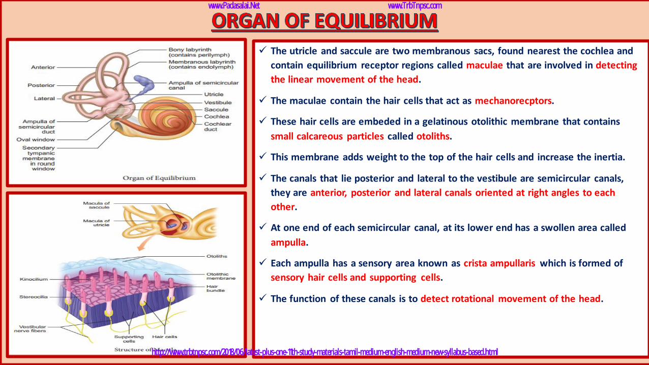
✓ The utricle and saccule are two membranous sacs, found nearest the cochlea and
contain equilibrium receptor regions called maculae that are involved in detecting
the linear movement of the head.
✓ The maculae contain the hair cells that act as mechanorecptors.
✓ These hair cells are embeded in a gelatinous otolithic membrane that contains
small calcareous particles called otoliths.
✓ This membrane adds weight to the top of the hair cells and increase the inertia.
✓ The canals that lie posterior and lateral to the vestibule are semicircular canals,
they are anterior, posterior and lateral canals oriented at right angles to each
other.
✓ At one end of each semicircular canal, at its lower end has a swollen area called
ampulla.
✓ Each ampulla has a sensory area known as crista ampullaris which is formed of
sensory hair cells and supporting cells.
✓ The function of these canals is to detect rotational movement of the head.
www.Padasalai.Net www.TrbTnpsc.com
http://www.trbtnpsc.com/2018/06/latest-plus-one-11th-study-materials-tamil-medium-english-medium-new-syllabus-based.html
www.Padasalai.Net www.TrbTnpsc.com
http://www.trbtnpsc.com/2018/06/latest-plus-one-11th-study-materials-tamil-medium-english-medium-new-syllabus-based.html
www.Padasalai.Net www.TrbTnpsc.com
http://www.trbtnpsc.com/2018/06/latest-plus-one-11th-study-materials-tamil-medium-english-medium-new-syllabus-based.html
www.Padasalai.Net www.TrbTnpsc.com
http://www.trbtnpsc.com/2018/06/latest-plus-one-11th-study-materials-tamil-medium-english-medium-new-syllabus-based.html
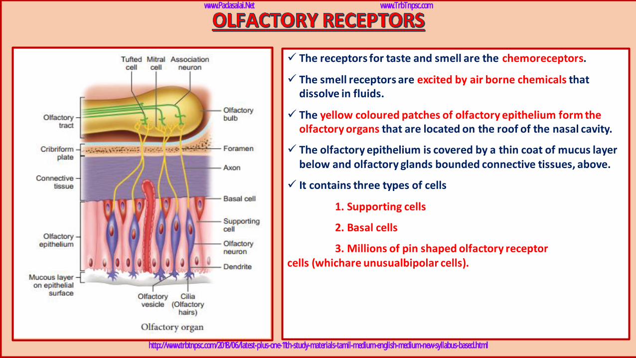
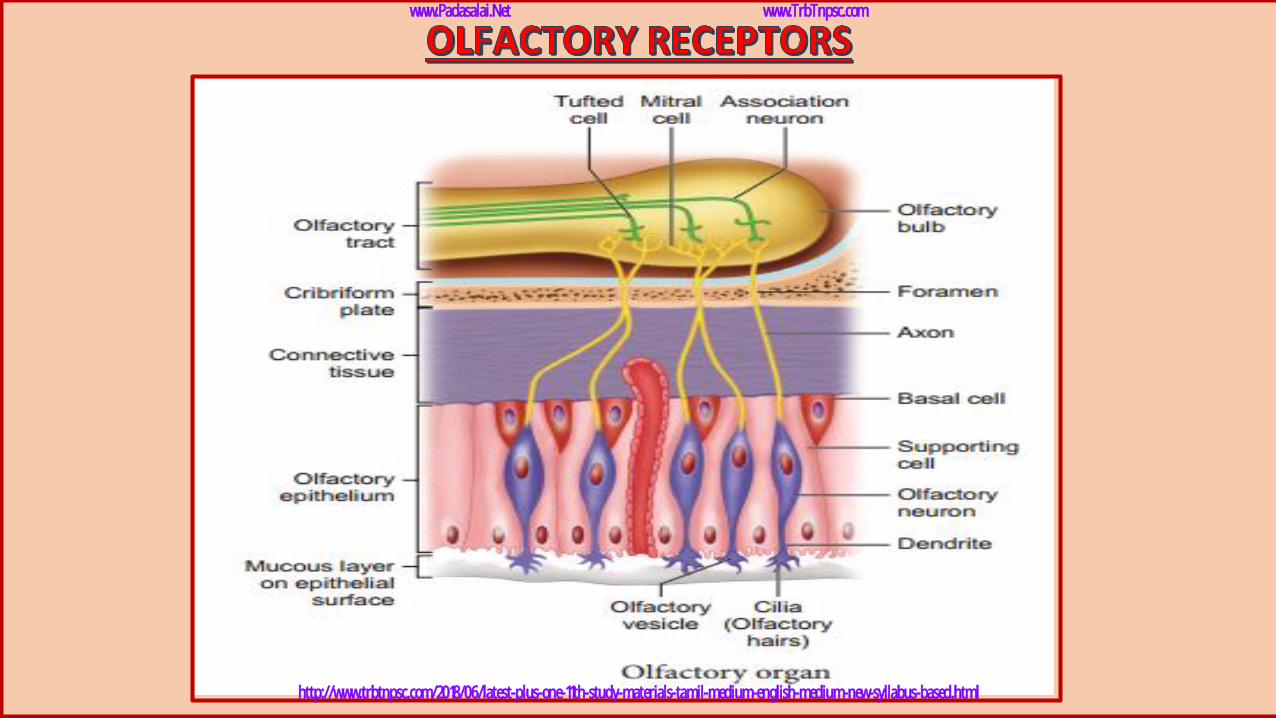
✓ The receptors for taste and smell are the chemoreceptors.
✓ The smell receptors are excited by air borne chemicals that dissolve in fluids.
✓ The yellow coloured patches of olfactory epithelium form the olfactory organs that are located on the roof of the nasal cavity.
✓ The olfactory epithelium is covered by a thin coat of mucus layer below and olfactory glands bounded connective tissues, above.
✓ It contains three types of cells
1. Supporting cells
2. Basal cells
3. Millions of pin shaped olfactory receptor cells (whichare unusualbipolar cells).
www.Padasalai.Net www.TrbTnpsc.com
http://www.trbtnpsc.com/2018/06/latest-plus-one-11th-study-materials-tamil-medium-english-medium-new-syllabus-based.html
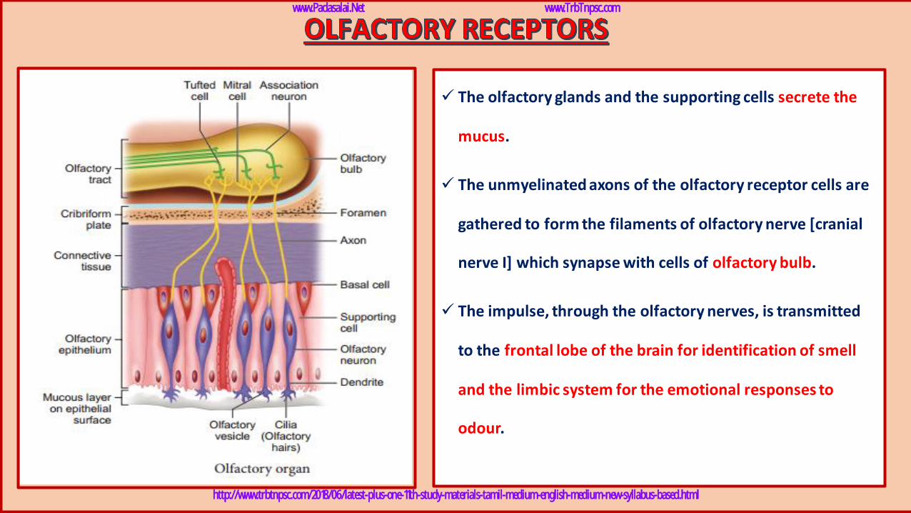
✓ The olfactory glands and the supporting cells secrete the
mucus.
✓ The unmyelinated axons of the olfactory receptor cells are
gathered to form the filaments of olfactory nerve [cranial
nerve I] which synapse with cells of olfactory bulb.
✓ The impulse, through the olfactory nerves, is transmitted
to the frontal lobe of the brain for identification of smell
and the limbic system for the emotional responses to
odour.
www.Padasalai.Net www.TrbTnpsc.com
http://www.trbtnpsc.com/2018/06/latest-plus-one-11th-study-materials-tamil-medium-english-medium-new-syllabus-based.html
www.Padasalai.Net www.TrbTnpsc.com
http://www.trbtnpsc.com/2018/06/latest-plus-one-11th-study-materials-tamil-medium-english-medium-new-syllabus-based.html
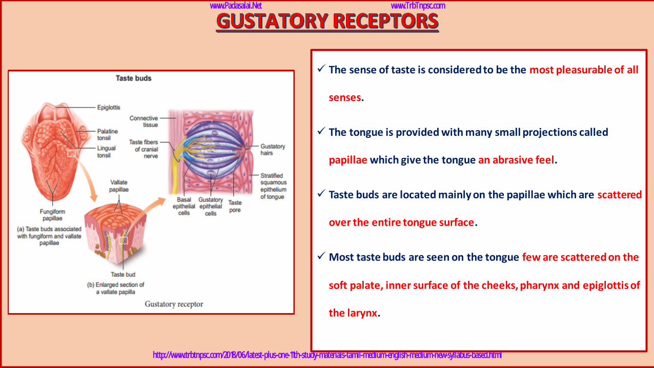
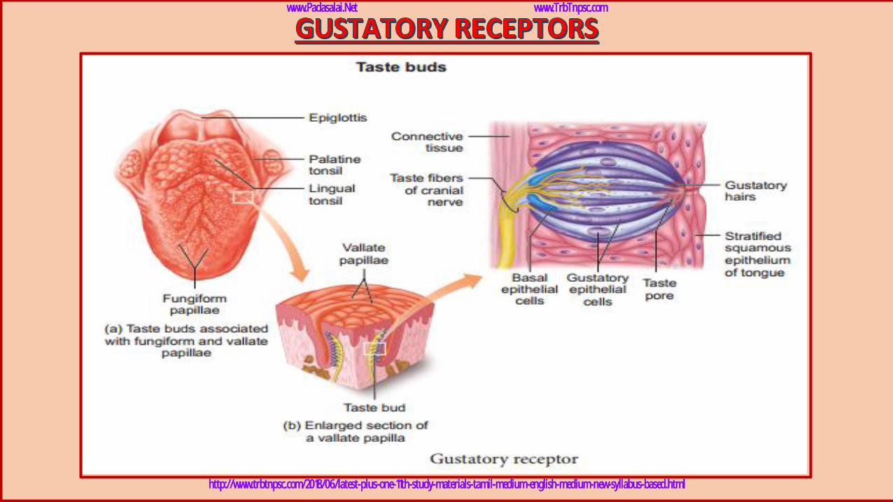
✓ The sense of taste is considered to be the most pleasurable of all
senses.
✓ The tongue is provided with many small projections called
papillae which give the tongue an abrasive feel.
✓ Taste buds are located mainly on the papillae which are scattered
over the entire tongue surface.
✓ Most taste buds are seen on the tongue few are scattered on the
soft palate, inner surface of the cheeks, pharynx and epiglottis of
the larynx.
www.Padasalai.Net www.TrbTnpsc.com
http://www.trbtnpsc.com/2018/06/latest-plus-one-11th-study-materials-tamil-medium-english-medium-new-syllabus-based.html
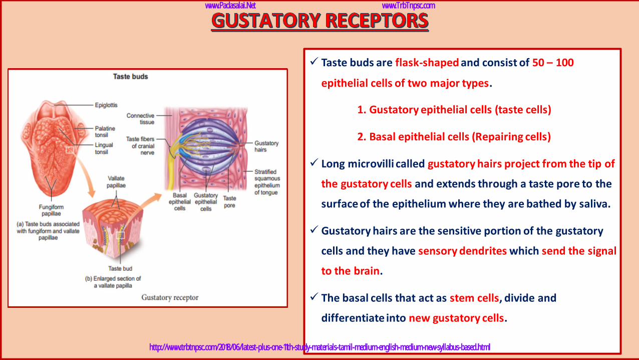
✓ Taste buds are flask-shaped and consist of 50 – 100
epithelial cells of two major types.
1. Gustatory epithelial cells (taste cells)
2. Basal epithelial cells (Repairing cells)
✓ Long microvilli called gustatory hairs project from the tip of
the gustatory cells and extends through a taste pore to the
surface of the epithelium where they are bathed by saliva.
✓ Gustatory hairs are the sensitive portion of the gustatory
cells and they have sensory dendrites which send the signal
to the brain.
✓ The basal cells that act as stem cells, divide and
differentiate into new gustatory cells.
www.Padasalai.Net www.TrbTnpsc.com
http://www.trbtnpsc.com/2018/06/latest-plus-one-11th-study-materials-tamil-medium-english-medium-new-syllabus-based.html
www.Padasalai.Net www.TrbTnpsc.com
http://www.trbtnpsc.com/2018/06/latest-plus-one-11th-study-materials-tamil-medium-english-medium-new-syllabus-based.html
www.Padasalai.Net www.TrbTnpsc.com
http://www.trbtnpsc.com/2018/06/latest-plus-one-11th-study-materials-tamil-medium-english-medium-new-syllabus-based.html
www.Padasalai.Net www.TrbTnpsc.com
http://www.trbtnpsc.com/2018/06/latest-plus-one-11th-study-materials-tamil-medium-english-medium-new-syllabus-based.html
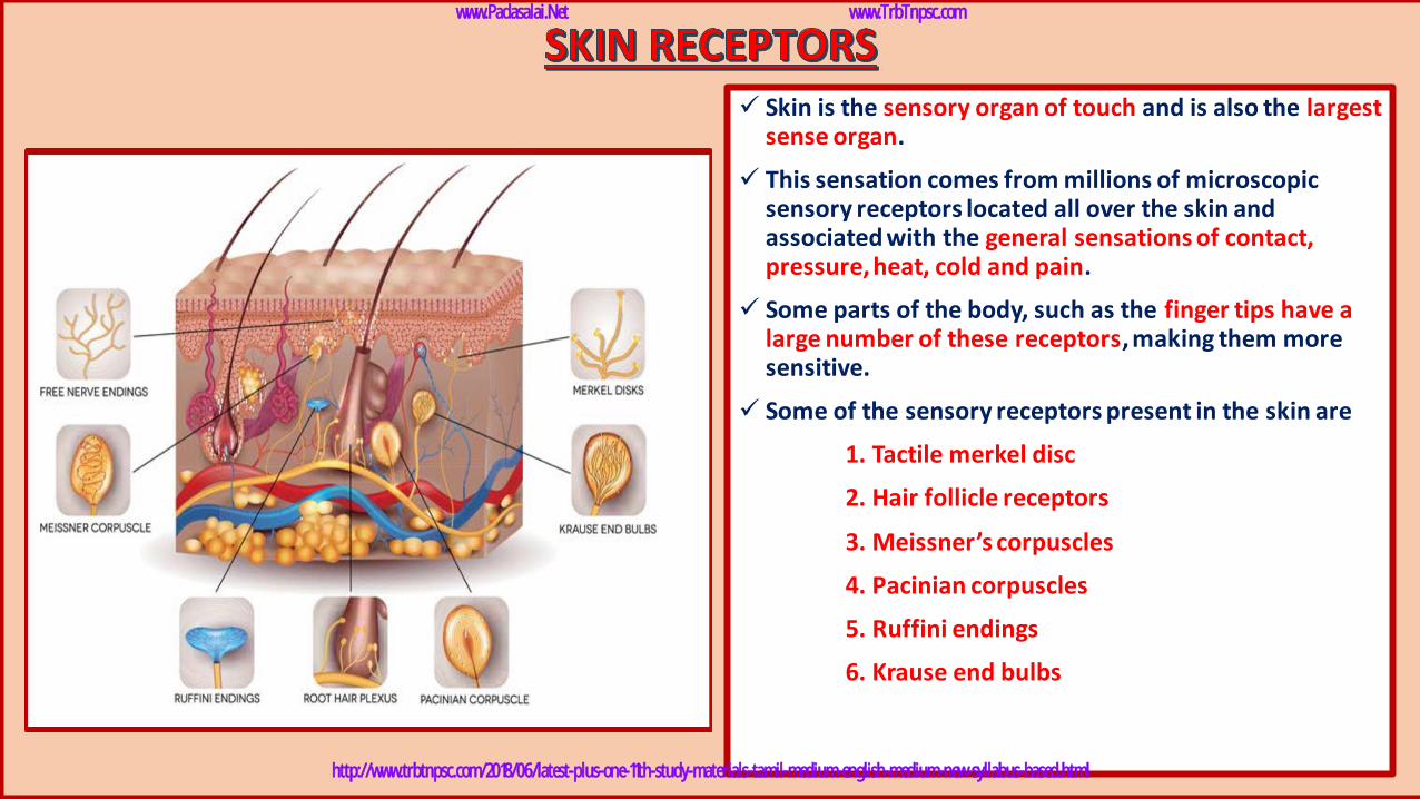
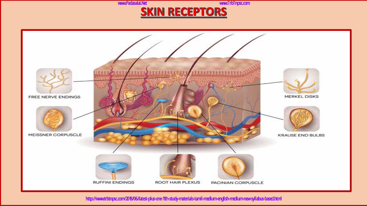
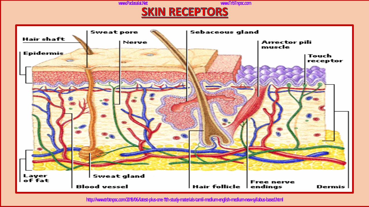
✓ Skin is the sensory organ of touch and is also the largest sense organ.
✓ This sensation comes from millions of microscopic sensory receptors located all over the skin and associated with the general sensations of contact, pressure, heat, cold and pain.
✓ Some parts of the body, such as the finger tips have a large number of these receptors, making them more sensitive.
✓ Some of the sensory receptors present in the skin are
1. Tactile merkel disc
2. Hair follicle receptors
3. Meissner’s corpuscles
4. Pacinian corpuscles
5. Ruffini endings
6. Krause end bulbs
www.Padasalai.Net www.TrbTnpsc.com
http://www.trbtnpsc.com/2018/06/latest-plus-one-11th-study-materials-tamil-medium-english-medium-new-syllabus-based.html

✓ Tactile merkel disk is light touch receptor lying in the
deeper layer of epidermis.
✓ Hair follicle receptors are light touch receptors lying
around the hair follicles.
✓Meissner’s corpuscles are small light pressure receptors
found just beneath the epidermis in the dermal
paoillae. They are numerous in hairless skin areas such
as finger tips and soles of the tips.
www.Padasalai.Net www.TrbTnpsc.com
http://www.trbtnpsc.com/2018/06/latest-plus-one-11th-study-materials-tamil-medium-english-medium-new-syllabus-based.html
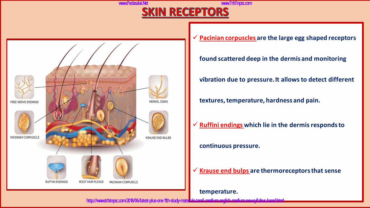
✓ Pacinian corpuscles are the large egg shaped receptors
found scattered deep in the dermis and monitoring
vibration due to pressure. It allows to detect different
textures, temperature, hardness and pain.
✓ Ruffini endings which lie in the dermis responds to
continuous pressure.
✓ Krause end bulps are thermoreceptors that sense
temperature.
www.Padasalai.Net www.TrbTnpsc.com
http://www.trbtnpsc.com/2018/06/latest-plus-one-11th-study-materials-tamil-medium-english-medium-new-syllabus-based.html

www.Padasalai.Net www.TrbTnpsc.com
http://www.trbtnpsc.com/2018/06/latest-plus-one-11th-study-materials-tamil-medium-english-medium-new-syllabus-based.html

பா.சனீிவாசன்முதுகலை விைங்கியல் ஆசிரியர்
நடராசன் தமயந்தி மமல் நிலைப் பள்ளிநாகப்பட்டினம்
லகமபசி எண் : 9994383274
நன்றி வணக்கம்www.Padasalai.Net www.TrbTnpsc.com
http://www.trbtnpsc.com/2018/06/latest-plus-one-11th-study-materials-tamil-medium-english-medium-new-syllabus-based.html


![TNPSC GROUP – II PRELIMINARY EXAM[11.11.2018] GENERAL](https://img.pdfslide.us/doc/110x75/624d497fba8125054339d1ae/tnpsc-group-ii-preliminary-exam11112018-general-.jpg)