Embed Size (px)
Citation preview

RBMOnline - Vol 17. No 6. 2008 866-880 Reproductive BioMedicine Online; www.rbmonline.com/Article/3415 on web 23 October 2008
866
© 2008 Published by Reproductive Healthcare Ltd, Duck End Farm, Dry Drayton, Cambridge CB23 8DB, UK
Ashok Agarwal is a Professor in the Lerner College of Medicine at Case Western Reserve University and the Director of Center for Reproductive Medicine, and the Clinical Andrology Laboratory at The Cleveland Clinic, Cleveland, Ohio, United States. He has published over 400 scientific articles, reviews and book chapters in different areas of andrology, male/female infertility and fertility preservation. His research program is known internationally for its focus on disease-oriented cutting edge research in the field of human reproduction. His team has presented over 700 papers at national and international meetings and more than 150 scientists, clinicians and biologists have received their training in his laboratory.
Dr Ashok Agarwal
Alex C Varghese1, Stefan S du Plessis1,2, Ashok Agarwal1,3
1Reproductive Research Centre, Glickman Urological and Kidney Institute, Cleveland Clinic, Cleveland, Ohio, USA; 2Division of Medical Physiology, University of Stellenbosch, Tygerberg, South Africa3Correspondence: Andrology Laboratory and the Reproductive Research Centre, Glickman Urological and Kidney Institute and Department of Obstetrics-Gynecology, Cleveland Clinic, 9500 Euclid Avenue, Desk A19.1, Cleveland, Ohio 44195, USA; Tel: (216) 444–9485; Fax: (216) 445–6049; e-mail: [email protected]
Abstract
Over the years, the development of assisted reproductive technology to bypass male factor infertility has improved drastically. Considered one of the most perplexing disorders in the reproductive field, male factor infertility is prevalent and may be on the rise. Unfortunately, its aetiology remains elusive. One of the main reasons lies in the complex machinery and structure of the hydrodynamic sperm cell. Its polyunsaturated fatty acid cell membrane, the protamines in its genetic material and the absence of antioxidants in its cytoplasm ensure that the spermatozoon is highly susceptible to environmental effects. The spermatozoon’s genesis, storage, and transport through the male reproductive tract are also susceptible, genetically and pathologically, to environmental effects. This review aims to include all the possible causes of disruption to this unique cell and their probable solutions, in the hope of clearing up the ambiguity that surrounds male factor infertility.
Keywords: assisted reproduction, endocrine disruption, male fertility, sperm chromatin, spermatozoa
Assisted reproductive technologies have made major strides since the birth of Louise Brown in 1978, the first baby resulting from the revolutionary work of Steptoe and Edwards (1978). From IVF to the introduction of intracytoplasmic sperm injection (ICSI) in 1992, physicians have used assisted reproductive technologies to treat infertility.
Approximately 8–10% of couples of reproductive age suffer from infertility (World Health Organization, 1992). Male infertility, isolated or combined with female infertility, is a contributing factor in about 50% of these cases (de Kretser, 1997). ICSI helped many infertile males, such as those with non-obstructive azoospermia or idiopathic male factor infertility, who became able to father offspring through this new technology (Ford, 2001). Of greatest concern are reports that suggest male subfertility is on the increase (Sharpe and Irvine, 2004). However, more precise evidence is needed to
substantiate the claim that male factor infertility is rising globally.
Owing to the prevalence of families with more than one infertile member, the genetic component of male infertility has become clear (Lilford et al., 1994). Assisted reproduction treatments are successful, but problems can nonetheless arise from interfering with the systematic nature of human reproduction. With the advent of ICSI, dysfunctional spermatozoa that normally cannot overcome the fertilizing barriers provided by the female reproductive tract can be used to fertilize an oocyte. The concern lies in the epigenetic transfer of a dysfunctional genome within the spermatozoa. Treatment with ICSI thus raises the moral dilemma that these disorders can be passed on to the next generation (Ford, 2001). Therefore, treating the underlying cause of infertility could prove to be more potent than evading it.
Review
Male gamete survival at stake: causes and solutions
Introduction
Unfortunately the aetiology of male infertility has eluded andrologists for decades. Despite the improvements in assisted reproduction treatments that serve to bypass the difficulties induced by male infertility, these procedures are just a means to circumvent the underlying causes. Until the causes of male infertility are elucidated, the prevalence of male subfertility will continue to rise (Sharpe and Irvine, 2004).
The complex metabolic machinery of spermatozoa is one of the reasons why the aetiology of male infertility is so difficult to decipher. Many crucial cell systems govern the viability and function of the spermatozoon so that failure or disruption in any of these systems can hinder its syngamy potential (Holt and Van Look, 2004). Previous studies have shown the deleterious effects of leukocytes, oxidative stress and metabolites on spermatozoa. However, those are only a few of the many factors that can affect male infertility. In the hope of shedding light on the elusive aetiology of male factor infertility, this review provides a comprehensive synopsis of all the aspects that can affect the highly susceptible spermatozoa and its fertilizing potential and discusses some of the recent studies on assessing spermatozoa and utilizing the results to treat male infertility.
Sperm dysfunction: causes and susceptibility
Sperm dysfunction is usually associated with poor semen parameters; however, a small population of men with normal semen parameters can exhibit a form of sperm dysfunction (Lefievre et al., 2007). Sperm dysfunction can be the result of endocrine disruption, defects in sperm production and maturation, and malfunctions associated with the transport and fertilization process of each spermatozoon. Sperm dysfunction is a prevalent factor, because the spermatozoon is a highly susceptible cell in which disruption of any of the cell systems hinders its fertilization potential and role in embryonic development (Holt and Van Look, 2004).
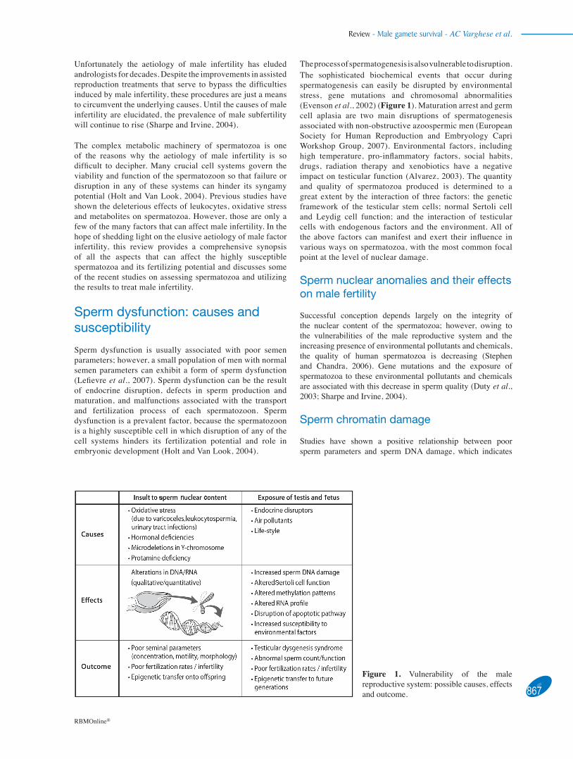
The process of spermatogenesis is also vulnerable to disruption. The sophisticated biochemical events that occur during spermatogenesis can easily be disrupted by environmental stress, gene mutations and chromosomal abnormalities (Evenson et al., 2002) (Figure 1). Maturation arrest and germ cell aplasia are two main disruptions of spermatogenesis associated with non-obstructive azoospermic men (European Society for Human Reproduction and Embryology Capri Workshop Group, 2007). Environmental factors, including high temperature, pro-inflammatory factors, social habits, drugs, radiation therapy and xenobiotics have a negative impact on testicular function (Alvarez, 2003). The quantity and quality of spermatozoa produced is determined to a great extent by the interaction of three factors: the genetic framework of the testicular stem cells; normal Sertoli cell and Leydig cell function; and the interaction of testicular cells with endogenous factors and the environment. All of the above factors can manifest and exert their influence in various ways on spermatozoa, with the most common focal point at the level of nuclear damage.
Sperm nuclear anomalies and their effects on male fertility
Successful conception depends largely on the integrity of the nuclear content of the spermatozoa; however, owing to the vulnerabilities of the male reproductive system and the increasing presence of environmental pollutants and chemicals, the quality of human spermatozoa is decreasing (Stephen and Chandra, 2006). Gene mutations and the exposure of spermatozoa to these environmental pollutants and chemicals are associated with this decrease in sperm quality (Duty et al., 2003; Sharpe and Irvine, 2004).
Sperm chromatin damage
Studies have shown a positive relationship between poor sperm parameters and sperm DNA damage, which indicates
867
Review - Male gamete survival - AC Varghese et al.
RBMOnline®
Figure 1. Vulnerability of the male reproductive system: possible causes, effects and outcome.

Review - Male gamete survival - AC Varghese et al.
disruptions in spermatogenesis in certain patients (Lopes et al., 1998). Aoki et al. (2005) demonstrated that 17% of patients with infertility had abnormal genes, while 17% of severe male infertility patients in a study by Carrell et al. had an abnormal amount of protamine 2 in their spermatozoa (Carrell and Liu 2001; Carrell et al., 2007). These studies indicate that genetic disorders might account for a significant percentage of male factor infertility. In addition, severe oligozoospermia and azoospermia are known consequences of microdeletions in the azoospermia factor (AZF) regions of the Y chromosome or other chromosomes (Vogt et al., 1996; Escalier 2001). Deletions in the AZFa and AZFb region are associated with impairment of spermatogenesis (Vogt et al., 1996), which causes lower sperm concentrations or a lack of spermatozoa. Currently, the fertilizing capacity of an individual patient’s sperm population is evaluated by conventional semen analysis, in which motility and morphology, along with sperm concentration parameters are assessed under a light microscope. It is assumed by many clinics that sperm DNA is sufficiently intact if the semen analysis produces relatively normal results.
While pertinent in the evaluation of infertility for males, simple semen analysis remains inadequate to detect subtle defects within the spermatozoa (Agarwal and Allamaneni, 2005). An example of this arises in the use of ICSI. A pivotal goal of ICSI is to select good quality spermatozoa to be injected into an oocyte. If poor quality spermatozoa with abnormal DNA are selected, fertilization failure (Lopes et al., 1998) or even abnormal fetal development (Cummins and Jequier, 1995) may occur. Further research assessing DNA damage, rather than basic semen analysis to predict fertilizing capacity, is required because it might result in more efficient and beneficial results for in-vivo and in-vitro fertilization (Agarwal and Said, 2003). There is substantial evidence that the spermatozoa of infertile men contain considerably more DNA damage than those of fertile men (Kodama et al., 1997; Spano et al., 2000). Other studies have suggested that the presence of ≥30% of spermatozoa with damaged DNA signifies reduced natural pregnancy rates (Evenson et al., 1999, 2002). Equally important are the protamines, which replace histones and transition proteins in the latter stages of the haploid phase in spermatogenesis and are adequate indicators of sperm nuclear maturity (Mengual et al., 2003). Unlike the male germ cells of other mammals, the replacement of histones by protamines is incomplete in human spermatozoa. Human spermatozoa retain roughly 15% of histones (Gatewood et al., 1987). This nucleo-protamine/nucleo-histone organization of sperm chromatin with histone-enriched regions is thought be a prerequisite for special gene activity during early embryogenesis (Wykes and Krawetz, 2003). Fertilization rates are also affected by lower levels of protamines within spermatozoa (Nasr-Esfahani et al., 2004a) and abnormal protamine expression has been associated with low sperm counts, reduced motility and morphology (Aoki et al., 2005). Complete protamine deficiency is found in approximately 15–20% of infertile men (Carrell and Liu, 2001; Oliva, 2006) in whom it has been associated with the increased susceptibility of spermatozoa to premature chromatin condensation. This is the second most prevalent cause of fertilization failure after aneuploidy (Nasr-Esfahani et al., 2004a).
In contrast to most mammals, whose spermatozoa contain only one type of protamine (P1), mouse, human and certain other mammalian sperm chromatin include a second type of protamine
(P2), which is deficient in cysteine residues. Therefore, human sperm chromatin has a lower level of free-sulphhydryl groups available for disulphide bonding, resulting in a potentially less stable chromatin structure than in species that contain P1 alone (Jager, 1990). Both protamines are expressed in roughly equal quantities (Balhorn et al., 1988). Elevated or diminished P1/P2 ratios have been observed in some infertile patients, indicating severe defects in spermatogenesis (Carrell et al., 2007). Nasr-Esfahani et al. (2004b) showed a significant negative correlation of ICSI fertilization rate with sperm protamine deficiency and P1/P2 ratio. However, no significant correlation was observed between protamine deficiency assessed by chromomycine A3 staining and P1/P2 ratio. This study proves that an altered P1/P2 ratio affects the fertilization rate and embryo quality, which may affect implantation and pregnancy outcome.
Regarding the importance of chromatin structure, McCarthy and Ward (2000) described the sperm nucleus as an ‘ordered library of DNA organized into functional zones’. Variations in the highly defined nuclear architecture of sperm chromatin may influence the initiation and regulation of paternal gene activity in early embryo development (Haaf and Ward, 1995). An increase in the histone to protamine ratio and DNA damage in ejaculated spermatozoa have been correlated with a febrile illness (Evenson et al., 2000).
RNA content in ejaculated spermatozoa
The concept that human spermatozoa are only carriers of highly condensed chromatin material (DNA–protamine complex) is fast changing with the recent evidence proving that spermatozoa contain an array of RNA molecules (approximately 3000). This confirms that spermatozoa contain a wealth of both known and unknown protein-coding and non-coding RNAs (Ostermeier et al., 2002, 2005). The consensus is that the pool of RNAs may represent a historical record and act as a fingerprint of spermatogenesis/spermiogenesis or probably a repertoire of epigenetic signals for early embryo development after fertilization. Recently, a serial analysis of gene expression tag map of human sperm RNA has identified 2712 and 2459 unique transcripts from pooled and individual semen samples, respectively (Zhao et al., 2006). Profiling the sperm RNA will probably have important clinical significance in the near future in the assessment of male fertility status.
There are still controversies regarding the transcriptional and translational activity of ejaculated spermatozoa. In-vitro radio-labelling experiments indicate that mature human spermatozoa do not transcribe novel RNAs (Grunewald et al., 2005). However, the fact that even the compact DNA in spermatozoa contain isolated domains that are in a more open conformation resembling a transcriptionally ready (histone-bound and DNase sensitive) state, raises speculation regarding the possibility of transcription under some circumstances. Novel evidence regarding active translation of stored mRNA in capacitated spermatozoa has been reported (Gur and Breitbart, 2006). Recent studies confirm that the mRNA profile varies between spermatozoa showing high and low motility patterns (Lambard et al., 2004; Wang et al., 2004). This finding is relevant to assisted reproductive technologies. Moreover, a study using real-time polymerase chain reaction (RT-PCR) found a 44% decrease in the amount of transcripts for aromatase among asthenozoospermic infertile men compared with normozoospermic men (Galeraud-Denis et al., 2007).868
RBMOnline®

These observations and evidence of aromatases and oestrogens in human male germ cells warrant further investigation on the role of these molecules in sperm capacitation/survival. A novel suit of ‘antisense’ RNAs has also been identified in human spermatozoa, which may provide a new level of control that confers transcriptional silencing by methylation. This is an imprinting mechanism during transition from the maternal to the embryonic genome after the fertilization process (Ostermeier et al., 2005). Experimental evidence also confirmed the role of paternally derived mRNA in embryo growth (Ostermeier et al., 2004; Rassoulzadegan et al., 2006).
Apoptosis
Apoptosis is a mechanism of programmed cellular death initiated at the molecular level by enzymes, such as caspases. This can be induced by a wide variety of cofactors, such as heat, irradiation, ischaemia, toxins, and withdrawal or increased levels of hormones such as prolactin, oestrogen, testosterone/FSH and antiandrogens (Said et al., 2004). The clonal expansion of germ cells through mitosis produces an excessive number of germ cells for possible differentiation into mature spermatozoa so apoptosis is required to restrict the number of germ cells to match the capacity of the Sertoli cells (Agarwal and Said, 2003). The majority of spermatogonial cells (±75%) undergo apoptosis during spermatogenesis (Allan et al., 1987). Apoptosis is also necessary in regulating the production of abnormal sperm. If conditions are unfavourable for spermatogenesis, apoptosis ensures that abnormal spermatozoa cease to proliferate (Agarwal and Said, 2003). Therefore, over-expression of apoptotic events could lead to oligozoospermia or azoospermia (Takagi et al., 2001), while under-expression could give rise to the survival of abnormal sperm. This could lead to fertilization by these abnormal sperm, which could potentially result in early embryonic loss or unhealthy offspring (Zini et al., 2001; Singh et al., 2003). Using Fas as a marker of apoptosis in human sperm, Sakkas et al. (1999) found that <10% apoptotic sperm was present in normospermic men, whereas approximately 60% of oligospermic men exhibited >10% apoptotic sperm. These findings are a major concern in the light of current assisted reproduction techniques such as ICSI where sperm selection is made arbitrarily.
Recent studies also show an age-related increase in DNA damage and an age-related decrease in the apoptotic rates of human sperm (Singh et al., 2003). While the incidence of DNA double-strand breaks increases with age, the frequency of apoptosis decreases. The correlation between age-related damage to genes and the apoptotic pathway may be a result of DNA damage to the genes involved in apoptosis. An impairment in the apoptotic pathway combined with the loss of free radical scavenging during ageing and continuous exposure to damaging environmental factors triggers the age-related increase of DNA double-strand breaks (Singh et al., 2003). An association between parental age and decreased lifespan of female offspring has also been reported (Gavrilov et al., 1997). Overall, the majority of evidence points to the association between the effects of parental age and decreased germ-cell genetic integrity (Walter et al., 2003).
Endocrine disruptors
The homeostasis of the endocrine and paracrine network determines the synchrony of the reproductive potential of the species. Its perturbation by endocrine disruptors during prenatal, perinatal or adult life are major causes of concern. Current research points to a newer dimension of these issues in which the consequence of developmental exposures to these endocrine disruptors can be propagated in multiple generations with a high degree of fidelity and suggests a potential epigenetic aetiology and molecular basis of adult onset disease.
Emerging cues from animal studies and fields of research as diverse as evolutionary ecology, behavioural development, life-history theory, molecular biology and medical epidemiology have converged on the key finding that a given genotype can give rise to different phenotypes, depending on environmental conditions (Sultan, 2003). Rats treated with the oestrogenic pesticide methoxychlor or the antiandrogenic fungicide vinclozolin during pregnancy produce male offspring that have decreased sperm capacity and fertility (Anway et al., 2005; Chang et al., 2006). It has also been demonstrated that the compromised fertility is passed through the adult male germline for four generations with high penetrance. Altered patterns of DNA methylation have been observed in the germ cells of generations two and three.
Analysis of the transgenerational epigenetic effects on the male germline (i.e. sperm) identified 25 candidate DNA sequences with altered methylation patterns in the vinclozolin-generation sperm (Chang et al., 2006). The authors provide detailed evidence of specific patterns of methylation that are unique to vinclozolin-exposed lineages and show that some of the mRNAs regulated by these promoter/DNA sequences are expressed at significantly lower levels in the vinclozolin-exposed generations. Thus, exposing the mother to vinclozolin can affect the abundance of specific mRNAs in her offspring, and this abnormal expression pattern is transmitted through the next three generations. It has been shown that treating pregnant mice with vinclozolin leads to adult onset of diseases such as tumours, prostate lesions, renal lesions and an increased incidence of infections. These effects were also seen in F2–F4 generations, in similar proportions. The authors report that disease prevalence is transmitted to the next generation through the male germline.
This study demonstrates the effects of two types of endocrine disrupting chemicals (EDC) on the reproductive function of males through epigenetic alterations when placed in an age-dependent environment. The target of these epigenetic alterations is the male germline, and they occur during the sex determination stage of cell differentiation (Chang et al., 2006). The type of EDC that the male germline is most susceptible to is the antiandrogenic endocrine disruptor; recent discoveries show androgen receptor (AR) transcripts and AR proteins being present in gonocyte extracts (Merlet et al., 2007).
A study by Anway et al. (2005) investigated the effects of the same endocrine disruptors, methoxychlor and vinclozolin, on humans involving male sex determination. Interestingly, even brief in-vivo exposure to these endocrine disruptors at the time 869
Review - Male gamete survival - AC Varghese et al.
RBMOnline®
Review - Male gamete survival - AC Varghese et al.
of male sex determination caused a transgenerational phenotype of spermatogenic cell apoptosis and subfertility.
Organochlorides
Certain organic chlorides, which can be considered to be a subset of EDC (Spano et al., 2005), can also disrupt the reproductive system. Polychlorinated biphenyls (PCB) are a group of highly lipophilic organic chlorines that are established persistent environmental pollutants in a variety of ecological systems across the world (Oskam et al., 2005). In a recent study using goats the percentage of sperm DNA damage tested by the sperm chromatin structure assay in young male goats increased with long-term maternal exposure to low doses of PCB126 and PCB153 (Oskam et al., 2005). Both PCB affect the reproductive system and related hormones through alterations in the reproductive endpoints. These results are in accordance with the concept that exposure to EDC during fetal development may be toxic to male reproductive health later in adult life (Oskam et al., 2005).
The adverse effects of another widely prescribed organochloride on the reproductive potential of sperm was studied by Silvestroni and Palleschi (1999). They determined that lindane (used in topical medications) diffuses into the lipid bilayer of the sperm membrane and alters the molecular dynamics of the bilayer. Even in minute amounts, lindane reduced the fertility potential of the sperm.
Effects of endocrine disrupting chemicals on Sertoli cell function
One of the most important stages in fetal and neonatal development occurs during the differentiation of reproductive organs and the proliferation of Sertoli cells (Sharpe et al., 2000). Sertoli cells, which are the principal site of spermatogenesis, determine the capacity of sperm production. Therefore, perinatal exposure to EDC could reduce Sertoli cell proliferation, which would subsequently lead to reduced sperm counts (Sharpe et al., 2000).
There has been speculation that certain phenolic plasticizing agents, such as p-nonylphenol, which are now prevalent in the environment, may affect Sertoli cell development and function. Some studies have shown that rats fed with fish taken from nonylphenol-contaminated sites have altered spermatogenesis and decreased sperm count. Evidence indicates that nonylphenol impairs intercellular communication between Sertoli cells in the gap junction regions. This effect is, in part, the result of a decrease in the expression and phosphorylation of connexin 43 (Cx43). The effects of nonylphenol on testicular gap junctional intercellular communication are mediated via an oestrogen receptor-independent mechanism and an inhibition of the p38- mitogen-activated protein kinase pathway (Aravindakshan and Cyr, 2005). The lower levels of oestradiol in males, compared with females, could then be considered a contributing factor.
Some studies have reported alterations in sperm quality and a higher incidence of genital malformations, cryptorchidism, and testicular cancer in boys born of women treated during their pregnancy with diethylstilbestrol (DES) (Strohsnitter et al., 2001). Epidemiological studies indicate that DES, a
xenoestrogen, seems to have a negative effect on sperm count only if administered at high dose during the first trimester of pregnancy (Storgaard et al., 2006). Xenoestrogens are synthetic substances that imitate the endogenous effects of oestrogen; therefore, they could be considered to be an EDC. However, concentrations of endocrine disruptors in the body are not usually sufficiently high to have a deleterious effect on human health. Nevertheless, due to the lipophilic characteristics of EDC, individuals exposed to them may accumulate them in adipose tissues. Furthermore, since maternal lipid stores are metabolized during pregnancy, the developing fetus may be subjected to higher levels of xenoestrogens (Delbes et al., 2006).
Recent findings, in accordance with the epidemiological studies of xenoestrogens, reiterate that any intervention in perinatal life involving altered hormone exposure of the infant is likely to have adult repercussions. Data from marmoset monkeys fed as infants with soy formula milk suggest that this feeding attenuates the neonatal testosterone rise and may have long-term consequences for testis composition and cell function (Sharpe et al., 2002; Tan et al., 2006). Soy formula milk contains high levels of plant oestrogens, and it is well established that oestrogens can suppress FSH secretion and potentially inhibit testosterone production by the testis, based on a range of laboratory animal studies (Badger et al., 2002). It is recommended that any hormonal exposures of the infant male, especially during the period of the neonatal testosterone rise (0–6 months of age), should be avoided whenever possible. The fact that as many as one-third of all infants in the USA are now fed with soy formula milk during this neonatal period should give pause for thought, if not concern (Merritt and Jenks, 2004; Sharpe, 2006b).
Swan et al. (2007) reported that high beef consumption by the mother during pregnancy could have adverse effects on the sperm count and overall fertility of her male offspring. American men whose mothers had consumed numerous beef meals (more than seven times per week) during their pregnancy had a 24.3% lower sperm concentration than the control group consisting of mothers who ate less than seven beef meals per week (Swan et al., 2007). Similarly, Jensen et al. (2004b) discovered that smoking during pregnancy correlated with reduced semen quality and testis development in adulthood due to the in-utero exposure of the fetus to the toxic chemicals.
These findings indicate that a developing embryo is most susceptible to environmentally induced disruptions during the earliest stages of pregnancy because, during this period, complex epigenetic programming occurs in which the slightest perturbation can negatively influence the embryo (Sinclair and Singh, 2007). One of the most susceptible aspects in an individual is the reproductive potential (Bateson et al., 2004) and since the prevalence of male and female infertility is apparently increasing, environmentally induced perturbations should be a major focus of study. Lambrot et al. (2006) developed an in-vitro organ culture system to study the development of human fetal testis and the potential effects of toxic substances on testis development. Hopefully, a better understanding of the reproductive effects induced by toxic environmental chemicals will lead to further developments in preventing and treating the genetic and epigenetic modifications involved in male fertility.870
RBMOnline®
Ethnic influence with regard to endocrine disrupting chemicals and polychlorinated biphenyls
The genetic differences in populations from different geographical and ethnic origins also account for the differential adverse effects of chemicals toxic to human health and disease. A positive and significant association between the percentage DNA fragmentation index (DFI) and CB-153 (2,2'4,4'5,5'-hexachlorobiphenyl) was observed in Swedish and Ukrainian cohorts, while no association emerged for Polish and Greenland populations (Spano et al., 2005). Surprisingly, the percentage DFI levels were low in Inuit men compared with European men. It is suggested that Inuit men are less susceptible to the toxic effects of PCB because of genetic differences compared with other European populations. However, little is known about possible differences in gene polymorphisms between Caucasians (Europeans) and populations of Asian origin (Inuits) for genes involved in the metabolism of persistent organochlorine pollutants (POP). The recent report by Giwercman et al. (2006) shows that the association between chemical exposure and the outcomes measured varies significantly among different populations. They studied the impact of POP exposure on the male hypothalamus-pituitary–gonadal axis among three European and Greenland Inuit cohorts. The group found that gonadotrophin levels and sex hormone binding globulin seem to be affected by POP exposure. However, patterns of endocrine response appear to have considerable geographical variation. This difference might contribute to the regional differences in semen quality and risk of testicular cancer cited in the literature.
The study by Giwercman et al. (2007) indicated that the androgen receptor gene CAG repeat length might modify the susceptibility of an individual to the adverse effects of persistent organohalogen pollutant exposure on semen quality. They found a statistically significant interaction between the CB-153 (2,2',4,4',5,5'-hexachlorobiphenyl) group and CAG repeat category in relation to sperm concentration and total sperm count (P = 0.03 and 0.01, respectively).
The diversity in past and present ecological conditions of humans is also likely to introduce complexity into the relationship between developmental prediction and later health outcome (Bateson et al., 2004). For example, some populations may have adapted genetically to conditions of nutritional stress, especially seasonal food shortages, over a long time span, while others will have been buffered from such local evolutionary effects. It is postulated that the sharp increase in glucose intolerance leading to type 2 diabetes might arise from genetic differences between populations (Neel, 1962; Diamond, 2003).
Testicular dysgenesis syndrome
Many studies have implicated testicular dysgenesis syndrome (TDS) as the consequence of fetal gonadal developmental anomalies induced by endocrine disruptors. TDS is commonly considered to correlate with a wide array of male reproductive disorders, such as cryptorchidism, hypospadias and testicular cancer (Norgil Damgaard et al., 2002). A range of genetic defects have been proposed as the reason for TDS, but only a few positive cases developed when testing for genetic and
chromosomal defects (Norgil Damgaard et al., 2002). However, the rapid proliferation of the occurrence of TDS suggests that environmental and lifestyle factors are involved (Norgil Damgaard et al., 2002).
High levels of organochlorides have been reported by Sharpe and Irvine (2004) in mothers of men with testicular cancer. Additionally, phthalates, which are plasticizing agents (Parks et al., 2000) ubiquitous in nature, have been pivotal in reviving the notion that environmental chemicals have an aetiological involvement in TDS. Phthalates have been shown to induce TDS in male offspring of female rats exposed during pregnancy (Fisher et al., 2003). Global gene expression in the fetal testis of the rat following in-utero exposure to a panel of phthalate esters shows disruption in gene pathways, including cholesterol transport and steroidogenesis, as well as newly identified pathways involved in intracellular lipid and cholesterol homeostasis, insulin signalling, transcriptional regulation, and oxidative stress (Liu et al., 2005). The phthalate di(2-ethylhexyl)phthalate (DEHP) has been found to be related to decreased testosterone levels in fetal male rats during reproductive tract differentiation (Parks et al., 2000). Further studies on humans are in accordance with the results found in the testing of rats. Contamination of breast milk by phthalates caused Leydig cells in 3-month-old infants to be susceptible to developmental and functional impairment (Main et al., 2006).
Further research is required in the area of cryptorchidism, since increased occurrences of testicular cancer have been associated with cryptorchidism (Norgil Damgaard et al., 2002). The first phases of cellular descent are controlled by both a hormone, insulin-like factor 3, and a receptor, LGR8 (leucine-rich repeat-containing G protein-coupled receptor 8) (El Houate et al., 2007). More studies are required to determine whether the effect of EDC on insulin-like factor 3 and LGR8 increases the occurrence of cryptorchidism.
Additional gene targets include alpha inhibin, essential for normal Sertoli cell development, and genes involved with communication between Sertoli cells and gonocytes. The common targeting of these genes by a select group of phthalates indicates a role for their associated molecular pathways in testicular development and offers new insight into the molecular mechanisms of testicular dysgenesis (Liu et al., 2005). An ethnic difference in the risk of TDS disorders has also been indicated (Sharpe, 2006a).
Lifestyle and environmental influences
Human fertility rates appear to be declining worldwide (Skakkebaek et al., 2006). Lutz reported that in western countries in particular the rates are far below the point at which the population can be maintained (Lutz et al., 2003). These trends are generally assumed to be the result of changed behaviour related to altered socioeconomic policies, but the possible contribution of synchronous adverse lifestyle and environmental factors causing a deterioration of reproductive health cannot be ruled out (Rosenfield and Schwartz, 2005; Skakkebaek et al., 2006). Along with modern civilization, industrialization and its accompanying environmental effects and many other aspects of 871
Review - Male gamete survival - AC Varghese et al.
RBMOnline®
Review - Male gamete survival - AC Varghese et al.
life, such as eating habits have changed. All of these factors also directly or indirectly place pressure on the survival of male gametes.
Obesity
Obesity is a menace that is reaching epidemic proportions in western society. The impact of body mass index (BMI) values and obesity on the outcome of the reproductive potential of women has been well documented (Zaadstra et al., 1993; Carrell and Liu, 2001). Recent reports indicate obesity is a potentially major cause of deteriorating semen quality and male fertility as well. Jensen et al. (2004a) studied 1558 young Danish military recruits and found an association of high or low BMI with reduced semen quality. A substantial decrease in serum testosterone, sex hormone binding globulin and inhibin B were found with increasing BMI (Jensen et al., 2004a). It is speculated that reduced total testosterone production in obese men could result in decreased intratesticular testosterone levels, thereby affecting the function of the seminiferous epithelium and the synchrony of spermatogenesis.
Recent data point to the negative impact of obesity on Sertoli cell proliferation during puberty that might substantially compromise male reproductive function for the next generation (Winters et al., 2006). The results reveal that inhibin-B levels are lower in younger adult obese men than in normal-weight men, making the increase in the prevalence of obesity among US adolescents from 5% to 15.5% between the 1960s and 1999–2001 (Ogden et al., 2006) a matter for thought. Results from the study by Kort et al. (2006) emphasize that males with a BMI >25 kg/m2 present with fewer normal chromatin-intact motile sperm cells per ejaculate. Previous studies also indicate that men from infertile couples showing a high DFI and BMI > 30 kg/m2 will have reduced fertility with more chance of miscarriage (Gopalkrishnan et al., 2000; Bungum et al., 2004).
The increasing occurrence of obesity in the western world might prove to be not only a major health hazard but also contribute to an epidemic of poor reproductive potential in males. The prevalence of obesity among children and adolescents and obesity among men increased significantly from 1999 to 2004, according to a recent study by Ogden et al. (2006). Among adults aged 20–39 years, 28.5% were obese, while 36.8% of adults aged 40–59 years and 31.0% of those aged 60 years or older were obese in 2003–2004. Estimates are that 33% of the daily caloric intake of the American diet is in the form of fats and oils (Bialostosky et al., 2002), comprising saturated fatty acids and mono- and polyunsaturated fatty acids (PUFAs). The ratio of n-6 to n-3 PUFAs, which are an absolute requirement for normal growth, reproduction, vision and brain development, is generally >10:1 in westernized diets. In contrast, man and livestock species are believed to have evolved on a diet with an n-6 to n-3 PUFA ratio of 1:1 (Simopoulos, 1991; Wathes et al., 2007). Although PUFAs play a relevant physiological role in the male reproductive potential, an excess can be detrimental. In animal models a higher intake of PUFA has been shown to result in reduced antioxidant status and decreased semen quality (Zanini et al., 2003). PUFAs therefore have both harmful and beneficial effects, depending on dosage and time of intake. Obesity, when combined with exposure to endocrine disruptors, environmental toxins, hazardous lifestyle factors such as smoking and excess alcohol intake, exposure to man-
made electromagnetic radiation and other factors may prove harmful to human fertility.
Air pollution
Heavy smoking and high magnitude air pollution can cause substantial damage to sperm nuclear materials and the deterioration of seminal quality (Potts et al., 1999; Sanchez-Pena et al., 2004; Rubes et al., 2005). In a study on Mexican agricultural workers, sperm aneuploidy rates were correlated with urinary organophosphate pesticide levels. It was found that 75% of the study group had abnormally high sperm DNA fragmentation indices compared with 12% of urban, non-exposed, controls (Recio et al., 2001; Sanchez-Pena et al., 2004). Similarly, exposure to the common pesticide carbaryl has been correlated with increased sperm aneuploidy, DNA fragmentation, and abnormal gross morphology in pesticide factory workers in China (Xia et al., 2005). A regional study of semen parameters in the USA found significantly lower sperm concentrations and motility in agricultural-dominant regions, compared with more urban locations (Swan, 2006).
Rapid industrialization, with the accompanying increase in human population and increased use of fossil fuels for transportation and electricity generation, is believed to be responsible for the increased release of toxic metals into the environment. Evidence of an association between exposure to ambient air pollution and altered semen quality is accumulating (Selevan et al., 2000; Rubes et al., 2005). Selevan et al. (2000) reported deterioration of sperm morphology and sperm chromatin integrity in men during episodic air pollution. This was further confirmed by Rubes et al. (2005) who showed the adverse effect of episodes of relatively high air pollution on sperm chromatin integrity as assessed by sperm chromatin structure assay (SCSA). Interestingly, neither study found any substantial changes in classical semen parameters such as sperm concentration or motility. In an extension of their study, Rubes et al. (2007) found a statistically significant association between glutathione-S-transferase M1 (GSTM1) null genotype and increased SCSA-defined percentage DFI. It provides novel evidence for a gene-environment interaction between GSTM1 and air pollution. It is said that men who are homozygous null for GSTM 1 are less able to detoxify reactive metabolites of carcinogenic polycyclic aromatic hydrocarbons (c-PAHs) found in air pollution, and thereby are more prone to sperm chromatin damage.
A 2-year longitudinal study of men living in a valley town with a reported abnormal level of infertility and spontaneous miscarriages and seasonal atmospheric smog pollution, showed that SCSA measurements of human sperm DNA fragmentation were detectable and correlated to dosage of air pollution, while the classical semen parameters were not correlated (Evenson and Wixon, 2005). A recent occupational health study in Italy found changes in semen quality in motorway tollgate workers exposed continuously to automobile exhausts (De Rosa et al., 2003). These men had reduced sperm viability, motility and velocity and normal DNA integrity.
The endocrine-disrupting activity of chemicals in diesel exhaust particles has been studied by Takeda et al. (2004). Low levels of diesel exhaust have been shown to reduce the expression of several genes known to play key roles in gonadal 872
RBMOnline®

development. A recent study in São Paulo analysed the correlation of air pollution and male-to-female ratio in human and mice (Lichtenfels et al., 2007). A significant negative association between airborne particulate matter and secondary sex ratio was observed, suggesting that ambient air pollution may interfere with sex distribution by altering the X:Y sperm population in pollution-exposed males. Persistent POP may be involved in changing the proportion of ejaculated Y-bearing sperm. The androgen receptor, aryl hydrocarbon receptor and aryl hydrocarbon receptor repressor have been implicated in modulating the effect of POPs with regard to previously observed sperm Y:X ratio changes (Tiido et al., 2007). The study by Tiido et al. indicates that the endocrine-disrupting action of POPs, in relation to the observed changes in sperm Y:X ratio, may be modulated by the genes involved in sex steroid and dioxin-mediated pathways (Tiido et al., 2007).
In a comparison study of germline expanded simple tandem repeat (ESTR) mutation rates in laboratory mice exposed to ambient air at an industrial site near integrated steel mills and those exposed at a rural reference location, heritable mutation frequency at tandem-repeat DNA loci was 1.5–2.0-fold elevated (Somers et al., 2002). In another study by the same group, significant reduction in heritable mutation rates at repetitive DNA loci were found when the mice were provided with filtered ambient air and housed outdoors near a major highway and two integrated steel mills (Somers et al., 2004). These findings suggest an urgent need to investigate the genetic consequences associated with exposure to chemical pollution through the inhalation of urban and industrial air.
Male factor infertility: laboratory diagnosis and possible solutions
Genomics and post-genomics in andrology
Despite a wide array of technologies to treat male infertility and a vast knowledge of testicular structure and associated cell biology, the relationships between the numerous types of testicular cells and their individual molecular pathways are still ambiguous, owing to the complexity of genetic defects underlying male infertility (He et al., 2006). The publication of the first draft of the human genome sequence (Lander et al., 2001) heralded the true beginning of genome-wide genetic exploration. It is hoped that the ability to profile gene and protein expression on a global scale in germinal cells and tissues will begin to unravel the molecular mechanisms underlying the disease state and also generate useful diagnostic biomarkers for use in a clinical setting.
Knowledge of the differences in gene expression between normal men and those with male infertility is essential for understanding male-factor infertility. Microarray technology offers a powerful tool for detecting the changes in gene expression between normal and infertile men. DNA microarrays are used as a high-throughput platform for the simultaneous quantification of differences in relative RNA transcript abundance for a very large number of genes. Using RNA amplification or PCR-based c-DNA amplification it is possible to start with a few cells, a fine biopsy (about 1 mm × 1 mm or less in size) or a minimum of 200 ng total RNA and obtain sufficient labelled targets for hybridization to an array (Fox et al.,
2003). If the accuracy of using differences in gene expression profiles to distinguish patients with male infertility from normal men can be established, microarray technology can be utilized to provide molecular signatures for the clinical diagnosis and gene therapy of male infertility (He et al., 2006).
There are two main factors that limit male fertility. The first is the inability of the spermatozoa to fertilize oocytes, and the second is the inability of the male gamete to initiate zygotic, embryonic or fetal development. Since the RNA profiles of spermatozoa coincide with those observed in the testis (in other words, they echo spermatogenic gene expression) these concordant profiles should permit the development of a non-invasive testing protocol to assess the functional capacity of human spermatozoa (Moldenhauer et al., 2003).
Some experts consider that fertilization-oriented physiological processes and promising technology (e.g. sperm capacitation and sperm proteomics) will expand our knowledge of the functionality of the male germ cell and pave the way for improvements in diagnosis and the development of rational therapy for male infertility. A wealth of new information on sperm proteomics is accumulating from different laboratories. The report by Johnston et al. (2005) represents the most comprehensive sperm proteome study to date. The researchers identified 1760 proteins representing 76% of the proteins predicted to be in sperm. Stein et al. (2006) have identified more than 100 proteins that are expressed on mature sperm at the site of sperm–oocyte interaction. In a recent study utilizing surface-enhanced laser desorption/ionization-time of flight (SELDI-TOF), Martinez-Heredia identified 98 proteins, of which 23% belonged to transcription, protein synthesis, transport, folding and turnover pathways. These novel findings, along with reports of trancriptosomes in sperm cells, indicate the physiological activeness of these cells and their possible involvement in the fertilization process and early embryogenesis.
The protein profile of seminal plasma by the proteomic approach has also been documented. Pilch and Mann (2006) identified 923 proteins with high confidence. The most abundant proteins based on the gene ontology analysis were those involved in coagulam formation, metabolism and protection of the sperm cell. Identification of accessory sex gland proteins by proteomics and correlation with the fertility index of dairy bulls have been reported (Moura et al., 2007). Proteomic analysis of seminal fluid proteins hold promise for the discovery of novel biomarkers for prostate and testis cancers, as well as the identification of markers of male infertility.
A comprehensive first draft of the whole-sperm proteome for the mature male germ cell is expected to be available in 2008. Once a subset of protein markers is available, performing protein chip diagnoses as used for cancer may be feasible (Ciordia et al., 2006). Redox proteomics are expected to change the course of antioxidant therapies for male infertility.
In addition to genomic and proteomic approaches, metabolomics has shown promise in male factor infertility evaluation and assisted reproductive technologies. Approximately 3000 small-molecule metabolites make up the human metabolome. The potential role of metabolomics profiling of biomarkers of oxidative stress (OS) as a diagnostic tool to evaluate semen function and quality has recently been verified (Agarwal 873
Review - Male gamete survival - AC Varghese et al.
RBMOnline®

Review - Male gamete survival - AC Varghese et al.
et al., 2006). This study revealed that different levels of OS biomarkers are uniquely associated with seminal plasma of normal men compared with different forms of male factor infertility. Metabolomic profiling of semen using near-infrared spectroscopy and proprietary chemometrics and bioinformatics may provide a rapid, non-invasive, cost-effective diagnostic tool for male factor evaluation (Deepinder et al., 2007).
Assess the whole sperm population or just competent cells?
The ubiquitous occurrence of sperm selection mechanisms throughout nature, some depending upon the autoselective tendency of spermatozoa, and others depending on the antagonistic approach imposed by the female genital tract, would also provide valuable insights that are essential for useful semen quality tests. Despite many years of developments and refinements, current sperm quality tests remain indistinct in assessing fertility potential (Holt and Van Look, 2004). Accumulating data from the literature suggest that only three potential tests of sperm function have sufficient data to support their routine use (Lefievre et al., 2007): assessment of the penetration ability of spermatozoa through the cervical mucus or its substitutes (Ivic et al., 2002); the measurement of reactive oxygen species production/lipid peroxidation levels (Williams and Ford, 2005); and the estimate of sperm chromatin/DNA damage (Agarwal and Allamaneni, 2005; Carrell et al., 2007).
The stringent selection procedure that potentially wards off a large proportion of spermatozoa commences once sperm is deposited into the female reproductive tract. Eventually, only a select few reach the site of fertilization and only one spermatozoon finds its way through the oocyte vestments to commence the origin of new life with a reshuffling of genetic material. Spermatozoa are groomed for the arduous journey through the female reproductive tract from the moment spermatogenesis begins. Hence, examination of a single sperm characteristic will not accurately predict male fertility potential because of the complex nature of sperm production, storage and transport through the male genital tract and the complexity of the fertilization process (Oehninger, 2000).
Currently, a search is underway to find those competitive sperm cells that have the potential to fuse with oocytes, thereby narrowing the search for the ideal sperm function
tests in andrology. Conventional semen tests look at the general population’s characteristics, probably missing the characteristics of those ‘few’ cells that are crucial for fertilization. Understanding that a minority of spermatozoa are the functionally significant population should, in theory, provide a warning that most spermatozoa are somehow functionally flawed (Holt and Van Look, 2004). Harrison et al. (1998) used porcine IVF as a model system to estimate that the number of competent spermatozoa in an IVF culture dish (i.e. an unselected population) might be as low as 0.036%, with the maximum estimate rising only to 0.88%. Rather than having a gross estimate of the sperm population that provides overwhelming data on ineffective spermatozoa, it is ideal to concentrate on the significantly competent population.
In Figure 2, a model test system that can be incorporated easily into any andrology laboratory is proposed. Although millions of spermatozoa are being deposited in the female reproductive tract during intercourse, only a select fraction, based on rapid linear forward progression, continue the journey to face the next selection hurdle – the cervical mucus. Once beyond the cervical mucus, the millions of spermatozoa that began the journey are now reduced to a countable amount. Clearly, cervical mucus, or a substitute, can be incorporated into in-vitro tests for assessing and selecting the sperm subfractions that are functionally competent.
As shown in Figure 2, a cryostraw fitted with a tuberculin syringe has three alternate columns containing semen, cervical mucus (or its substitutes) and culture media (none capacitating). Once it has migrated to the culture media following incubation, the sperm fraction can be subjected to various tests of competence or efficacy. These may include an estimate of sperm protamine/DNA status, sperm kinematics (including hyperactivation), sperm morphology, capacitation (CTC/tyrosine phosphorylation) and acrosome status. For asthenozoospermic samples, a prior density gradient centrifugation to remove cells producing reactive oxygen species and seminal plasma of low antioxidant capacity, which may further deteriorate sperm motion upon incubation, could be incorporated. The authors’ preliminary study using such a device shows a better concentration of spermatozoa with high motility kinetics after penetration through a column of hyaluronic acid as assessed by Sperm Class Analyser (Microptic SA, Barcelona, Spain) a computer-assisted sperm analyser (unpublished observation, Varghese et al.).
874
RBMOnline®
Figure 2. Proposed in-vitro model for the selection and testing of only competent sperm cells.

Early diagnosis of infertility
Much attention has been focused on the developmental origin of adult health and diseases and perturbations of spermatogenesis and sperm functional competence by environmental risk factors, which probably account for the leading cause of male infertility around the globe. The escalating cost of assisted reproduction procedures, the impact of infertility scenarios on social and familial welfare, and the financial implications for governments and insurance companies have been less discussed. In light of the global trend of increasing male infertility and its possible devastating effects on future generations, the diagnostic approach should be redefined. Novel and sophisticated technologies in the post-genomic era can be of use in this approach. What should the diagnostic strategy be?
If men can be screened at the age of 19–21 years (post-pubertal age), those falling within a potentially vulnerable group can be identified at an early stage. The World Health Organization, non-governmental organizations, parents, educational institutions and other social agencies should play an instrumental role in this model system. Adolescent boys should be screened for testicular size, volume, toxic chemicals in seminal plasma, and sperm functional and genetic integrity. Individual male fertility signatures thus obtained can be of great help in planning treatment options and also in identifying the risk factors in each geographical region that identify the population at greatest risk of infertility. Detection of toxic chemical in the seminal plasma of this group can also lead to changes in lifestyle patterns and the development of preventive measures to reverse the initiating pathologies at an early age. This strategy opens up the option of cryostorage (fertility insurance) of samples from individuals with subnormal seminal parameters. In an era of delaying fatherhood, this screening procedure also can help those individuals in deciding their time of fatherhood.
How the vulnerable male gametes could be rescued and used in assisted reproduction
Since the birth of Louise Brown in 1978, more than 3 million babies have been born through some form of assisted reproduction treatment (European Society for Human Reproduction and Embryology, 2006). However, many disorders and complications are associated with assisted reproduction techniques, a fact that makes many infertile patients reluctant to use it. Hence, the study by Caperton et al. (2007) was such a relief for the assisted reproduction community. Using a transgenic mouse model as the study subject, this group demonstrated no significant difference in the frequency of de novo point mutations between naturally conceived fetuses and fetuses conceived through assisted reproduction treatment.
The interest in male factor infertility has encouraged andrological research to understand the physiology of spermatozoa better and to develop more effective empirical therapies, surgical options and sophisticated techniques to separate functional spermatozoa from those that are non-competent in the context of the fertilization process.
In general, most assisted conception programs manage patients with seminal pathologies with in-vitro selection procedures such as oligospermia with ICSI, asthenospermia with IVF or intrauterine insemination. Although there are limitations to medical and surgical therapies for male factor infertility to increase sperm fertilizing potential and normalize seminal characteristics, when these are systematically combined with in-vitro manipulations, they can yield fruitful results (Figure 3). These hypotheses are corroborated by recent research findings. A combination therapy of recombinant FSH (rFSH) and human chorionic gonadotrophin has initiated sperm production in azoospermic men with hypogonadotrophic hypogonadism, which was followed by ICSI and resulted in successful pregnancies (Bakircioglu et al., 2007).
Patients with oligo-astheno-teratozoospermia undergoing ICSI are reported to have a higher rate of aneuploid embryos (Pfeffer et al., 1999). Preimplantation genetic screening (PGS) has been introduced to decrease the incidence of embryos with aneuploidy (European Society for Human Reproduction and Embryology Capri Workshop Group, 2007). Unfortunately, a threshold level for DNA damage in DNA screening has not been determined to ensure that the best selected sperm are used for fertilization. Consequently, the application of DNA tests during PGS is debatable (Palermo et al., 2002; Gianaroli et al., 2005; Petit et al., 2005). However, a recent publication showed a general reduction in the percentage of sperm chromosome abnormalities (total aneuploidy, diploidy and disomy of XY chromosomes) after 90 days of rFSH treatment (De Leo et al., 2006). Zini et al., (2005) have reported the beneficial effect of microsurgical varicocelectomy on human sperm DNA integrity. Pretreatment of male ART patients before commencement of ICSI can probably yield better quality embryos and healthy offspring. Moreover, a study in 2007 supports the use of the hypo-osmotic swelling test (HOST) and fluorescence in-situ hybridization (FISH) (Pang et al., 2007). The study shows that utilizing both HOST and FISH can assist in choosing viable sperm with a lower frequency of aneuploidy.
The past 25 years have brought enormous advances and uses for andrology testing and application. Treatment options for male infertility currently include a large number of surgical and non-surgical urological procedures and medical-pharmacological interventions, as well as low complexity and advanced assisted reproductive technologies. ICSI, which made a dramatic impact on the treatment of male factor infertility (Palermo et al., 1992), standardization of routine semen analysis, and the impact of sperm nuclear chromatin on fertilization and pregnancy outcome are a few of those milestones.
A recent upsurge of studies on sperm chromatin and the role of reactive oxygen species and lipid peroxidation patterns in subfertile male gametes opens the way for initial in-vivo therapies to normalize, at least partly, seminal pathologies before embarking on aggressive ART modalities. It has been shown that iatrogenic damage to already compromised human sperm can lead to negative outcomes in assisted conception. In-vitro and in-vivo antioxidant therapies have been studied extensively in animals and humans with some encouraging results (Agarwal et al., 2004). Occupational exposure and lifestyle (smoking, alcohol consumption) have been correlated with sperm pathologies. Current research focusing on genetic polymorphism, especially in endocrine-related genes, such 875
Review - Male gamete survival - AC Varghese et al.
RBMOnline®

Review - Male gamete survival - AC Varghese et al.
as the androgen, oestrogen and FSH receptors, along with the efforts to analyze their interaction with the endocrine-disrupting agents might bring new clues to the deterioration of semen quality (Foresta et al., 2006). Patients presenting with subnormal semen samples for assisted conception treatments not only need a superior in-vitro sperm selection procedure but also a thorough lifestyle change, at least until they procreate. Further research on the causes and impact of sperm genomic integrity and translation of this knowledge from the research bench to clinical applications with more co-operation between the disciplines of andrology and gynaecology is needed. Moreover, pre-emptive measures need to be adopted by couples seeking to reproduce, and healthier lifestyle changes need to be adopted by the general population to reduce the release of toxic chemicals into the environment (Royal Commission on Environmental Pollution, 2003; Sharpe and Irvine, 2004). The aetiology of male factor infertility remains a focal point for andrological studies. Until the aetiology is deciphered, everything else is just a detour.
Acknowledgements
The authors thank Andrew Luu for his help with the literature search, review and assistance with the article. The work for
this review was supported in part by funds from the Cleveland Clinic’s Reproductive Research Centre.
References
Agarwal A, Said TM 2003 Role of sperm chromatin abnormalities and DNA damage in male infertility. Human Reproduction Update 9, 331–345.
Agarwal A, Allamaneni SS 2005 Sperm DNA damage assessment: a test whose time has come. Fertility and Sterility 84, 850–853.
Agarwal A, Sharma R, Prabakaran S et al. 2006 Assessment of oxidative stress levels in semen using spectroscopy-based metabolomic profiling: implications in male infertility. Fertility and Sterility 86, S180.
Agarwal A, Nallella KP, Allamaneni SS, Said TM 2004 Role of antioxidants in treatment of male infertility: an overview of the literature. Reproductive BioMedicine Online 8, 616–627.
Allan DJ, Harmon BV, Kerr JFR 1987 Cell death in spermatogenesis In: Porte CS, ed. Perspective on Mammalian Cell Death, Oxford University Press, Oxford, pp. 229–258.
Alvarez JG 2003 Nurture vs nature: How can we optimize sperm quality? Journal of Andrology 24, 640–648.
Anway MD, Cupp AS, Uzumcu M, Skinner MK 2005 Epigenetic transgenerational actions of endocrine disruptors and male fertility. Science 308, 1466–1469.
Aoki VW, Liu L, Carrell DT 2005 Identification and evaluation of a 876
RBMOnline®
Figure 3. A systemic approach to the management of male infertility. IUI, intrauterine insemination; ROS, reactive oxygen species; TAC, total antioxidant capacity; WHO, World Health Organization.

novel sperm protamine abnormality in a population of infertile males. Human Reproduction 20, 1298–306.
Aravindakshan J, Cyr DG 2005 Nonylphenol alters connexin 43 levels and connexin 43 phosphorylation via an inhibition of the p38-mitogen-activated protein kinase pathway. Biology of Reproduction 72, 1232–1240.
Badger TM, Ronis MJJ, Hakkak R et al. 2002 The health consequences of early soy consumption. Journal of Nutrition 132, 559S–565S.
Bakircioglu ME, Erden HF, Ciray HN et al. 2007 Gonadotrophin therapy in combination with ICSI in men with hypogonadotrophic hypogonadism. Reproductive BioMedicine Online 15, 156–160.
Balhorn R, Reed S, Tanphaichitr N 1988 Aberrant protamine 1/protamine 2 ratios in sperm of infertile human males. Experientia 44, 52–55.
Bateson P, Barker D, Clutton-Brock T et al. 2004 Developmental plasticity and human health. Nature 430, 419–421.
Bialostosky K, Wright JD, Kennedy-Stephenson J et al. 2002 Dietary intake of macronutrients, micronutrients, and other dietary constituents: United States 1988–94. Vital Health Statistics 11, 1–158.
Bungum MP, Humaidan, Spano PM et al. 2004 The predictive value of sperm chromatin structure assay (SCSA) parameters for the outcome of intrauterine insemination, IVF and ICSI. Human Reproduction 19, 1401–1408.
Caperton LP, Murphey P, Yamazaki Y et al. 2007 Assisted reproductive technologies do not alter mutation frequency or spectrum. Proceedings of the National Academy of Sciences Online (US) 104, 5085–5090.
Carrell DT, Liu L 2001 Altered protamine 2 expression is uncommon in donors of known fertility, but common among men with poor fertilizing capacity, and may reflect other abnormalities of spermiogenesis. Journal of Andrology 22, 604–610.
Carrell DT, Emery BR, Hammoud S et al. 2007 Altered protamine expression and diminished spermatogenesis: what is the link? Human Reproduction Update 13, 313–327.
Chang HS, Anway MD, Rekow SS et al. 2006 Transgenerational epigenetic imprinting of the male germline by endocrine disruptor exposure during gonadal sex determination. Endocrinology 147, 5524–5541.
Ciordia SV, de Los Rios, Albar JP et al. 2006 Contributions of advanced proteomics technologies to cancer diagnosis. Clinical and Translational Oncology 8, 566–580.
Cummins JM, Jequier AM 1995 Concerns and recommendations for intracytoplasmic sperm injection (ICSI) treatment. Human Reproduction 10 (Suppl. 1), 138–143.
de Kretser DM 1997 Male infertility. Lancet 349, 787–790.De Rosa M, Zarrilli S, Paesano L et al. 2003 Traffic pollutants affect
fertility in men. Human Reproduction 18, 1055–1061.Deepinder F, Chowdary HT, Agarwal A 2007 Role of metabolomic
analysis of biomarkers in the management of male infertility. Expert Review of Molecular Diagnostics 7, 351–358.
Delbes G, Levacher C, Habert R 2006 Estrogen effects on fetal and neonatal testicular development. Reproduction 132, 527–538.
De Leo V, La Marca A, Piomboni P et al. 2006 Sperm chromosome aneuploidy and gonadotropins treatment. Human Reproduction 21 (Suppl. 1), 212.
Diamond J 2003 The double puzzle of diabetes. Nature 423, 599–602.Duty SM, Silva MJ, Barr DB et al. 2003 Phthalate exposure and
human semen parameters. Epidemiology 14, 269–277.El Houate B, Rouba H, Sibai H et al. 2007 Novel mutations involving
the insl3 gene associated with cryptorchidism. Journal of Urology 177, 1947–1951.
Escalier D 2001 Impact of genetic engineering on the understanding of spermatogenesis. Human Reproduction Update 7, 191–210.
European Society for Human Reproduction and Embryology Capri Workshop Group 2007 Intracytoplasmic sperm injection (ICSI) in 2006: evidence and evolution. Human Reproduction Update 13, 515–526.
European Society for Human Reproduction and Embryology 2006 Three million babies born using assisted reproductive technologies
http://www.eurekalert.org/pub_releases/2006-06/esfh-tmb062106.php [accessed 13 October 2008].
Evenson DP, Wixon R 2005 Environmental toxicants cause sperm DNA fragmentation as detected by the sperm chromatin structure assay (SCSA). Toxicology and Applied Pharmacology 207(Suppl. 2), 532–537.
Evenson DP, Larson KL, Jost LK 2002 Sperm chromatin structure assay: Its clinical use for detecting sperm DNA fragmentation in male infertility and comparisons with other techniques. Journal of Andrology. 23, 25–43.
Evenson DP, Jost LK, Marshall D et al. 2000 Characteristics of human sperm chromatin structure following an episode of influenza and high fever: A case study. Journal of Andrology 21, 739–746.
Evenson DP, Jost LK, Marshall D et al. 1999 Utility of the sperm chromatin structure assay as a diagnostic and prognostic tool in the human fertility clinic. Human Reproduction 14, 1039–1049.
Fisher JS, Macpherson S, Marchetti N et al. 2003 Human ‘testicular dysgenesis syndrome’: a possible model using in-utero exposure of the rat to dibutyl phthalate. Human Reproduction 18, 1383–1394.
Ford WC 2001 Biological mechanisms of male infertility. Lancet 357, 1223–1224.
Foresta C, Selice R, Ferlin A et al. 2007 Hormonal treatment of male infertility: FSH. Reproductive BioMedicine Online 15, 666–672.
Fox MS, Ares VX, Turek PJ et al. 2003 Feasibility of global gene expression analysis in testicular biopsies from infertile men. Molecular Reproduction and Development 66, 403–421.
Galeraud-Denis I, Lambard S, Said L et al. 2007 Relationship between chromatin organization, MRNAS profile and human male gamete quality. Asian Journal of Andrology 9, 587–592.
Gatewood JM, Cook GR, Schmid CW et al. 1987 Sequence-specific packaging of DNA in human sperm chromatin. Science 236, 962–964.
Gavrilov LA, Gavrilova NS, Kroutko VN et al. 1997 Mutation load and human longevity. Mutation Research 377, 61–62.
Gianaroli L, Magli MC, Ferraretti AP et al. 2005 The beneficial effects of preimplantation genetic diagnosis for aneuploidy support extensive clinical application. Reproductive BioMedicine Online 10, 633–640.
Giwercman A, Rylander L, Rignell-Hydbom A et al. 2007 Androgen receptor gene cag repeat length as a modifier of the association between persistent organohalogen pollutant exposure markers and semen characteristics. Pharmacogenetics and Genomics 17, 391–401.
Giwercman AH, Rignell-Hydbom A, Toft G et al. 2006 Reproductive hormone levels in men exposed to persistent organohalogen pollutants: A study of Inuit and three European cohorts. Environmental Health Perspectives 114, 1348–1353.
Gopalkrishnan K, Padwal V, Meherji PK et al. 2000 Poor quality of sperm as it affects repeated early pregnancy loss. Archives of Andrology 45, 111–117.
Grunewald S, Paasch U, Glander HJ et al. 2005 Mature human spermatozoa do not transcribe novel RNA. Andrologia 37, 69–71.
Gur Y, Breitbart H.2006 Mammalian sperm translate nuclear-encoded proteins by mitochondrial-type ribosomes. Genes and Development 20, 411–416.
Haaf T, Ward DC 1995 Higher order nuclear structure in mammalian sperm revealed by in situ hybridization and extended chromatin fibers. Experimental Cell Research 219, 604–611.
Harrison RA 1998 Sperm evaluation: what should we be testing? In The 6th MAFF International Workshop on Genetic Resources. Genetic Diversity and Conservation of Animal Genetic Resources, Ibaraki, Japan: National Institute of Agrobiological Resources.
He Z, Chan WV, Dym M 2006 Microarray technology offers a novel tool for the diagnosis and identification of therapeutic targets for male infertility. Reproduction 132, 11–19.
Holt WV, Van Look KJ 2004 Concepts in sperm heterogeneity, sperm selection and sperm competition as biological foundations for laboratory tests of semen quality. Reproduction 127, 527–535.
Human Fertilization and Embryology Authority 2005, www.hfea.gov.uk [accessed 07/05/08].
Ivic AH, Onyeaka H, Girling A et al. 2002 Critical evaluation of 877
Review - Male gamete survival - AC Varghese et al.
RBMOnline®

Review - Male gamete survival - AC Varghese et al.
methylcellulose as an alternative medium in sperm migration tests. Human Reproduction 17, 143–149.
Jager S 1990 Sperm nuclear stability and male infertility. Archives of Andrology 25, 253–259.
Jensen TK, Andersson AM, Jorgensen N et al. 2004a Body mass index in relation to semen quality and reproductive hormones among 1,558 Danish men. Fertility and Sterility 82, 863–870.
Jensen TK, Jorgensen N, Asklund C et al. 2004b Association of in utero exposure to maternal smoking with reduced semen quality and testis size in adulthood: a cross-sectional study of 1,770 young men from the general population in five European countries. American Journal of Epidemiology 159, 49–58.
Johnston DS, Wooters J, Kopf GS et al. 2005 Analysis of the human sperm proteome. Annals of the New York Academy of Sciences 1061, 90–202.
Kodama H, Yamaguchi R, Fukuda J et al. 1997 Increased oxidative deoxyribonucleic acid damage in the spermatozoa of infertile male patients. Fertility and Sterility 68, 519–524.
Kort HI, Massey JB, Elsner CW et al. 2006 Impact of body mass index values on sperm quantity and quality. Journal of Andrology 27, 450–452.
Lambard S, Galeraud-Denis I, Martin G et al. 2004 Analysis and significance of mRNA in human ejaculated sperm from normozoospermic donors: relationship to sperm motility and capacitation. Molecular Human Reproduction 10, 535–541.
Lambrot R, Coffigny H, Pairault C et al. 2006 Use of organ culture to study the human fetal testis development: effect of retinoic acid. Journal of Clinical Endocrinology and Metabolism 91, 2696–2703.
Lander ES, Linton LM, Birren B et al. 2001 Initial sequencing and analysis of the human genome. Nature 409, 860–921.
Lefievre L, Bedu-Addo K, Moseley FL et al. 2007 Counting sperm does not add up any more: time for a new equation? Reproduction 133, 675–684.
Lichtenfels AJ, Gomes JB, Pieri PC et al. 2007 Increased levels of air pollution and a decrease in the human and mouse male-to-female ratio in São Paulo, Brazil. Fertility and Sterility 87, 230–232.
Lilford R, Jones AM, Thornton J et al. 1994 Case–control study of whether subfertility in men is familial. British Medical Journal 309, 570–573.
Liu K, Lehmann KP, Kim P et al. 2005 Gene expression profiling following in utero exposure to phthalate esters reveals new gene targets in the etiology of testicular dysgenesis. Biology of Reproduction 73, 180–192.
Lopes S, Jurisicova A, Sun JG et al. 1998 Gamete-specific DNA fragmentation in unfertilized human oocytes after intracytoplasmic sperm injection. Human Reproduction 13, 703–708.
Lutz W, O’Neill BC, Scherbov S et al. 2003 Demographics. Europe’s population at a turning point. Science 299, 1991–1992.
Main KM, Mortensen GK, Leffers H et al. 2006 Human breast milk contamination with phthalates and alterations of endogenous reproductive hormones in infants three months of age. Environmental Health Perspectives 114, 270–276.
Martinez-Heredia J, Estanyol JM, Ballesca JL, Oliva R 2006 Proteomic identification of human sperm proteins. Proteomics 6, 4356–4369.
McCarthy S, Ward WS 2000 Interaction of exogenous DNA with the nuclear matrix of live spermatozoa. Molecular Reproduction and Development 56 (Suppl. 2), 235–237.
Mengual L, Ballesca JL, Oriola J et al. 2003 Marked differences in protamine content and P1/P2 ratios in sperm cells from percoll fractions between patients and controls. Journal of Andrology 24, 438–447.
Merlet J, Racine C, Moreau E et al. 2007 Male fetal germ cells are targets for androgens that physiologically inhibit their proliferation. Proceedings of the National Academy of Sciences Online (US) 104, 3615–3620.
Merritt RJ, Jenks BH 2004 Safety of soy-based infant formulas containing isoflavones: the clinical evidence. Journal of Nutrition 134, 1220S-1224S.
Moldenhauer JS, Ostermeier GC, Dix D et al. 2003 Diagnosing male
factor infertility using microarrays. Journal of Andrology 24, 783–789.
Moura AA, Chapman DA, Koc H et al. 2007 A comprehensive proteomic analysis of the accessory sex gland fluid from mature Holstein bulls. Animal Reproduction Science 98, 169–188.
Nasr-Esfahani MH, Razavi S, Mardani M et al. 2004a Relationship between protamine deficiency with fertilization rate and incidence of sperm premature chromosomal condensation post-ICSI. Andrologia 36, 95–100.
Nasr-Esfahani MH, Salehi M, Mardani M et al. 2004b Effect of protamine-2 deficiency on ICSI outcome. Reproductive BioMedicine Online 9, 652–658.
Neel JV 1962 Diabetes mellitus: A “thrifty” genotype rendered detrimental by progress? American Journal of Human Genetics 14, 353–362.
Norgil Damgaard IN, Main KM, Toppari J et al. 2002 Impact of exposure to endocrine disrupters in utero and in childhood on adult reproduction. Best Practice & Research Clinical Endocrinology 16, 289–309.
Oehninger S 2000 Clinical and laboratory management of male infertility: an opinion on its current status. Journal of Andrology 21, 814–821.
Ogden CL, Carroll MD, Flegal KM et al. 2006 Prevalence of overweight and obesity in the united states, 1999–2004. Journal of American Medical Association 295, 1549–1555.
Oliva R 2006 Protamines and male infertility. Human Reproduction Update 12, 417–435.
Oskam IC, Lyche JL, Skaare JU et al. 2005 Effects of long-term maternal exposure to low doses of PCB126 and PCB153 on the reproductive system and related hormones of young male goats. Reproduction 130, 731–742.
Ostermeier GC, Goodrich RJ, Moldenhauer JS et al. 2005 A suite of novel human spermatozoal RNAs. Journal of Andrology 26, 70–74.
Ostermeier GC, Miller D, Diamond MP et al. 2004 Reproductive biology: Delivering spermatozoan RNA to the oocyte. Nature 429, 154.
Ostermeier GC, Dix DJ, Krawetz SA et al. 2002 Spermatozoal RNA profiles of normal fertile men. Lancet 360, 772–777.
Palermo G, Joris H, Devroey P et al. 1992 Pregnancies after intracytoplasmic injection of single spermatozoon into an oocyte. Lancet 340, 17–18.
Palermo GD, Colombero LT, Schlegel PN et al. 2002 Chromosome analysis of epididymal and testicular sperm in azoospermic patients undergoing ICSI. Human Reproduction 17, 570–575.
Pang MG, You YA, Oh SA, Kim YJ 2007 P-524 Novel sperm selection method for minimization of cytogenetically abnormal outcomes. Human Reproduction i204
Parks LG, Ostby JS, Lambright CR et al. 2000 The plasticizer diethylhexyl phthalate induces malformations by decreasing fetal testosterone synthesis during sexual differentiation in the male rat. Toxicological Science 58, 339–349.
Petit FM, Frydman N, Vekemans M et al. 2005 Could sperm aneuploidy rate determination be used as a predictive test before intracytoplasmic sperm injection? Journal of Andrology 26, 235–241.
Pfeffer J, Pang MG, Hoegerman SF et al. 1999 Aneuploidy frequencies in semen fractions from ten oligoasthenoteratozoospermic patients donating sperm for intracytoplasmic sperm injection. Fertility and Sterility 72, 472–478.
Pilch B, Mann M 2006 Large-scale and high-confidence proteomic analysis of human seminal plasma. Genome Biology 7, R40.
Potts RJ, Newbury CJ, Smith G et al. 1999 Sperm chromatin damage associated with male smoking. Mutation Research 423, 103–111.
Rassoulzadegan M, Grandjean V, Gounon P et al. 2006 RNA-mediated non-Mendelian inheritance of an epigenetic change in the mouse. Nature 441, 69–74.
Recio R, Robbins WA, Borja-Aburto V et al. 2001 Organophosphorous pesticide exposure increases the frequency of sperm sex null aneuploidy. Environmental Health Perspectives 109, 1237–1240.878
RBMOnline®

Rosenfield A, Schwartz K 2005 Population and development -shifting paradigms, setting goals. New England Journal of Medicine 352, 647–649.
Royal Commission on Environmental Pollution 2003 Chemicals in Products: Safeguarding the Environment and Human Health www.rcep.org.uk/chreport.htm [accessed 22 October 2008).
Rubes J, Selevan SG, Evenson DP et al. 2007 Gstm1 genotype influences the susceptibility of men to sperm DNA damage associated with exposure to air pollution. Mutant Research 625, 20–28.
Rubes J, Selevan SG, Evenson DP et al. 2005 Episodic air pollution is associated with increased DNA fragmentation in human sperm without other changes in semen quality. Human Reproduction 20, 2776–2783.
Said TM, Paasch U, Agarwal A et al. 2004 Role of caspases in male infertility. Human Reproduction Update 10, 39–51.
Sakkas D, Mariethoz E, Manicardi G et al. 1999 Abnormal sperm parameters in humans are indicative of an abortive apoptotic mechanism linked to the FAS-mediated pathway. Experimental Cell Research 251, 350–355.
Sanchez-Pena LC, Reyes BE, Lopez-Carrillo L et al. 2004 Organophosphorous pesticide exposure alters sperm chromatin structure in Mexican agricultural workers. Toxicology and Applied Pharmacology 196, 108–113.
Selevan SG, Borkovec L, Slott VL et al. 2000 Semen quality and reproductive health of young Czech men exposed to seasonal air pollution. Environmental Health Perspectives 108, 887–894.
Sharpe RM 2006a Pathways of endocrine disruption during male sexual differentiation and masculinization. Best Practice & Research Clinical Endocrinology & Metabolism 20, 91–110.
Sharpe RM 2006b Perinatal determinants of adult testis size and function. Journal of Clinical Endocrinology and Metabolism 91, 2503–2505.
Sharpe RM, Irvine DS 2004 How strong is the evidence of a link between environmental chemicals and adverse effects on human reproductive health? British Medical Journal 328, 447–451.
Sharpe RM, Martin B, Morris K et al. 2002 Infant feeding with soy formula milk: Effects on the testis and on blood testosterone levels in marmoset monkeys during the period of neonatal testicular activity. Human Reproduction 17, 1692–1703.
Sharpe RM, Walker M, Millara MR et al. 2000 Effect of neonatal gonadotropin-releasing hormone antagonist administration on Sertoli cell number and testicular development in the marmoset: Comparison with the rat. Biological Reproduction 62, 1685–1693.
Silvestroni L, Palleschi S 1999 Effects of organochlorine xenobiotics on human spermatozoa. Chemosphere 39, 1249–1252.
Simopoulos AP 1991 Omega-3 fatty acids in health and disease and in growth and development. American Journal of Clinical Nutrition 54, 438–463.
Sinclair KD, Singh R 2007 Modelling the developmental origins of health and disease in the early embryo. Theriogenology 67, 43–53.
Singh NP, Muller CH, Berger RE 2003 Effects of age on DNA double-strand breaks and apoptosis in human sperm. Fertility and Sterility 80, 1420–1430.
Skakkebaek NE, Jorgensen N, Main KM et al. 2006 Is human fecundity declining? International Journal of Andrology 29, 2–11.
Somers CM, Yauk CL, White PA et al. 2002 Air pollution induces heritable DNA mutations. Proceedings of the National Academy of Sciences of the United States of America 99, 15904–15907.
Somers CM, McCarry BE, Malek F et al. 2004 Reduction of particulate air pollution lowers the risk of heritable mutations in mice. Science 304, 1008–1010.
Spano M, Bonde JP, Apostoli P et al. 2000 Sperm chromatin damage impairs human fertility. The Danish first pregnancy planner study team. Fertility and Sterility 73, 43–50.
Spano MG, Toft G, Hagmar L et al. 2005 Exposure to PCB and P, P’-DDE in European and Inuit populations: Impact on human sperm chromatin integrity. Human Reproduction 20, 3488–3499.
Stein KK, Go JC, Lane WS et al. 2006 Proteomic analysis of sperm regions that mediate sperm–egg interactions. Proteomics 6, 3533–3543.
Stephen EH, Chandra A 2006 Declining estimates of infertility in the United States: 1982–2002. Fertility and Sterility 86, 516–523.
Steptoe PC, Edwards RG 1978 Birth after the reimplantation of a human embryo. Lancet 2, 366.
Storgaard L, Bonde JP, Olsen J 2006 Male reproductive disorders in humans and prenatal indicators of estrogen exposure. A review of published epidemiological studies. Reproductive Toxicology 21, 4–15.
Strohsnitter WC, Noller KL, Hoover RN et al. 2001 Cancer risk in men exposed in utero to diethylstilbestrol. Journal of the National Cancer Institute 93, 545–551.
Sultan SE 2003 Commentary: The promise of ecological developmental biology. Journal of Experimental Zoology Part B Molecular and Developmental Evolution 296, 1–7.
Swan SH 2006 Semen quality in fertile us men in relation to geographical area and pesticide exposure. International Journal of Andrology 29, 62–68; discussion 105–108.
Swan S H, Liu F, Overstreet JW, Skakkebaek NE 2007 Semen quality of fertile us males in relation to their mothers’ beef consumption during pregnancy. Human Reproduction 22, 1497–1502.
Takagi S, Itoh N, Kimura M et al. 2001 Spermatogonial proliferation and apoptosis in hypospermatogenesis associated with nonobstructive azoospermia. Fertility and Sterility 76, 901–907.
Takeda K, Tsukue N, Yoshida S et al. 2004 Endocrine-disrupting activity of chemicals in diesel exhaust and diesel exhaust particles. Environmental Science 11, 33–45.
Tan KA, Walker M, Morris K et al. 2006 Infant feeding with soy formula milk: effects on puberty progression, reproductive function and testicular cell numbers in marmoset monkeys in adulthood. Human Reproduction 21, 896–904.
Tiido TA, Rignell-Hydbom, Jönsson BAG et al. 2007 Modifying effect of the ar gene trinucleotide repeats and snps in the ahr and ahrr genes on the association between persistent organohalogen pollutant exposure and human sperm Y:X ratio. Molecular Human Reproduction 13, 223–229.
Vogt PH, Edelmann A, Kirsch S et al. 1996 Human Y chromosome azoospermia factors (AZF) mapped to different subregions in Yq11. Human Molecular Genetics 5, 933–943.
Walter CA, Walter RB, McCarrey JR et al. 2003 Germline genomes - a biological fountain of youth? Science of Aging Knowledge Environment 8, PE4.
Wang H, Zhou Z, Xu M et al. 2004 A spermatogenesis-related gene expression profile in human spermatozoa and its potential clinical applications. Journal of Molecular Medicine 82, 317–324.
Wathes DC, Abayasekara DR, Aitken RJ et al. 2007 Polyunsaturated fatty acids in male and female reproduction. Biology of Reproduction 77, 190–201.
Williams AC, Ford WC 2005 Relationship between reactive oxygen species production and lipid peroxidation in human sperm suspensions and their association with sperm function. Fertility and Sterility 83, 929–936.
Winters SJ, Wang C, Abdelrahaman E et al. 2006 Inhibin-B levels in healthy young adult men and prepubertal boys: Is obesity the cause for the contemporary decline in sperm count because of fewer Sertoli cells? Journal of Andrology 27, 560–564.
World Health Organization 1992 Recent advances in medically assisted conception. WHO Technical Report Series 820, 2.
Wykes SM, Krawetz SA 2003 The structural organization of sperm chromatin. Journal of Biological Chemistry 278, 29471–29477.
Xia Y, Cheng S, Bian Q et al. 2005 Genotoxic effects on spermatozoa of carbaryl-exposed workers. Toxicological Sciences 85, 615–623.
Zaadstra BM, Seidell JC, Van Noord PA et al.1993 Fat and female fecundity: Prospective study of effect of body fat distribution on conception rates. British Medical Journal 306, 484–487.
Zanini SF, Torres CA, Bragagnolo N et al. 2003 Evaluation of the ratio of omega6:omega 3 fatty acids and vitamin E levels in the diet on the reproductive performance of cockerels. Archiv für Tierernährung 57, 429–442.
Zhao Y, Li Q, Yao C et al. 2006 Characterization and quantification of mrna transcripts in ejaculated spermatozoa of fertile men by serial analysis of gene expression. Human Reproduction 21, 1583–1590. 879
Review - Male gamete survival - AC Varghese et al.
RBMOnline®

Review - Male gamete survival - AC Varghese et al.
Zini A, Blumenfeld A, Libman J 2005 Beneficial effect of microsurgical varicocelectomy on human sperm DNA integrity. Human Reproduction 20, 1018–1021.
Zini A, Bielecki R, Phang D 2001 Correlations between two markers of sperm DNA integrity, DNA denaturation and DNA fragmentation, in fertile and infertile men. Fertility and Sterility 75, 674–677.
Declaration: The authors report no financial or commercial conflicts of interest.
Received 15 January 2008; refereed 13 March 2008; accepted 25 June 2008.
880
RBMOnline®