Embed Size (px)
Citation preview
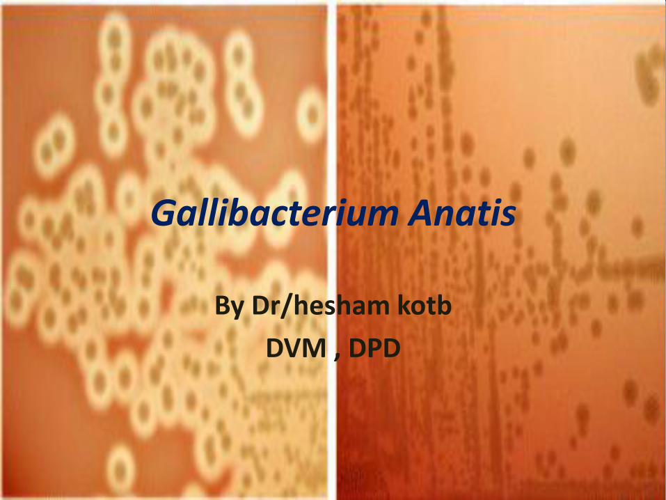
Gallibacterium Anatis
By Dr/hesham kotb
DVM , DPD
Gallibacterium anatis
• is a common organism of the upper respiratory and lower genital tract
of poultry. The bacterium has been reported worldwide from a broad
host range among farmed
and wild birds. The bacterium has no public health significance.
• It is potentially pathogenic for poultry and is mainly associated with
lesions in the reproductive tract, including the ovary.
• Disease associated this microorganism is related to lowered egg
production and occasionally an increase in mortality.
• may cause sudden mortality and septicaemia or purulent (pus
forming) salpingitis, oophoritis and even peritonitis.
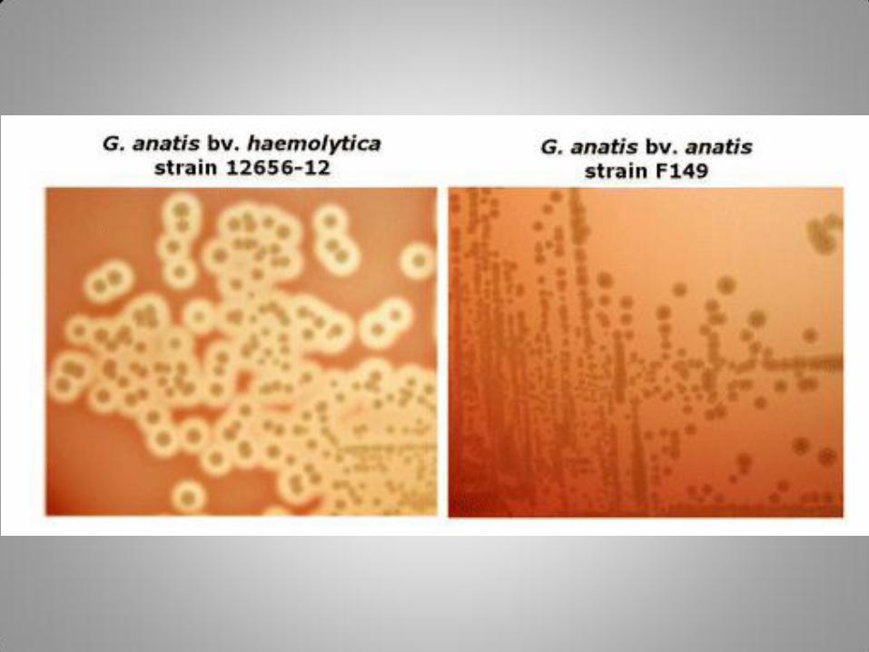
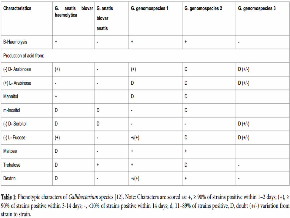
• G. anatis has too biovars which are: (a) G. anatis haemolytica: It produces haemolysis of 1-2 mm diameter on blood agar. (b) G. anatis biovar anatis: This biovar produces mild disease (respiratory; in ducks and geese (non haemolytic).
• G. anatis is a Gram negative, nonmotile, rod shaped or pleomorphic organism. Sometimes they are in pairs since it belongs to family Pasteurellaceae (Bojesen A.M. et al, 2007 Syst. Appl. Microbiol. 30: 119-127). The bacteria produce semitransparent colonies of 1.2 mm diameter in 24 hours at 37°C.
• Hosts it affects domestic poultry and also wild birds. Among domestic species ducks, geese, turkeys and pheasants are important. The disease has been reported from Europe, America, Australia, Asia and Africa.
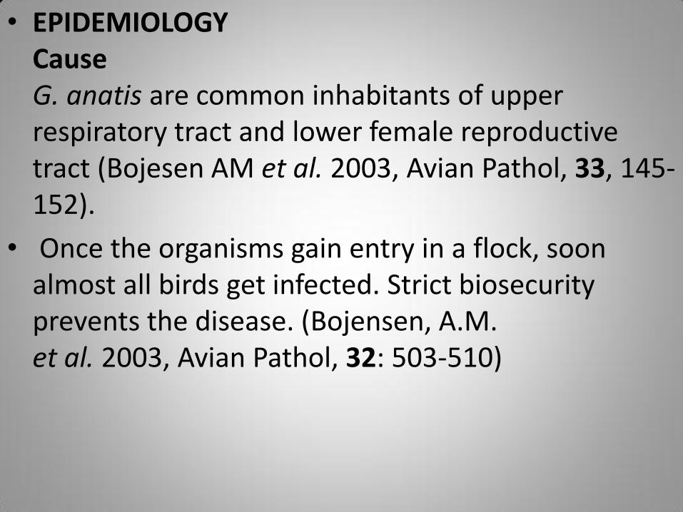
• EPIDEMIOLOGY Cause G. anatis are common inhabitants of upper respiratory tract and lower female reproductive tract (Bojesen AM et al. 2003, Avian Pathol, 33, 145-152).
• Once the organisms gain entry in a flock, soon almost all birds get infected. Strict biosecurity prevents the disease. (Bojensen, A.M. et al. 2003, Avian Pathol, 32: 503-510)
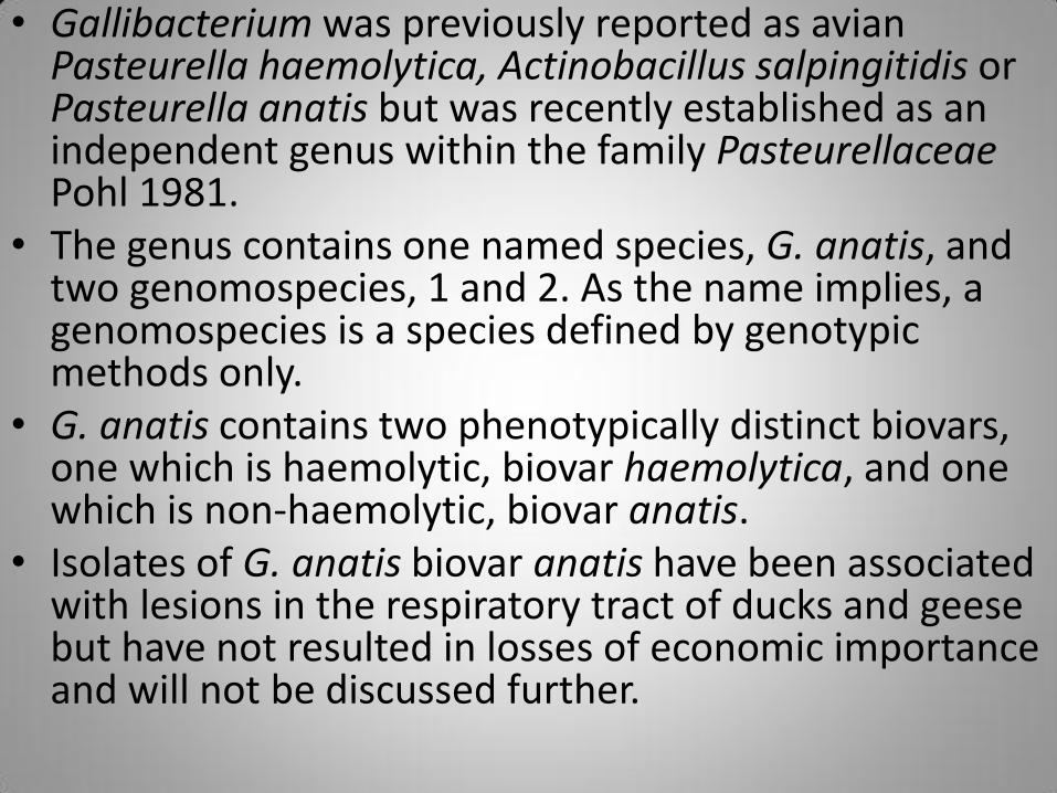
• Gallibacterium was previously reported as avian Pasteurella haemolytica, Actinobacillus salpingitidis or Pasteurella anatis but was recently established as an independent genus within the family Pasteurellaceae Pohl 1981.
• The genus contains one named species, G. anatis, and two genomospecies, 1 and 2. As the name implies, a genomospecies is a species defined by genotypic methods only.
• G. anatis contains two phenotypically distinct biovars, one which is haemolytic, biovar haemolytica, and one which is non-haemolytic, biovar anatis.
• Isolates of G. anatis biovar anatis have been associated with lesions in the respiratory tract of ducks and geese but have not resulted in losses of economic importance and will not be discussed further.
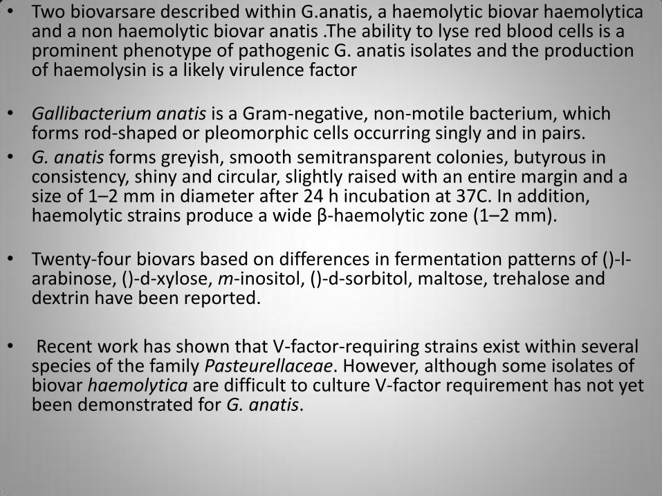
• Two biovarsare described within G.anatis, a haemolytic biovar haemolytica and a non haemolytic biovar anatis .The ability to lyse red blood cells is a prominent phenotype of pathogenic G. anatis isolates and the production of haemolysin is a likely virulence factor
• Gallibacterium anatis is a Gram-negative, non-motile bacterium, which forms rod-shaped or pleomorphic cells occurring singly and in pairs.
• G. anatis forms greyish, smooth semitransparent colonies, butyrous in consistency, shiny and circular, slightly raised with an entire margin and a size of 1–2 mm in diameter after 24 h incubation at 37C. In addition, haemolytic strains produce a wide β-haemolytic zone (1–2 mm).
• Twenty-four biovars based on differences in fermentation patterns of ()-l-arabinose, ()-d-xylose, m-inositol, ()-d-sorbitol, maltose, trehalose and dextrin have been reported.
• Recent work has shown that V-factor-requiring strains exist within several species of the family Pasteurellaceae. However, although some isolates of biovar haemolytica are difficult to culture V-factor requirement has not yet been demonstrated for G. anatis.
• in case study Gallibacterium anatis biovar heamolytica with a percentage 24 % and Gallibacterium anatis biovar anatis 76 %
• Spread G. anatis has been reported from many countries in Europe, Africa and Asia, and in Australia and American states, underlining its widespread occurrence. • Although chickens have been suggested as the main host for haemolytic
isolates Gallibacterium organisms do seem to have a wide.
• The occurrence of haemolytic strains of G. anatis in commercial flocks has been investigated recently and it has been shown that these organisms are very common inhabitants of the upper respiratory and lower genital tract of healthy chickens.
• Furthermore the occurrence of G. anatis appeared to be highly influenced by farm biosecurity level such that only flocks kept under very high levels of biosecurity were likely to be free of G. anatis.
• In addition, once the microorganisms were present in a flock, nearly all individuals in the flock were infected.
• Bird-to-bird transmission is considered to be the main mode of infection. Evidence for vertical transmission has not been shown so far.
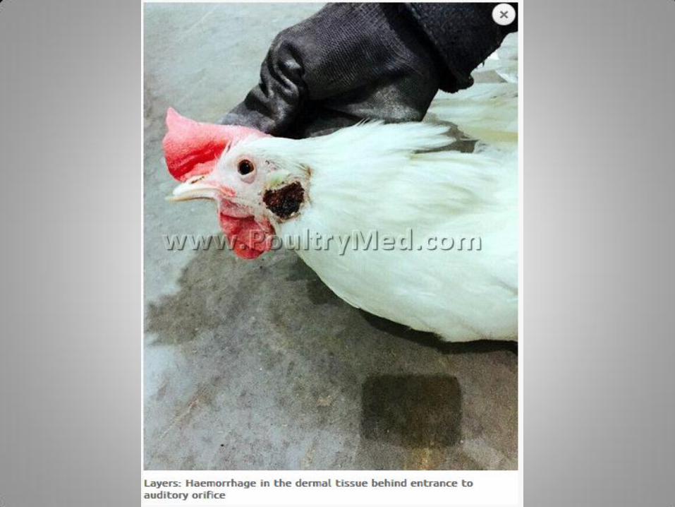
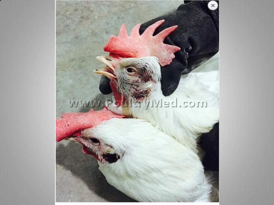
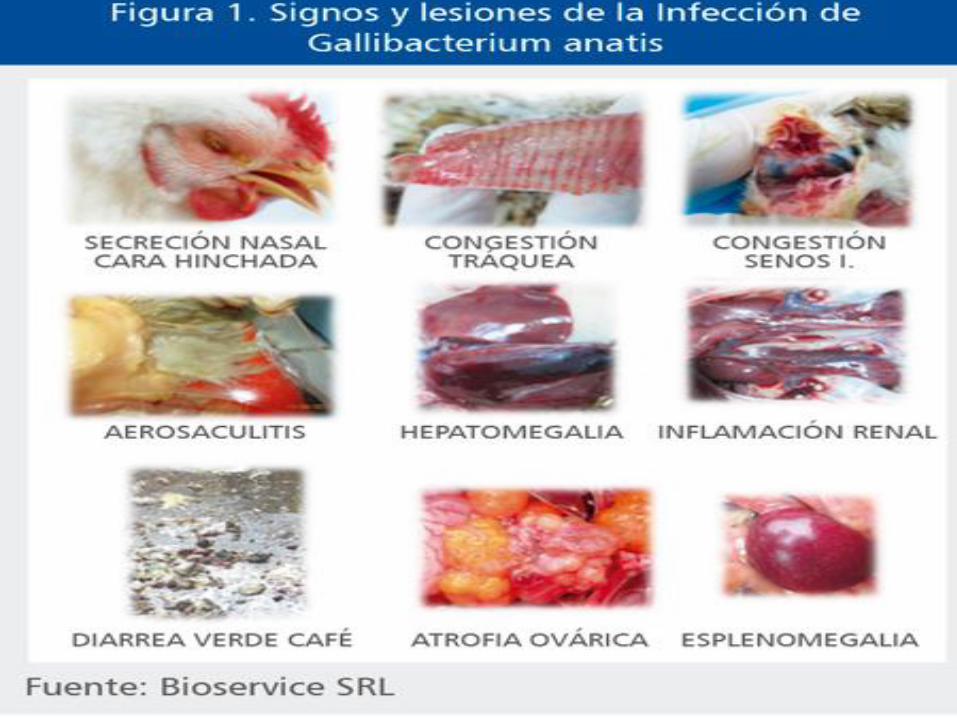
• Clinical signs and lesions Normally the signs and symptom of diseases caused by G. anatis infection in chicken are not pathgnomonic leading to creation of confusion between the different similar symptomatic disease like Newcastle, fowl cholera and bird flu.
• There is accumulated evidence of haemolytic isolates of G. anatis being able to act as a primary disease-causing agent. However, other factors possibly contribute to the clinical and pathological manifestations.
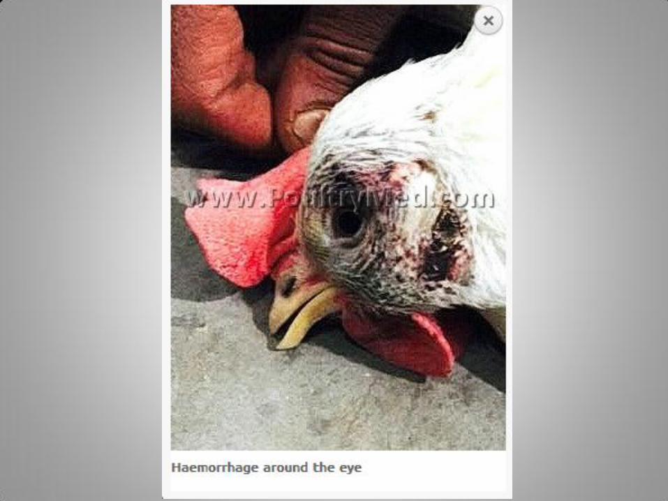
• The birds are unthrifty, dull. Sometimes the disease takes acute septicaemic condition with sudden mortality in healthy layer’s..
• The clinical signs are unspecific but will usually include depression, diarrhea and pasting around the vent and reduced egg production around peak of lay.
• the lesions typically involve the reproductive tract and the ovary, exhibiting purulent salpingitis and oophoritis.
• Chronic cases tend to include local or generalized purulent peritonitis, often with simultaneous growth of Escherichia coli.
• Sudden mortality associated with acute septicaemia has occasionally been recorded from table-egg-producing flocks in good body condition from which G. anatis has been isolated in pure culture from various organs.
• Clinical signs and pathological lesions have been reproduced experimentally in specific pathogen-free birds as well as in conventional layers.
• The outcome of such infections varies and seems to depend strongly on the strain, the route of inoculation and secondary factors, including immune suppression. In addition, at onset, peak and late in the production period chickens appear more susceptible to infection.
• G. anatis seems to have different virulence factors influencing its pathogenicity.
• Recently, whole genome sequencing has revealed possession of a capsule locus, and the haemolytic phenotype appears to be encoded by a RTX-like toxin also known in other members of the family Pasteurellaceae.
• The direct role of these putative virulence factors in the pathogenesis remains to be demonstrated.
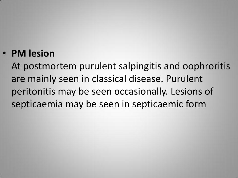
• PM lesion At postmortem purulent salpingitis and oophroritis are mainly seen in classical disease. Purulent peritonitis may be seen occasionally. Lesions of septicaemia may be seen in septicaemic form
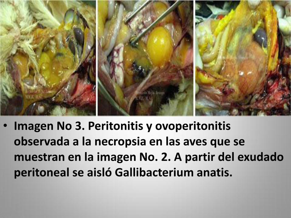
• Imagen No 3. Peritonitis y ovoperitonitis observada a la necropsia en las aves que se muestran en la imagen No. 2. A partir del exudado peritoneal se aisló Gallibacterium anatis.
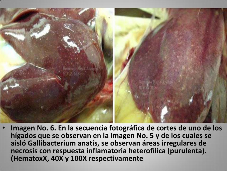
• Imagen No. 6. En la secuencia fotográfica de cortes de uno de los hígados que se observan en la imagen No. 5 y de los cuales se aisló Gallibacterium anatis, se observan áreas irregulares de necrosis con respuesta inflamatoria heterofílica (purulenta). (HematoxX, 40X y 100X respectivamente
• DIAGNOSIS Signs and lesions cannot be used to distinguish infection by G. anatis from other bacterial infections affecting the salpinx, ovary and abdominal cavity.
• Furthermore, the fact that most birds carry hemolytic G. antes as a part of their resident flora in the upper respiratory and lower genital tract makes careful identification and characterization crucial.
• The diagnosis should be confirmed by isolation and identification of the causal agent.
• This should be attempted by culture from the affected organs and, in the case of systemic disease,the liver and spleen.
• G. anatis grows readily on enriched media and in the case of the β-haemolytic biovar with acharacteristic haemolysis zone following 24 h of incubation.
• Identification based on morphology and biochemical tests will give a good indication but this should be combined with one or more genotypic culturing the organisms from ovary, salpinx peritoneum blood or spleen is important.
• Culture is possible on enriched blood agar, producing colonies as described above.
• Hints for confirmed diagnosis (a) haemolysis around 1-2 mm, shiny, translucent colonies grown at 37°C. (b) Pleomorphic, rods, or bipolar, Gram negative morphology, in the exudates. (c) Agglutination test, ELISA test, polymerase chain reaction (PCR) with a specific antiserum or probe.
• In Denmark latex agglutination test and enzyme linked immunosorbent assey (ELISA) are being developed (Bojensen, et al., 2008, Poultry Diseases 6th Edn., p. 162).
• Recent work has demonstrated that a specific polymerase chain reaction (PCR) test, based on the internal transcribed spacer (ITS) separating the 16S and 23S rRNA genes, can be used as a confirmatory test following cultural isolation of G. anatis or directly on material from the affected bird.
• Another culture independent technique, fluorescence in situ hybridization (FISH) using a Gallibacterium-specific probe, similarly allows specific detection of the bacteria.
• Tools to determine genetic diversity have also been developed and include amplified fragment length polymorphism and pulsed field gel electrophoresis.
• Currently, two serological tests based on latex agglutination and enzyme-linked immunosorbent assay (ELISA) are being developed and used for the examination of sera for specific antibodies against G. anatis. T e number and prevalence of serotypes within G. anatis remains to be investigated in detail, in addition to cross-reactivity and cross-protection of serotypes
• Control The success of treating Gallibacterium infections with antibiotics depends highly on the practices used in the individual flocks.
• The level of acquired resistance in G. anatis seems limited under conditions of limited exposure to antibiotics, enabling the use of narrow-spectrum antibiotics, however, it is also evident that G. anatis readily acquires resistance.
• This has been demonstrated in a number of strains recovered from outbreaks in Mexican poultry flocks where the majority of the isolates were multi resistant to a broad range of antibiotics including penicillins, trimethoprim–sulfamethoxazole and fluoroquinolones.
• The antimicrobial sensitivity pattern should therefore always be established in relation to treatment.
• Currently a commercial vaccine is available based on three of the more prevalent biovars, however, protection under field conditions remains to be investigated in further detail.
• 1. Immunosuppression may flare up the disease because G. anatist is an opportunistic pathogen.
• 2. Drug resistance is a problem with this organism. Narrow spectrum antibiotics generally are useful such as penicillin, sulfumathoxazole, trimethoprim etc.
• 3. Lowering of egg production must be watched to suspect and investigate the disease.
• 4. High level of farm biosecurity is essential.
• 5. Commercial vaccine is not available but an auto vaccine prepared from the affected flock may be attempted by an expert poultry
• Case study
• A recent investigation with clinically healthy chickens from different Danish layer production systems showed that hemolytic Gallibacterium was highly prevalent in birds from production systems with moderate or low levels of biosecurity
• Mirle et al. (24) examined496 hens with reproductive tract lesions and isolated Gallibacterium in pure culture from 23% of the diseased organs, whereas the second most prevalent agent, Escherichia coli, was isolated from 21% of the cases.
• In addition, others have isolated Gallibacterium in pure cultures of samples from animals with various pathological lesions or conditions, including salpingitis, oophoritis, peritonitis, pericarditis, hepatitis, enteritis, upper respiratory tract lesions, and septicemia (1, 6, 18–20, 27, 28, 31–34). Evidently, the bacteria are capable of causing serious, systemic infections affecting multiple organ systems, but the mechanisms of pathogenesis remain obscure
• The Gallibacterium-induced lesions that have been reported are not pathognomonic, and detection and identification are dependent on classical isolation and identification procedures, including phenotypic characterization (12).
• As is the case for most other genera of the family Pasteurellaceae Pohl 1981, the genus Gallibacterium is phenotypically a heterogeneous group (12, 30). Phenotypic characterization therefore involves laborious and time-consuming methods, which may also give ambiguous results due to variable outcomes. This in turn leads to difficulty in interpretation of the genus and species designations from some earlier studies, in which only a relatively few phenotypic characters have been investigated (7).
• Additionally, Gallibacterium may be underestimated as a cause of salpingitis and/or peritonitis due to the forementioned limitations of the diagnostic methods available
• a substantial antigenic diversity (Vazquez et al., 2006) make it difficult to prevent the negative effects of Gallibacterium anatis using traditional anti-microbial agents and vaccine
• Bacteriology • A-Bacterial investigations of Gallibacterium anatis:
1 - Trypticase soy agar enhanced by addition of 0.05% yeast extract and 5% newborn calf serum. (Sandhu and Richard, 1997). 2- Brain heart Infusion medium (Li et al., 2011). 3- Blood agar: within 24 hour, which is characterized as follows: circular, raised colonies with entire margin, shiny and semi-transparent with a β heamolytic zone. Such colonies were regarded as suspicious of Gallibacterium. Suspected colonies were subcultured on blood agar to obtain pure cultures. (Neubauer et al., 2009). 4- Brain heart Infusion broth: A- Propagation and maintenance of bacterial cultures for improved growth (Zepeda et al., 2009). B- Preservation (Bojesen et al., 2003). Seven hundred microlitres of Brain heart Infusion broth were mixed with 300μl sterile glycerol 50% and stored at – 80 ◦C until further use. 5-India ink 6- API 20 for differentiation of Gallibacterium anatis According to (Florence et al., 2008).
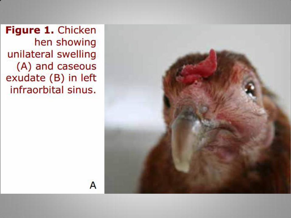
• Infra orbital sinus samples were taken and cultivated on 10% sheep blood agar plates with Staphylococcus epidermidis as colony feeder. Plates were incubated at 37° C into a candle jar.
• Gallibacterium forms grayish, semitransparent colonies, butyrous in consistence, smooth,shiny and circular with an entire margin and a size of 1-2 mm in diameter after 24 h at 37 oC. In addition, haemolytic strains produce a wide β-haemolytic zone (1-2 mm),
• for A nother study based on inoculation of Gallibacterium into the crop of day-old and 5-6 weeks old chickens did only result in weak or no signs of disease when performed in normal chickens, whereas chickens infected by the same procedure, but also “cold stressed” during the infection, experienced a mortality rate at 36% (Matthes and Löliger, 1976).
• Evidently, there seems to be significant differences as to virulence of different strains of Gallibacterium, but also characteristics related to the host seem to have a major impact on the course of infection.
• These characteristics suggest Gallibacterium as an opportunistic pathogen, which normally needs the influence of other adverse effects in order to promote disease in its host, a feature which is commonly encountered with members of the family Pasteurellaceae (Mannheim, 1984). However, further studies allowing insight into specific
• bacteria-host interactions by the use of a well characterized bacterial strain and a defined host are warranted.
• Most Gallibacterium isolates recovered from diseased chickens have had a haemolytic phenotype. Greenham and Hill (1962) demonstrated the haemolysis by Gallibacterium on nutrient agar containing bovine, horse, rabbit or chicken blood, indicating that the haemolysin(s) must work via a mechanism common to a broad range of target cells.
• The identification of haemolytic Gallibacterium in the present study relied on bacterial cultivation on bovine blood agar plates, which at the time was the only detection method available
• However, haemolytic Gallibacterium spp. grew readily on bovine blood agar plates and were easily identified by their characteristic wide β- haemolytic zone within 24 h of incubation
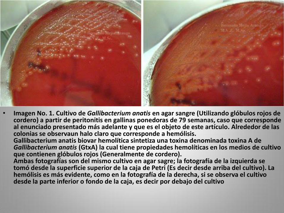
• Imagen No. 1. Cultivo de Gallibacterium anatis en agar sangre (Utilizando glóbulos rojos de cordero) a partir de peritonitis en gallinas ponedoras de 79 semanas, caso que corresponde al enunciado presentado más adelante y que es el objeto de este artículo. Alrededor de las colonias se observaun halo claro que corresponde a hemólisis. Gallibacterium anatis biovar hemolítica sintetiza una toxina denominada toxina A de Gallibacterium anatis (GtxA) la cual tiene propiedades hemolíticas en los medios de cultivo que contienen glóbulos rojos (Generalmente de cordero). Ambas fotografías son del mismo cultivo en agar sagre; la fotografía de la izquierda se tomó desde la superficie superior de la caja de Petri (Es decir desde arriba del cultivo). La hemólisis es más evidente, como en la fotografía de la derecha, si se observa el cultivo desde la parte inferior o fondo de la caja, es decir por debajo del cultivo
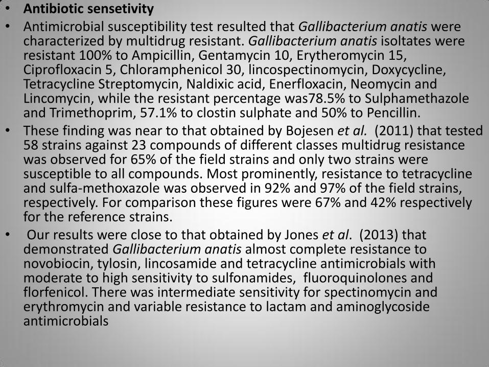
• Antibiotic sensetivity • Antimicrobial susceptibility test resulted that Gallibacterium anatis were
characterized by multidrug resistant. Gallibacterium anatis isoltates were resistant 100% to Ampicillin, Gentamycin 10, Erytheromycin 15, Ciprofloxacin 5, Chloramphenicol 30, lincospectinomycin, Doxycycline, Tetracycline Streptomycin, Naldixic acid, Enerfloxacin, Neomycin and Lincomycin, while the resistant percentage was78.5% to Sulphamethazole and Trimethoprim, 57.1% to clostin sulphate and 50% to Pencillin.
• These finding was near to that obtained by Bojesen et al. (2011) that tested 58 strains against 23 compounds of different classes multidrug resistance was observed for 65% of the field strains and only two strains were susceptible to all compounds. Most prominently, resistance to tetracycline and sulfa-methoxazole was observed in 92% and 97% of the field strains, respectively. For comparison these figures were 67% and 42% respectively for the reference strains.
• Our results were close to that obtained by Jones et al. (2013) that demonstrated Gallibacterium anatis almost complete resistance to novobiocin, tylosin, lincosamide and tetracycline antimicrobials with moderate to high sensitivity to sulfonamides, fluoroquinolones and florfenicol. There was intermediate sensitivity for spectinomycin and erythromycin and variable resistance to lactam and aminoglycoside antimicrobials
• Layer bacterial peritonitis • Peritonitis is an important disease of laying hens in commercial
table egg operations. Despite its importance, the etiology and pathogenesis of this disease have not been completely clarified. Two bacterial species are routinely isolated from cases of avian peritonitis: avian pathogenic Escherichia coli (APEC) and Gallibacterium anatis.
• Although APEC’s contributions to peritonitis have been well documented (7), the exact role played by G. anatis in the pathogenesis of this disease has only recently been recognized. Danish researchers have reported that G. anatis is frequently associated with peritonitis lesions (1-6), with or without concurrent APEC isolation.
• While current wisdom suggests that APEC and G. anatis infections are opportunistic in nature, co-infection studies involving these two species inoculated into the oviduct of laying hens were highly lethal towards chickens. Such research suggests that the pathogenesis of peritonitis might be quite complex, and could involve interplay between multiple microbial species (i.e., polymicrobial infection). A better understanding of APEC and G. anatis is needed in order to elucidate their pathogenesis.
• Although much effort has been devoted to understanding APEC’s virulence mechanisms, little effort has been directed towards the study of the virulence mechanisms of G. anatis. Since epidemiological and in vivo studies suggest that G. anatis plays an important role in avian peritonitis, lack of understanding of G. anatis virulence renders control of peritonitis problematic.
• In this study, we performed draft genome sequencing on two G. anatis strains, one classified as highly virulent and another classified as a virulent. We compared these two genomes to identify putative G. anatis virulence genes. We then determined the prevalence of some of these genes among G. anatis isolates from healthy hens and from peritonitis lesions, and used these data in an effort to examine possible correlations between clonality and virulence genotype.

• Disease pathology • The gross pathological lesions of salpingitis/peritonitis,
a typical manifestation of Gallibacterium infection (Bisgaard, 1977;Gerlach, 1977; Mirle et al., 1991), cannot easily be distinguished from a range of other Gram negative pathogens, especially E. coli, which is regarded as the most prevalent cause of this type of disease (Bisgaard & Dam, 1981). However, in cases where diagnosis is established solely by characteristics of the pathological lesions, Gallibacterium is not likely to be held responsible, which is why the importance of these bacteria may be underestimated.
• Detailedpost mortem investigations including natural cases of peritonitis/salpingitis carried out as soon as sick or dead birds are observed to prevent possible overgrowth by other bacterialspecies, may reveal a more prominent role for Gallibacterium as a pathogen
• Pathogenesis Repeated isolation of G. anatis from the trachea and cloaca of healthy birds indicates its commensal status in the upper respiratory tract and lower genital tract of healthy chickens [2,14,49,50,55,90].
• However, isolation of G. anatis in association with a wide range of different pathological lesions, including septicaemia, pericarditis, hepatitis, oophoritis, follicle degeneration, enteritis, upper respiratory tract lesions, salpingitis and peritonitis revealed its importance as an opportunistic pathogen [2,17,36,39-43,45,47,55,88,89,91,92].
• Recent investigations confrmed that G. anatis colonizes the upper respiratory tract without causing clinical signs, whereas it may cause severe lesions in the reproductive tract [54,56].
• Studies established G. anatis as the most common single bacterial infection in chickens causing reproductive tract disorders [36]. Simultaneous infection with other microorganisms [39,47,48], hormonal influences [42,43], age [2,45], seasonal changes [36], stress [15], cold stress [47], and compromised immunological status [46] are a few predisposing factors nurturing the infection of G. anatis.
• In experimental infections semen quality has been found to be reduced significantly due to decrease in sperm density, total motility with progressive motility, and membrane integrity [54].
• Disease associated with G. anatis infection Due to vast range of pathological manifestations of G. anatis infection it is diffcult to decide the exact disease condition caused by G. anatis.
• Incidences of infection to chicken increase during the peak and late phases of production period [41].
• In diseased birds’ mortality might take place mainly due to salpingitis, oophoritis and peritonitis.
• Respiratory tract infections might be responsible for major economic losses due to the rise in treatment cost and losses due to higher condemnation rates and mortality.
• Gallibacterium may be causing primary or secondary infections leading to fatal bacteremia, septicaemia and acute septicaemia [41].
• the severity of clinical signs, duration of the disease and mortality rate are variable and influenced by environmental factors, such as poor hygiene, inadequate management ventilation, ammonia levels in poultry premises and concurrent diseases.
• Study on pathogen-specifc genes of Gallibacterium populations [79] suggested the ability of the pathogen to cause lesions in reproductive organs such as folliculitis, ruptured and haemorrhagic follicles as well as a drop in egg production in adult hens [22,38,55,90]. Haemolytic G. anatis was associated with infection in birds kept in alternative husbandry systems and suffering from reproductive disorders [38].